Arq. Bras. Oftalmol. 200265
| DOI: 10.1590/S0004-27492002000600002
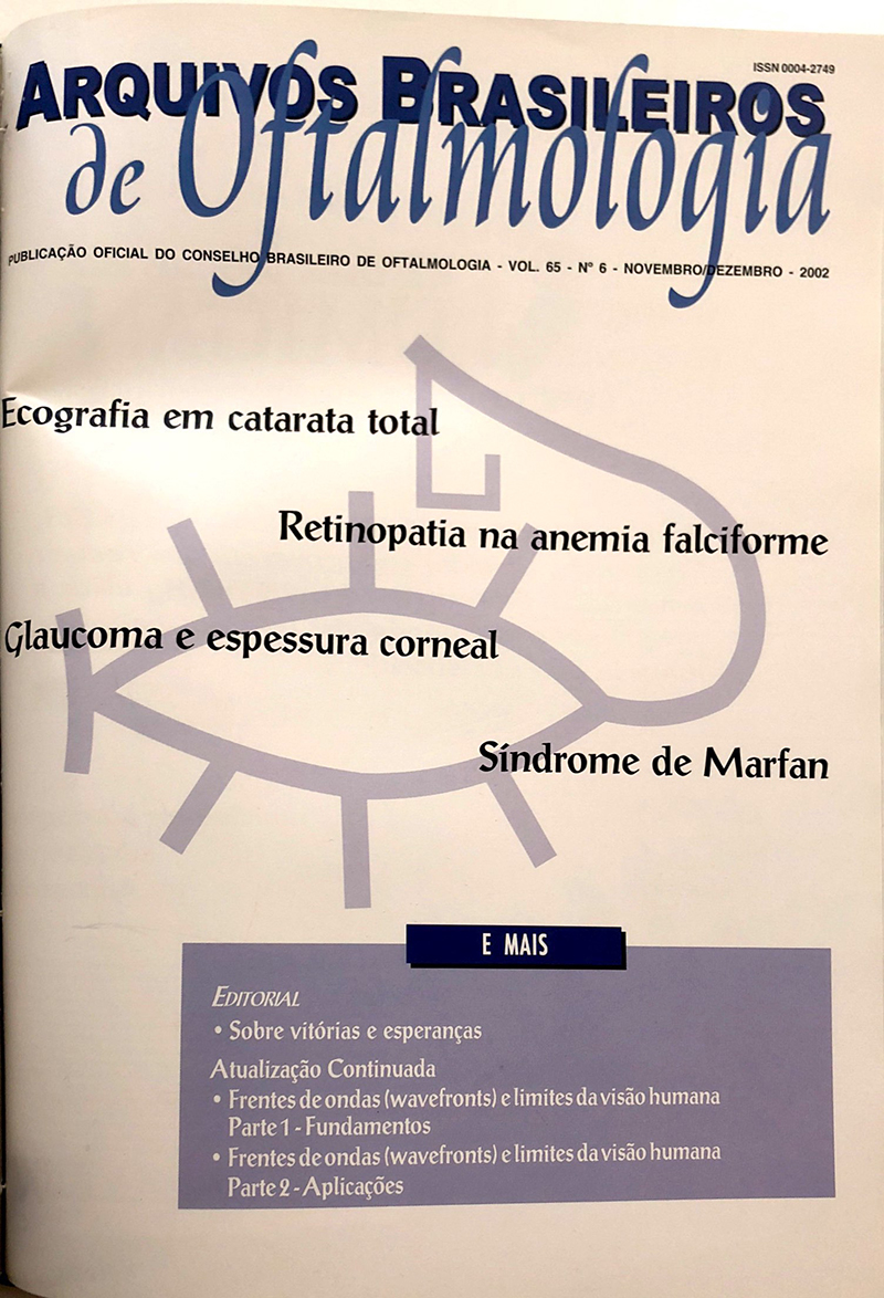
Purpose: To evaluate the prevalence and nature of intra-ocular abnormalities detected by conventional B-scan ultrasound in patients with dense cataracts and compare them to their postoperative findings.
Methods: Retrospective chart review of 724 B-scan ultrasound studies performed between January 1999 and July 2001. Two hundred and eighty-nine of these exams were referred due to a dense cataract that obscured any view of the fundus. The ultrasound findings were documented by prints and revised for the study. Follow-up was obtained in 131 patients to evaluate sensibility and specificity of B-scan ultrasound as a diagnostic method. Statistical analysis was performed by Student's "t" test using SPSS (Statistical Package for the Social Science) 8.0 for Windows.
Results: Two hundred and eighty-nine patients with dense cataracts were studied, all of them evaluated by B-scan ultrasound; 200 had abnormal examination. In patients without a history of ocular trauma (n=268), 82 eyes (30.6%) presented a normal posterior segment on ultrasound and 26 eyes (9.7%), retinal detachment. In patients with a prior history of ocular trauma (n=21), 8 eyes (38.1%) presented retinal detachment and 7 eyes (33.3%) were normal on ultrasound. The agreement between ultrasound findings and postoperative findings was 95.4% in the 131 patients followed after surgery. B-scan ultrasound presented sensitivity of 91.3% and specificity of 100%.
Conclusion: Preoperative evaluation of eyes with dense cataracts using B-scan ultrasound has shown to be efficient in diagnosing posterior segment abnormalities in this study. The sensibility and specificity of this exam found in the studied group confirm the importance of B-scan ultrasound in the preoperative evaluation of patients with dense cataracts.
Keywords: Cataract; Ultrasonography; Eye injuries; Preoperative care; Sensitivity and specificity; Follow-up
Arq. Bras. Oftalmol. 200265
| DOI: 10.1590/S0004-27492002000600003
Purpose: To point out the main fundoscopic findings in children with sickle cell trait hemoglobinopathies.
Methods: We studied 26 patients with sickle cell hemoglobinopathies in the ophthalmological ward at the Hospital Universitário Onofre Lopes, Natal, RN. The patients were submitted to a research questionnaire and the results were statistically evaluated by the chi² test.
Results: The average age was 10,6 years, with visual acuity equal or better than 20/25 in the majority, except for three eyes which presented other associated pathologies. The most frequent type was SS in 57.7% (15/26) of cases, followed by SC e SA in 15.4% (4/26) each, and by S-Thal in 11.5% (3/26). The frequency of sickle cell retinopathy was higher after 10 years of age, being more frequent, in relative values, in type S-Thal (100%) and, in absolute values, in type SS (9 cases). The two most common findings were venous tortuosity (12/26) and "black sunburst" (7/26).
Conclusion: We observed that the incidence of retinopathy in sickle cell increased with age over 10 years and no evidence of proliferative disease was found. Therefore, we emphasize the need for early ophthalmological examination in carriers of sickle cell disease as a form of preventing future complications.
Keywords: Sickle cell disease; Hemoglobinopathies; Fundus oculi; Hemoglobin C disease; Hemoglobin SC disease; Retinal diseases; Children
Arq. Bras. Oftalmol. 200265
| DOI: 10.1590/S0004-27492002000600004
Purpose: To determine if there is a significant difference in central corneal thickness (CCT) among patients with normal tension glaucoma (NTG), primary open angle glaucoma (POAG) and normal patients.
Methods: One hundred and thirteen patients were evaluated; 40 had NTG, 51 were normal and 22 had POAG. IOP (Goldmann) and CCT (Topcon SP-2000P) were measured in all patients.
Results: The CCT mean in NTG patients was 500.95 µ (SD = 28.65µ); in normal patients the CCT mean was 521.11µ (SD = 43.30µ); and in the POAG group the CCT mean was 522.23µ (SD = 27.87µ). By statistical analysis (ANOVA, 95% confidence interval) the CCT in NTG was significantly lower than in normal patients and in the POAG group (p= 0.01 and p=0.006), but no differences was found between normal patients and the POAG group (p= 0.91). Conclusions: In this study we concluded that in a Brazilian population sample the NTG patients tend to present significantly lower ECC measurements when compared with normal and POAG patients.
Keywords: Intraocular pressure; Glaucoma; Cornea
Arq. Bras. Oftalmol. 200265
| DOI: 10.1590/S0004-27492002000600005
Purpose: To identify the ocular abnormalities in Marfan´s syndrome patients.
Methods: Prospective study of 46 Marfan patients with complete ophthalmologic evaluation. Seventeen patients also underwent a clinical genetic and molecular analysis.
Results: Among the 46 patients included in this study, the following ocular abnormalities were found more frequently: ectopia lentis (67.3%), hypoplastic irides (67.3%), retinal detachment (7.6%), flattened cornea (2.2%), megalocornea (2.2%) and myopia (34.8%). Five patients (10.9%) presented normal ocular evaluation in both eyes. A pathogenic mutation different from those published in other studies was found in one patient. Conclusions: Ocular abnormalities in Marfan´s syndrome are frequent. The better understanding of the FBN-1 gene and its expression in the eye are very helpful tools for the diagnosis and treatment of the abnormalitites.
Keywords: Marfan's syndrome; Eye abnormalities; Eye diseases
Arq. Bras. Oftalmol. 200265
| DOI: 10.1590/S0004-27492002000600006
Purpose: To report the data obtained through a project based on visual screening of newborns from three reference public maternities of Pernambuco state, Brazil.
Methods: This investigation was a prospective study, according to a previously elaborated protocol and organogram. A multidisciplinary team of 37 professionals was involved. This team was trained and conferences were given to ophthalmologists, directors, pediatricians, neonatologists, obstetricians and nurses of each service. Ocular exams were performed in the selected maternity hospitals. The highly complex cases were referred to the Altino Ventura Foundation. Search for conjunctival flora in neonates and vaginal flora in their mothers was performed in 25 cases.
Results: Twenty ophthalmologists were trained to perform ocular examination in newborns. Visual screening was conducted in 3,280 of them (1,601 females and 1,679 males). Ocular abnormalities or risk factors were found in 701 cases (21.4%). Retinal hemorrhage was the most frequent ocular abnormality seen in 255 cases (7.8%). The more prevalent risk factor for eye disease was retinopathy of prematurity found in 325 cases (9.9%). These patients were referred to specialized services. Of these 65 (20.0%) were examined and 15 (23.1%) had some stage of retinopathy of prematurity. The examination of the conjunctival flora of the newborns (50 eyes) showed at least one microorganism in 13 eyes (26.0%). Babies of 18 mothers with positive vaginal culture for Staphylococcus epidermis 10 (40.0%) had the same bacterium in one or both eyes. Conclusions: The authors emphasize that this was a pioneer project and that it provided a greater interaction among ophthalmologists and other health professionals, ophthalmic training in performing eye examination in newborns, allowing a better knowledge of ocular morbidity in the region, providing adequate treatment and follow up of the cases at an earlier age.
Keywords: Neonatal screening; Conjunctiva; Vagina; Staphylococcus epidermidis; Retinal hemorrhage; Eye diseases; Retinopathy of prematurity; Practice guidelines; Risk factors; Pregnancy; Newborn infant; Prospective studies
Arq. Bras. Oftalmol. 200265
| DOI: 10.1590/S0004-27492002000600007
Purpose: To analyze the knowledge and attitude of the general population in order to identify the main deficiencies and to suggest measures to increase the number of cornea donations by the general population.
Methods: Study through questionnaire polls answered by 321 people at the São Paulo Hospital.
Results: Among the general population, 79.4% said to be cornea donors. The reason for the other 20.6% not to be donors was fear of trade of organ and not to receive proper treatment by the medical staff for being a potential donor. There was a statistically significant difference of knowledge about transplantation between those who said to be donors and those who did not.
Conclusion: The authors concluded that better information on corneal transplantation for the general population would be of great help in increasing the number of cornea donations.
Keywords: Corneal transplantation; Eye banks; Tissue donors; Organ procurement; Physician's role; Informed consent; Health education; Knowledge, attitudes, practice; Questionnaires
Arq. Bras. Oftalmol. 200265
| DOI: 10.1590/S0004-27492002000600008
Purpose: To compare the safety and efficacy of extracapsular cataract extraction (ECCE) combined with trabeculectomy and combined phacoemulsification/trabeculectomy.
Methods: The records of 46 patients (53 eyes) who underwent combined glaucoma and cataract surgery at the Santa Casa de São Paulo between January 1996 and November 1999 were reviewed.
Results: After a mean follow-up of 18 months, visual acuity improved and intraocular pressure decreased in both groups after surgery (P<0.05). In the phacofiltration group 55.5% of eyes achieved intraocular pressure < 22 mmHg without medication as compared to 46.1% in the extracapsular cataract extraction/trabeculectomy group (P=0.3).
Conclusion: Both techniques proved to be safe and efficacious in the treatment of glaucoma and cataract. However, the phacofiltration surgery seems to promote lower intraocular pressure without additional medication in a larger number of eyes.
Keywords: Glaucoma; Cataract; Trabeculectomy; Visual acuity; Mitomycin; Combined modality therapy; Follow-up; Comparative study
Arq. Bras. Oftalmol. 200265
| DOI: 10.1590/S0004-27492002000600009
Purpose: To evaluate the association between high doses of 4-aminoquinolone derivatives and loss of visual acuity, visual field defects, and fundus changes.
Methods: Prospective study of 86 patients in use of chloroquine or hydroxychloroquine (Plaquinol
â) whose initial visit consisted of visual acuity testing, anterior segment slit lamp examination, indirect ophthalmoscopy, computerized perimetry (red stimulus, program 10.2-central), calculation of the daily and cumulative dose used. Cumulative toxic doses were considered
³100 g for chloroquine and
³300 g for hydroxychloroquine.
Results: All examined eighty-six patients were women, the average age was 45.08 years in use of chloroquine (n=13) or hydroxychloroquine (n=73) (Plaquinol
â). There was a marked correlation between the use of medication and decrease in vision (p=0.05) with a relative risk of 0.3 (CI 95% 0.1-1.1). Slit lamp examination of the anterior segment was unremarkable in all patients. Fundus examination of chloroquine patients taking nontoxic doses failed to reveal any abnormalities and 41.7% (n=5) of the patients using toxic doses had fundus abnormalities. In the hydroxychloroquine group, 21.8% (n=13) had fundus abnormalities while using nontoxic doses and only 17.6% (n=3) using toxic doses presented fundus abnormalities. Comparing the patients with and without visual field defect in the chloroquine group (p=0.423) and hydroxychloroquine group (p=0.999), there was no statistical difference between the visual field defect and the drug dosage.
Conclusion: Loss of vision was greater in chloroquine patients. This information is relevant to stress the importance of visual acuity testing to follow-up patients treated with 4-aminoquinolone derivatives. The presence of visual field defects was not statistically different between patients on high or low doses of 4-aminoquinolone derivates.
Keywords: Retina; Visual acuity; Visual fields; Fundus oculi; Macula lutea; Retinal diseases; Scotoma; Hydroxychloroquine; Hydroxychloroquine; Chloroquine; Chloroquine; Drug toxicity; Comparative study
Arq. Bras. Oftalmol. 200265
| DOI: 10.1590/S0004-27492002000600010
Purpose: To study visual acuity of the candidates for driving licences in the city of Pelotas and its relationship with sociodemographic characteristics of the candidates.
Methods: One thousand and ten candidates for driving license were examined in a Detran approved medical service. To check the social, demographic and visual characteristics of candidates, visual acuity, with or without correction, as well as its relation to color, gender, age and profession of the interviewed persons was evaluated, through analysis of frequencies and association.
Results: In this study 72.5% were males, 33.8% were between 18 and 45 years old, 52.0% were in the service sector and 25.4% used some instrument of visual correction, 0.5% of the interviewed from the ophthalmological viewpoint, would not be able to drive automotor vehicles and 4.7% should only would get a driving license in the A and B categories. A low visual acuity has been associated with a higher longevity.
Conclusion: Based on the results, a shorter time period for visual reavaluation of the C, D and E categories drivers is suggest.
Keywords: Visual acuity; Vision tests; Vision disorders; Automobile driver examination; Color perception; Socioeconomic factors; Color vision defects; Sex factors
Arq. Bras. Oftalmol. 200265
| DOI: 10.1590/S0004-27492002000600011
Purpose: To analyze the factors related to primary corneal graft failure after penetrating keratoplasty in the Corneal and External Diseases Section of the Federal University of São Paulo, Brazil.
Methods: A retrospective study was performed using the charts of patients submitted to penetrating keratoplasty from July 1999 to March 2001. Cases of primary failure (group I) and patients submitted to the surgical procedure at the same period (group II) were selected and compared. Information collected from donors were age, cause of death, endothelial cell count, time between death and enucleation (T1), time between enucleation and preservation of the graft (T2) and time of tissue preservation until surgery (T3).
Results: 164 patients were analyzed with 21 cases of primary failure (12.8%). The mean age of the donors of group I was 43.1 years (±22.0) and from group II was 47.9 years (±18.9). There was no statistical difference between the two groups considering the time intervals (T1, T2, T3). Causes of death included trauma, cancer and heart diseases.
Conclusion: Differences in all analyzed factors were not significant between both groups. The difficulties in determining the causes of primary corneal graft failure suggests a multifactorial process involved in this problem. Well-trained Eye Bank staff and notifications about postoperative complications are important to decrease the primary failure incidence.
Keywords: Corneal transplantation; Eye banks; Corneal diseases; Tissue preservation
Arq. Bras. Oftalmol. 200265
| DOI: 10.1590/S0004-27492002000600012
Purpose: To analyze the prevalence of reduced visual acuity in students of elementary state public and private schools of Londrina's urban area; to compare the prevalence of reduced visual acuity in children of state public and private schools; to analyze and compare the prevalence of children wearing eye-glasses, and to describe the causes of reduced vision in this population.
Methods: The visual acuity test was performed in 1688 school children of state public schools and in 611 school children of private schools. The children with visual acuity 0.7 or worse in one of the eyes were directed to a ophthalmic examination.
Results: The reduced visual acuity prevalence in school children was 17.1% in state public schools, and 19.8% in private schools; this difference does not have statistical significance. The prevalence of children wearing eyeglasses was 2.4% in state public schools and 3.6% in private schools, without statistical significance between these results. The causes of reduced visual acuity were refractive errors, amblyopia, strabismus and other causes.
Conclusion: The prevalence of reduced visual acuity in students of elementary state public and private schools was statistically the same, as well as the prevalence of children wearing eyeglasses. The causes of reduced visual acuity were refractive errors, amblyopia, strabismus, congenital cataract aphakia, zonular congenital cataract, persistent of the hyperplastic primary vitreous, optic nerve atrophy and macular disorder.
Keywords: Visual acuity; Cross-sectional studies; School health; Health education
Arq. Bras. Oftalmol. 200265
| DOI: 10.1590/S0004-27492002000600013
The authors report a rare case of a diabetic patient with rubeosis iridis who had cataract extraction with intraocular lens implantation, in whom neovascularization developed within the posterior lens capsule.
Keywords: Neovascularization, pathologic; Diabetes mellitus; Lenses, intraocular; Postoperative complications; Cataract extraction; Iris diseases
Arq. Bras. Oftalmol. 200265
| DOI: 10.1590/S0004-27492002000600014
We herein describe a case of a patient whose only symptom was nyctalopia. Peculiar ophthalmoscopic findings of Oguchi's disease were observed. Classic changes in the electroretinogram (ERG) and Mizuo-Nakamura's phenomenon were also reported. To the best of our knowledge, there isn't another occurrence in our published literature. A brief bibliographic review is added.
Keywords: Night blindness; Dark adaptation; Electroretinography; Case report; Adult; Female
Arq. Bras. Oftalmol. 200265
| DOI: 10.1590/S0004-27492002000600015
Purpose: To report a case of Erdheim-Chester disease with bilateral orbital involvement and to inform ophthalmologists about this uncommon condition.
Methods: Case report. A 41-year-old man presented with proptosis, lid xanthelasmas, disc edema and extraocular muscle restrictions. He was suspected of having a systemic lymphoma and had already been submitted to a retroperitoneal biopsy, however, the diagnosis was still uncertain.
Results: The patient was evaluated by computed tomography and magnetic resonance imaging of the orbit. Orbital pseudotumor was suspected and a biopsy of the orbital lesion initially failed to provide the correct diagnosis which was only made after detailed analysis of the clinical and imaging findings, and review of the histopathologic studies. The systemic diagnosis was made after the orbital diagnosis although the patient had been extensively investigated for more than one year. Conclusions: Erdheim-Chester disease is a rare idiopathic systemic condition characterized by a xanthogranulomatous process involving retroperitoneum, heart, lungs, bone and other tissues. The condition is often fatal due to renal or cardiovascular complications. Ocular findings are rare but may be very helpful for the diagnosis. Therefore, ophthalmologists should be aware of the clinical manifestations and imaging findings of this rare disease in order to establish an early diagnosis.
Keywords: Langerhans-cell histiocytosis; Orbital diseases; Xanthomatosis; Orbit; X-ray computed tomography; Magnetic resonance imaging; Case report
Arq. Bras. Oftalmol. 200265
| DOI: 10.1590/S0004-27492002000600016
Light spreads out uniformly at the same speed in all directions. Its position at any given moment is a sphere that connects all the corresponding phase points, having the source at its center. Such imaginary spherical surfaces are called light fronts or wavefronts. There are three principal factors that limit the finest details an eye can see: optical (due to scattering, diffraction, chromatic and monochromatic aberration), retinal and neural factors (limiting visual acuity to an approximate maximum of 20/10 or 2.0). A mathematical system, the Zernike polynomials, can define geometrical surfaces in order to describe the monochromatic aberrations, both for the lower order aberrations ('prism', 'sphere' and 'astigmatism') and the higher order ones ('coma', 'spherical aberration' and others). The wavefront measures the performance of the whole optical system of the eye. Both systems described herein, the aberrometer based on the Tscherning principle and the one originated from the Hartmann-Shack sensor, start from the same logic: to compare the actual position of the wavefronts with the ideal one, calculate mathematically the geometrical surface that describes that discrepancy and represent it in the terms of the Zernike polynomials. Corneal topography measurement, with adequate software, can also express the wavefront, caused by the corneal irregularities, with the Zernike polynomials, but it still represents the anterior corneal surface only. Wavefront technology offers a new way to quantify and classify optical imaging errors of the human eye. The next article will deal with the peculiarities of the wavefront analysis, as well as with some of the clinical and surgical applications to the day-to-day ophthalmic practice.
Keywords: Light; Cornea; Corneal topography; Refractive errors; Visual acuity; Ocular refraction
Arq. Bras. Oftalmol. 200265
| DOI: 10.1590/S0004-27492002000600017
The mean values for all the Zernike coefficients are approximately zero, while the individual variability is very high, what means that humans as a species in general have a very good optical system, though individually imperfect. Some temporal instability of higher-order aberrations, whose largest source is accommodation has been described. There arises a new question: will the correction of all the aberrations for the non-accommodated state of the eye be as beneficial for the near sight also? It is possible to modify and decrease the optical aberrations by a process of photo-ablation carried out by a scanning spot LASER with the wavefront data serving as a basis for the customized ablation profile. Perfect alignment of the LASER beam on the eye by means of high frequency eye trackers is fundamental. The theoretical visual benefit by correcting the higher order aberrations is up to 12 times. The main goal of the wavefront-guided treatments is not to allow the decrease in visual acuity after refractive surgical treatments, as may occur nowadays with the traditional LASER treatments; the next step would be treatments aiming at the improvement of visual acuity, as well as the treatments of highly irregular corneas (primary irregular, small or decentered ablations, central islands or irregularities after corneal transplantation). Understanding the new terminology and the bases of the new technological evolution, together with the continuous and critical follow-up of the new results in the literature, is the key of success of the modern approach to the problems related to refraction and refractive surgery.
Keywords: Light; Corneal topography; Refractive errors; Visual acuity


 ABO is licensed under a Creative Commons Attribution-NonComercial 4.0 Internacional.

