Arq. Bras. Oftalmol. 200467
| DOI: 10.1590/S0004-27492004000100002
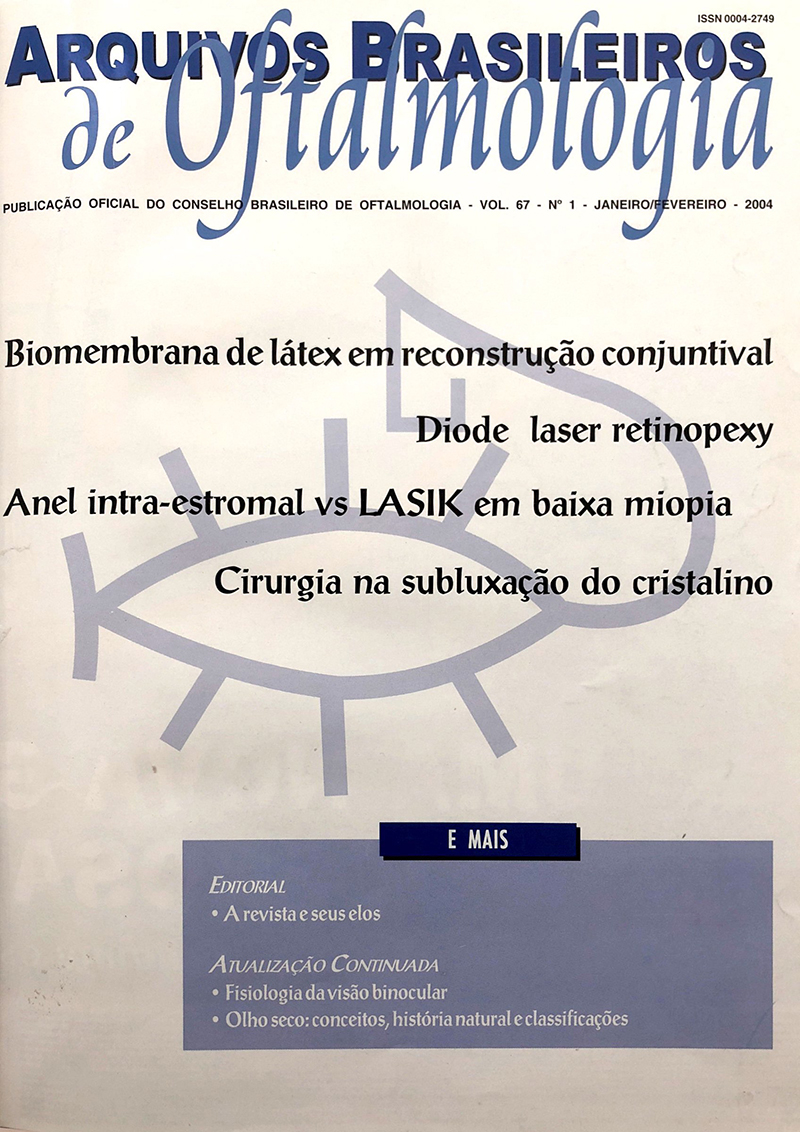
PURPOSE: To evaluate 10 cases of subluxated lens due to trauma or Marfan syndrome, focussing on their treatment and the results.
METHODS: Ten eyes of 9 patients were operated on due to lens subluxation. Six eyes were due to trauma and 4 eyes to Marfan syndrome. The mean age was 48.9 ± 18.2 years, and the follow-up period of 34.6 ± 11.6 months. Patients were submitted to intracapsular lens extraction (1 eye), extracapsular lens extraction (1 eye), phacoemulsification without IOL (1 eye), and phacoemulsification with IOL (7 eyes). Endocapsular rings were used in 7 eyes.
RESULTS: Best-corrected visual acuity improved in all patients, except for one, who had a regmatogeneous retinal detachment, 20 days after surgery.
CONCLUSION: Lens extraction with IOL implantation in subluxated lens patients has a good prognosis, especially with pha-coemulsification and endocapsular rings. With this approach, we were able to improve visual acuity and life quality in 90% of the cases.
Keywords: Lens subluxation; Eye injuries; Marfan syndrome; Cataract extraction
Arq. Bras. Oftalmol. 200467
| DOI: 10.1590/S0004-27492004000100003
PURPOSE: To evaluate the efficacy and safety of intrastromal corneal ring segments (ICRS) for the correction of low myopia, and to compare the obtained results with a control group submitted to laser in situ keratomileusis (LASIK).
METHODS: Prospective, nonrandomized and bilateral study. Fourteen patients with preoperative spherical equivalent ranging from -1.00 to -4.50 D received ICRS in one eye and LASIK in the other. Postoperative evaluations were performed at day one and seven and months 1, 3, 6, 12 and 24.
RESULTS: At the last follow-up, 64.29% of ICRS eyes (9/14) and 85.71% of LASIK eyes (12/14) were within ± 0.50 D of the predicted refractive outcome (p=0.385). Uncorrected visual acuity of 20/20 or better was observed in four of 14 ICRS eyes (28.57%), and in 12 of 14 LASIK eyes (85.71%) (p=0.002
²). No variances of more than 1.00 D in the manifest spherical equivalent refraction between two consecutive examinations or significant loss of best spectacle-corrected visual acuity were observed in both groups. Surgically induced astigmatism was greater in ICRS eyes than in LASIK eyes. At 24 months, the ICRS eyes scored lower in contrast sensitivity testing, especially at higher spatial frequencies (p=0.032
² at 18 cpd). The patients reported more subjective complaints in the early postoperative period for the ICRS eyes. CONCLUSIONS: ICRS were considered effective for the correction of low myopia, but their safety was limited by the observed surgically induced astigmatism, and their results were inferior compared to the control group.
Keywords: Myopia; Cornea; Prosthesis implantation; Laser in situ keratomileusis; Myopia
Arq. Bras. Oftalmol. 200467
| DOI: 10.1590/S0004-27492004000100004
PURPOSE: Transscleral diode retinal photocoagulation (diopexy) is becoming an accepted technique in the treatment of selected retinal diseases. The objective of this study is to evaluate diopexy technique in the production of adhesive chorioretinal lesions during the surgical treatment of the rhegmatogenous retinal detachment.
METHODS: 25 patients with rhegmatogenous retinal detachment were enrolled in a prospective clinical-surgical study to evaluate the technique of transscleral diode laser photocoagulation to obtain adhesive chorioretinal lesions during retinal reattachment surgery. The surgery consisted of the placement of an exoplant silicon to produce a buckle effect combined with a drainage of subretinal fluid in most cases.
RESULTS: By a mean follow-up of 10 months, 21 of 25 eyes had their retinas reattached after only one surgery with diopexy used in all cases.
CONCLUSION: Transscleral diode laser photocoagulation was a technically easy, controlled, effective, reproducible and safe means of obtaining chorioretinal adhesion in retinal reattachment surgery.
Keywords: Sclera; Retina; Choroid; Laser coagulation; Retinal detachment; Drainage
Arq. Bras. Oftalmol. 200467
| DOI: 10.1590/S0004-27492004000100005
INTRODUCTION: The biomembrane of natural latex is believed to promote neoformation of biological tissues and to be biocompatible. In animal studies, enhancement of wound healing in the esophagus, peritoniostomy, and pericardiostomy has been observed. It was used in chronic phlebopatic cutaneous ulcer and otologic surgeries in human subjects.
PURPOSE: To study the effect of biomembrane of natural latex in ocular surface.
METHODS: In fifteen New Zealand white rabbits, the superior nasal conjunctiva was removed from both eyes. In the right eyes, the biomembrane of natural latex was sutured to the conjunctiva with continuous 10.0 monofilament thread. In the left eye, the sclera was left bare. The rabbits were sacrified and the eyes were submitted to histological analysis on days five, seven, fourteen, twenty-one and twenty-eight. The eyes of a rabbit with no surgery were enucleated and sent for histological analysis of the normal conjunctiva.
RESULTS: Considering the whole period of the experiment, the number of eyes with satisfactory healing was greater in the biomembrane group than in the bare sclera group (p=0.06). The number of perilimbic vessels was significantly higher in the biomembrane group than in the bare sclera group (p=0.0284). The infection rate was similar in both groups.
CONCLUSION: Similar to prior studies, the biomembrane of natural latex seemed to benefit conjunctival reconstruction and neoangiogenesis. Depending on the results using human eyes, this material might be a new type of therapeutic resource for conjunctival replacement, especially in cases of desirable tissue revascularization.
Keywords: Conjunctiva; Cicatrix; Membranes, artificial; Regeneration; Rabbits
Arq. Bras. Oftalmol. 200467
| DOI: 10.1590/S0004-27492004000100006
With the new techniques of cataract surgery visual success has increased and it became necessary to foresee undesirable postoperative results regarding visual acuity.
PURPOSE: To verify the role of ocular ultrasonography in the preoperative evaluation of patients with mature cataract searching for pos terior segment pathologies that may affect surgical results, their prevalence and the main associated risk factors.
METHODS: A retrospective study was performed analyzing records of 262 patients indicated for ocular ultrasonography because the presence of mature cataract, thus preventing fundoscopic evaluation of the posterior segment. It was also tried to associate the presence of those changes with sex, age, race, history of ocular trauma, systemic and ocular diseases and the presence of cataract in the contralateral eye.
RESULTS: Ecographic changes were found in 24.8% of the examinations performed between 1996 and 2001, of which retinal detachment and vitreous condensations were the most common, with 9.9% of the findings for each. CONCLUSIONS: Among the risk factors pointed out as predisposing to findings in ocular ultrasonography, uveitis was the only statistically significant element. It was not possible to correlate positively the described posterior segment pathology to ocular trauma using obtained data.
Keywords: Cataract; Retinal detachment; Time factors; Preoperative care; Fundus oculi; Ophthalmoscopy
Arq. Bras. Oftalmol. 200467
| DOI: 10.1590/S0004-27492004000100007
OBJECTIVE: To evaluate the effectiveness of associating diagnostic tests for the detection of patients with glaucoma. METHOD: Forty-six eyes with cup/disc ratio >0.4 or disc asymmetry >0.2 were submitted to aplanation tonometry and automated static perimetry (screening and full threshold). The full threshold was adopted as gold standard for glaucoma diagnosis.
RESULTS: The positive predictive value (pretest probability) of disc parameters was 34.8%. The sensitivity, specificity, positive and negative likelihood ratios of screening perimetry were respectively 75%, 93%, 10.7 and 0.27. Using a cutoff of 20 mmHg the sensitivity, specificity, positive and negative likelihood ratios of tonometry were respectively 19%, 90%, 1.9 and 0.9. An abnormal screening test changes the probability of having a pathologic full threshold from 34.8% to 85% and a normal one changes this probability to 12%. The results of tonometry in changing the probability of having a pathologic full threshold were 34.8% to 50% when the test was positive (IOP>20 mmHg) and 34.8 to 32% when the test was negative (IOP<20 mmHg).
CONCLUSION: The results in identifying subjects with glaucoma are better when disc parameters are associated with screening visual field than with tonometry.
Keywords: Glaucoma; Tonometry; Perimetry; Blindness; Optic disc; Macular degeneration; Vision screening
Arq. Bras. Oftalmol. 200467
| DOI: 10.1590/S0004-27492004000100008
PURPOSE: To evaluate both the costs and the impact on the quality of life of essential blepharospasm and hemifacial spasm patients treated with botulinum toxin-A.
METHODS: 28 patients (16 essential blepharospasm and 12 hemifacial spasm), treated with BTX-A, were evaluated by means of two research tools, namely Medical Outcomes Study Short Form Healthy Survey, and Fahn Disability Rating Scale. The costs were mainly calculated on the basis of the toxin units used by each patient during a one-year period.
RESULTS: The total estimated direct cost was estimated at 1,081.62 reais per year (±89.39) for essential blepharospasm and 618.06 reais per year (±60.06) for hemifacial spasm, at an outpatient level (p<0.001). Before treatment all domains of SF-36 were significantly worse in both diseases. Under effective treatment with botulinum toxin-A the increases in the indices of SF-36 show changes in the general health rates of the population under study, with a statistically significant difference between the pre and post answers (p<0.05). In relating SF-36 with the Fahn Disability Rating Scale, the essential blepharospasm cases showed significant positive relationships (functional capacity, general health, emotional aspect and social aspects) while in the hemifacial spasm patients no correlation was found.
CONCLUSION: The present study demonstrates a remarkable negative impact of both diseases on the quality of life of essential blepharospasm and hemifacial spasm patients, as well as a significant improvement of both diseases after botulinum toxin-A treatment. Furthermore, the research data seem to justify the high costs of botulinum toxin-A treatment in view of its considerable benefits regarding the patients' quality of life.
Keywords: Botulinum toxin type A; Botulinum toxin type A; Hemifacial spasm; Blepharospasm; Blepharospasm; Health care costs; Life of quality; Drug costs
Arq. Bras. Oftalmol. 200467
| DOI: 10.1590/S0004-27492004000100009
PURPOSE: To compare the costs of local anaesthesia (retrobulbar block, peribulbar block, sub-Tenon and topical) to perform phacoemulsification cataract extraction.
METHODS: We studied the costs of the materials that are necessary to perform the local anaesthesia (retrobulbar block, peribulbar block, sub-Tenon and topical). We adopted the standard technique used for the Cataract Sector of the Department of Ophthalmology, ABC University. For each technique the total price of the each item to perform the above described local anaesthesia was analyzed. Prices were obtained from three different stores which sell hospitals materials and we considered for this study the item with a lower price.
RESULTS: The costs of topical anaesthesia were 0.06 reais, for the retrobulbar it was 7.42 reais, 24.96 reais for the peribulbar was and 48.12 reais for the sub-Tenon. CONCLUSIONS: 1. Topical anaesthesia showed the lowest cost. 2. Retrobulbar anaesthesia showed low cost when compared with the peribulbar and sub-Tenon 3. Sub-Tenon's anaesthesia showed the highest cost.
Keywords: Health care costs; Anesthesia, local; Cataract extraction; Phacoemulsification
Arq. Bras. Oftalmol. 200467
| DOI: 10.1590/S0004-27492004000100010
PURPOSE: To analyze the efficacy and safety of hyperopic laser in situ keratomileusis using the Ladar Vision excimer laser system.
METHODS: Twenty-eight eyes of 17 patients with hyperopia from +1.00 to +3.00 D (group 1), and 29 eyes of 18 patients with hyperopia from +3.25 to +6.00 D (group 2) that had LASIK for hyperopia with the Ladar Vision, were retrospectively analyzed. Uncorrected visual acuity, best spectacle-corrected visual acuity and cycloplegic refraction were evaluated 1 , 3 and 6 months after surgery.
RESULTS: In group 1, the mean preoperative cycloplegic spherical equivalent (SE) was +2.14 ± 0.64 D and 6-month postoperative SE was +0.44 ± 0.38 D. In group 2, the mean preoperative SE was +4.26 ± 0.75 D and the 6-month postoperative SE was +1.14 ± 0.63 D. 3.4% of the eyes in group 2 and none of the eyes in group 1 lost 2 or more lines of best spectacle-corrected visual acuity in the first postoperative month. CONCLUSIONS: LASIK with the Ladar Vision excimer laser system is an effective and safe procedure to correct hyperopia. Patients in group 2 appear to be at greater risk for loss of lines of best spectacle-corrected visual acuity.
Keywords: Keratomileusis; laser in situ; Hyperopia; Refractive errors; Laser surgery; Treatment outcome
Arq. Bras. Oftalmol. 200467
| DOI: 10.1590/S0004-27492004000100011
PURPOSE: 1) To check knowledge and actions developed by teachers of elementary schools working with low vision students, regarding signals and symptoms evidencing the students' visual difficulties: 2) To collect information useful for the design of preventing actions regarding eye health in school.
METHODS: A survey was applied to teachers of elementary schools in 23 public schools in the country of Campinas / SP, with low vision students, during the academic year of 1999. A self-applied questionnaire was used as tool for data obtention.
RESULTS: Of 84 teachers, 68 (81%) answered the questionnaire. The average teaching experience was 20.8 years. Most of those teachers (92.6%) did not report any previous specific training in visual deficiency. As concerns the recognition of signs and symptoms, most of the teachers indicated blackboard reading difficulty (94.1%), followed by headache (89.7%) and placement of subjects too close to the eyes (88.2%). A little over half of the teachers (55.9%) identified students showing visual difficulties. As regards developed actions 84.2% of the teachers declared to have oriented the students and 63.2% oriented parents and relatives and also informed the school about the fact. Only 26.3% oriented the student to look for an ophthalmologist.
CONCLUSION: Teachers did not show adequate knowledge about eye health and thus the developed actions were not comprehensive. Therefore, in order to develop actions regarding the prevention of visual impairment and to promote the enhancement of eye health, the implementation of a program aiming at the care of eye health in the public school is suggested.
Keywords: Vision, low; Vision disorders; School health; Eye health; Knowledge attitudes and practice; Health education; Students; Visual acuity; School health services
Arq. Bras. Oftalmol. 200467
| DOI: 10.1590/S0004-27492004000100012
PURPOSE: To evaluate the correlation between the parameters derived from the topographic scanning system (TOPSS) and visual field indices.
METHODS: One-hundred and twelve primary open-angle glaucoma patients and 88 normal controls were enrolled in the study. All individuals underwent a thorough ophthalmic examination, including visual field (Humphrey 24-2, Full Threshold mode), evaluating Mean Deviation (MD) and correct pattern standard deviation (CPSD) and TOPSS (average disc diameter, total disc area, cup area, cup shape, cup volume, average cup depth, average disc depth, neuroretinal rim (NRR) volume, NRR area, cup/disc area ratio, horizontal cup/disc ratio e vertical cup/disc ratio). The comparison between groups was done using the Mann-Whitney Test or the independent Student's t test. The correlation between the visual field indices and TOPSS parameters was performed with the Spearman's correlation.
RESULTS: The TOPSS parameters with better correlation were: cup area (MD: r= -0.538, p<0.001; CPSD: r= 0.512, p<0.001), vertical C/D ratio (MD: r= -0.506, p<0.001; CPSD: r= 0.483, p<0.001) e cup/disc area ratio (MD: r= -0.458, p<0.001; CPSD: r= 0.453, p<0.001). However, there was considerable variability between groups with different levels of visual field loss. CONCLUSIONS: The TOPSS variables presented, in general, substantial correlation with visual field indices. Although some of these parameters showed highly significant correlations, considerable variability of the structural parameters for the same level of functional damage may limit the usefulness of TOPSS isolated parameters in the diagnosis of glaucoma.
Keywords: Glaucoma; Optic disc; Perimetry; Diagnostic techniques, ophthalmological
Arq. Bras. Oftalmol. 200467
| DOI: 10.1590/S0004-27492004000100013
PURPOSE: To study the long-term behavior of induced astigmatism in a series of patients who underwent extracapsular extraction in which a superoposterior limbal incision sutured with interrupted 10-0 mononylon sutures was performed.
METHODS: 38 eyes of 35 patients were studied, 21 females and 17 males, with ages varying from 47 to 85 year (average = 70.17 year). All cases were operated by the same surgeon. The postoperative follow-up ranged from 15 to 33 months (average = 22.07 months). None of the cases had any of the sutures sectioned or removed. All patients recovered a corrected visual acuity equal or superior to 0.5. To calculate the induced astigmatism in each case, the preoperative and postoperative keratometric readings were considered and a program developed by Jaffe was used.
RESULTS: The induced astigmatism ranged from 0.13 diopters (D) to 2.74D (average = 1.19D). Regarding the axis, there was induction of with-the-rule astigmatism in the majority of cases (60.52%). A significant shift in the dioptric power of the induced astigmatism was observed when the preoperative keratometric readings were compared with the keratometric readings recorded at the second postoperative week, between the first and the third postoperative months, between the third and the sixth postoperative months and after the twelfth postoperative month.
CONCLUSION: When mononylon 10-0 is used as suture material in extracapsular extraction, the initially observed values of with-the-rule induced astigmatism tend to decrease during the post operative period.
Keywords: Astigmatism; Cataract extraction; Ophthalmologic surgical procedures; suture techniques; Follow-up studies; Adult
Arq. Bras. Oftalmol. 200467
| DOI: 10.1590/S0004-27492004000100014
PURPOSE: To study the evolution of hyperopia during childhood.
METHODS: A retrospective study through the analysis of 67 patients' charts. All patients were checked before the 3rd year of age and were followed for at least 5 years. The mean age at the first examination was 18.5 ± 6.9 months and the mean age at the end of the follow-up was 8.4 ± 1.7 years, providing an average time of the 6.8 ± 1.8 years of follow-up.
RESULTS: There was an increase in hyperopia when comparing the 1st with the 3rd year of life (p < 0.05). This was maintained without statistically significant variation until the 10th year of life. There was no difference in the variation of the hyperopia when the children were separeted into groups with low and high ametropia.
CONCLUSION: The present study demonstrates that hyperopia presents small variations during the first years of life, however it tends to be statistically stable from the 3rd to the 10th year of age.
Keywords: Hyperopia; Children; Retrospective studies; Evolution
Arq. Bras. Oftalmol. 200467
| DOI: 10.1590/S0004-27492004000100015
PURPOSE: To describe the Ocular Health Campaign ("Olho no Olho" - 2000), as perceived by parents of schoolchildren who study at Alagoas State public schools, in the city of Maceió.
METHODS: Questionnaires were applied to 263 parents of the school children, chosen at random from a group of 1996 children referred to attendance. Interviews were done between August and October 2001 in order to collect data and the opinions about the stages of the Campaign, ocular health and the social aspects of the parents.
RESULTS: The parents of 263 children referred to appointment were interviewed, of which 89.35% were present at the appointment and 82.98% were accompanied by the interviewed parent; 82.13% of the examinations were performed through a special gratuitous work regimen in a school of the region; 73.85% considered satisfactory the information given about the prescription or non-prescription of glasses, at time of appointments. Glasses were prescribed for 47.23% of the children, of which 87.39% received the glasses; 65.77% of the interviewed parents stated that there was no delay in the delivery of the glasses; 69.07% stated that their child liked the received glasses and 79.38% of the children were wearing the glasses. The Campaign was approved by 91.64% of the interviewed parents; 69.96% were the children's mothers and 66.92% could read. The family income was up to R$ 300 in 71.86% of the interviewed parents; 15.59% of the interviewed wore glasses. Of the 10.65% of subjects who were not present at the appointment, 28.57% alleged lack of money for transportation. CONCLUSIONS: Good presence of children and their parents at examinations in the special gratuitous work regimen. The parents were satisfied with the Campaign. The production and delivery of glasses was accomplished with satisfactory results. The target population is destitute and they have a low educational level. We suggest that the families of the schoolchildren are better advised about ocular health and the prescription of glasses at the examination.
Keywords: School health; Health education; Health promotion; Eye health; Visual tests; Visual acuity; School health services; Vision disorders; Vision disorders
Arq. Bras. Oftalmol. 200467
| DOI: 10.1590/S0004-27492004000100016
OBJECTIVE: The aim of this study was to compare the sensitivity of three methods of central scotoma localization.
METHODS: Twenty-nine patients (range 10-78 years) were referred to the Service of Subnormal Vision of the São Geraldo Hospital (Minas Gerais Federal University, School of Medicine) with previous diagnosis of diseases with central scotoma. The Asmler grid, face observation and near acuity test observation methods were used.
RESULTS: Among these methods, the one which presented the highest percentile of patients that reported scotoma presence was the Asmler grid (63.6%), followed by face observation (47.3%) and near acuity test observation (30.9%) (p<0.05). The three methods presented low sensitivity, possibly explained by the plasticity of the visual system trying to compensate the scotoma area.
CONCLUSION: The present result demonstrates that new techniques should be developed to improve the sensitivity of scotoma localization methods.
Keywords: Scotoma; Vision, low; Observation; Vision tests
Arq. Bras. Oftalmol. 200467
| DOI: 10.1590/S0004-27492004000100017
PURPOSE: The aim of this study was performing a comparative evaluation of cost in small ocular perforation repair using cyanoacrylate adhesive technique Super BonderTM (methyl-2-cyanoacrylate) as compared with corneal suture technique.
METHODS: In order to compare evaluations of cost a cost analysis in "reais" (R$) was conducted for both techniques according to the materials used in cyanoacrylate glue technique and the corneal suture.
RESULTS: The analysis of comparative cost of the cyanoacrylate adhesive technique in small perforations versus corneal suture showed a lower cost of the former.
CONCLUSION: Lower cost was found for the cyanoacrylate glue technique for corneal perforation repair when compared to the corneal suture technique.
Keywords: Cornea; Health care costs; Sutures; Cyanocrilates; Cola
Arq. Bras. Oftalmol. 200467
| DOI: 10.1590/S0004-27492004000100018
PURPOSE: To evaluate the percentage of agreement between the findings of the ophthalmic ultrasonography in preoperative cases with vitreous opacities and the intraoperative findings in pars plana vitrectomy.
METHODS: In a prospective study, 21 eyes with previous indication for pars plana vitrectomy underwent A- and B-scan ophthalmic ultrasonography by an experienced ultrasonographer and were graded into groups according to the findings. The accuracy of preoperative ultrasonography was determined by the agreement between the preoperative findings and those observed during surgery.
RESULTS: The most frequent indication for vitrectomy was hemorrhage secondary to proliferative diabetic retinopathy (16 eyes). The overall accuracy of ultrasound examination in determining the retinal status in these cases was 80%. The agreement was more precise for the cases of isolated vitreous opacity and total retinal detachment. The best visual prognosis was observed in the group with only vitreous hemorrhage and no vitreous or retinal detachment. CONCLUSIONS: Ophthalmic ultrasonography has good accuracy in demonstrating structural changes in the vitreous body and retina.
Keywords: Ultrasonography; Vitrectomy; Vitreous body; Vitreous body; Retina; Retina; Technology reliability; Preoperative care; Prospective studies
Arq. Bras. Oftalmol. 200467
| DOI: 10.1590/S0004-27492004000100019
PURPOSE: To compare the effectiveness of two forms of treatment of corneal abrasion secondary to removal of corneal foreign body: eyepad and no eyepad.
METHODS: Fifty-four patients with corneal abrasion secondary to removal of foreign body were randomized into two groups: the eyepad group and the no eyepad group. The patients were followed up daily, until complete healing, to evaluate: size of corneal abrasion, pain, photophobia, tearing, foreign body sensation, blurred vision.
RESULTS: There was no statistical difference between the two groups comparing: size of corneal abrasion, days until complete healing and amount of pain, photophobia, tearing, foreign body sensation or blurred vision. There were no systemic or ocular complications during the study.
CONCLUSION: Corneal abrasion secondary to removal of foreign body, smaller than 9 mm² , can be treated with large spectrum topical antibiotic and cicloplegic drops alone without the need for eyepad, making treatment simpler and less expensive.
Keywords: Eye injuries; Cornea; Eye foreign bodies; Curatives occlusive; Occlusive dressings; Wound healing
Arq. Bras. Oftalmol. 200467
| DOI: 10.1590/S0004-27492004000100020
PURPOSE: To verify the prevalence of diabetic retinopathy found in a type I diabetic population, accompanied at the Pediatric Endocrinology Outpatient Clinic of the "Santa Casa de Misericórdia de São Paulo", between August 29, 2000 and August 17, 2001, and it's relationship with sex, the patient's age on occasion of the diagnosis of diabetes and the duration of diabetes.
METHODS: Retinal mappings, biomicroscopic fundus and retinography were accomplished in 81 patients, 41 female and 40 male, in order to detect and to classify the diabetic retinopathies in that population.
RESULTS: The patients' age varied between 4 and 23 years (average = 12.0 ± 12.0), the patients' age on occasion of the diagnosis, between 6 months and 15 years (average = 3.4 ± 5.8) and the duration of the diabetes, between 7 months and 20 years (average = 5.8 ± 4.4). Diabetic retinopathy was observed in 14 patients (17.3%), in 7 (8.6%) with very mild and non-proliferative form, in 5 (6.2%) with mild and non-proliferative form, in 1 (1.2%) with high risk proliferative form and in 1 (1.2%) with advanced proliferative form. CONCLUSIONS: The prevalence of diabetic retinopathy in our sample is 17.3%. There is no difference between those with and those without of diabetic retinopathy regarding sex and the patient's age on occasion of the diagnosis. The longer the duration of the diabetes, the higher the prevalence of diabetic retinopathy.
Keywords: Diabetic retinopathy; Diabetes melittus, insulin-dependent; Prevalence; Hospital, community; Brazil
Arq. Bras. Oftalmol. 200467
| DOI: 10.1590/S0004-27492004000100021
PURPOSE: To evaluate the outcomes of intraocular lens (IOL) exchange in eyes with opacified hydrogel IOLs.
METHODS: We analyzed retrospectively the charts of 12 patients (15 eyes) who had been submitted to opacified intraocular lens exchange.
RESULTS: The opacified hydrogel intraocular lens caused decreased visual acuity in 13 eyes (86,7%). The opacified intraocular lens exchange improved visual acuity in 11 eyes (73,3%). Three eyes (20%) had the following complications during surgery: silicone oil loss, complete zonular dehiscence with loss of capsular bag and partial zonular dehiscence. During the intraocular lens exchange it was necessary to perform relaxing incisions in the anterior capsulorrhexis edge in three eyes (20%) and, in two eyes (13,3%) it was necessary to cut the haptics removing only the optical zone of the intraocular lens.
CONCLUSION: Intraocular lens exchange was an effective treatment in patients with opacified hydrogel intraocular lens (p > 0,05).
Keywords: Device removal; Lens implantation, intraocular; Lenses intraocular; Reoperation; Hydrogel; Phacoemulsification; Visual acuity
Arq. Bras. Oftalmol. 200467
| DOI: 10.1590/S0004-27492004000100022
PURPOSE: To analyze by impression cytology the presence of goblet cells on corneal surfaces with conjunctivalization.
METHODS: Corneal-conjunctival impression cytology was performed in 65 eyes of 65 patients who had clinical signs of conjunctivalization with or without previous ocular surgery. Patients were classified into two groups according to previous history of ocular surface reconstructive surgery associated with limbal transplantation and human amniotic membrane. In group I, 49 patients without previous ocular surgery were evaluated and in group II 16 patients were included after ocular surface reconstructive surgery with conjunctivalization recurrence. Samples were obtained in the affected eye between February 2000 and February 2002 at the UNIFESP's External Eye Disease Laboratory. Limbal deficiency was detected when one or more goblets cells were found on the corneal surface.
RESULTS: In group I one or more goblet cells were found on the corneal surface of 21 eyes (42.85%). In group II goblet cells were found on the corneal surface of 9 patients (56.25%).
CONCLUSION: Presence of goblet cells on the corneal surface detected by impression cytology in patients with conjunctivalization can confirm limbal stem cell deficiency, however, its absence does not exclude the diagnosis.
Keywords: Limbus corneae; Conjunctiva; Goblet cells; Epithelial cells; Cytological techniques; Diagnostic techniques opthalmological
Arq. Bras. Oftalmol. 200467
| DOI: 10.1590/S0004-27492004000100023
INTRODUCTION: Intraocular pressure (IOP) is a major risk factor for the development of open angle glaucoma (OAG). Intraocular pressure control is the main target of glaucoma therapy at the moment. The 24-hour intraocular pressure curve (circadian curve-CC) is of great importance for open angle glau-coma diagnosis and follow-up. Because of practical facilities, simplified methods such as the diurnal curve (DC) have been used.
PURPOSE: Comparison between circadian curve and diurnal curve as regards intraocular pressure peak detection and the verification of the influence of postural variation on 6 a.m. measurement.
METHODS: Sixty-four open angle glaucoma or suspected glaucoma patients (126 eyes) were submitted to circadian curve. Diurnal curves considered 9 a.m. - noon - 3 p.m. 6 p.m. measurements of the circadian curve. At 6 a.m., measurements were performed in the dark, while patients were lying, with the Perkins tonometer. Then, another measurement was taken, with the Goldmann tonometer with the patients seated. Mean intraocular pressure and intraocular pressure peaks in the circadian and diurnal curves were compared, as well as the time when the peaks occurred.
RESULTS: Mean IOP was higher in the open angle glaucoma patients in both the circadian curve and diurnal curve. When the circadian curve and diurnal curve were compared, mean intraocular pressure was higher in the 6 a.m. circadian curve IOP measurement was higher when taken in bed. The diurnal curve did not detect 60.42% of the peaks in open angle glaucoma patients and 88.24% of the peaks in suspected glaucoma patients. CONCLUSIONS: 1. The circadian curve detects more intraocular pressure peaks than the diurnal curve; 2. Six a.m. intraocular pressure is higher when taken in bed; 3. 6 a.m. intraocular pressure in bed is higher than the mean intraocular pressure in both circadian curve and diurnal curve.
Keywords: Intraocular pressure; Glaucoma open-angle; Tonometry; Circadian rhythm; Ocular hypertension; Visual fields
Arq. Bras. Oftalmol. 200467
| DOI: 10.1590/S0004-27492004000100024
PURPOSE: To evaluate the evolution of the patients with hyphema resulting from blunt ocular trauma associated with initial and final visual acuity, elevated intraocular pressure, rebleeding, clot absorption and surgical intervention.
METHODS: Fifty-four patients aged more than fifteen years, with blunt ocular trauma attended on the Ophthalmology Emergency Room of São Paulo Hospital were studied, between December 2000 to January 2002. The hyphema was classified in to five groups: microscopic; grade I; grade II; grade III; and grade IV (total hyphema). The patients were divided into two groups according to the involvement or not of the posterior segment. The data were compared by Mann-Whitney and the exact Fisher test and the final visual acuity was evaluated by multiple linear regression.
RESULTS: Ninety-one percent of the patients were men with an average age of thirty two years. At admission, 37% of patients showed intraocular pressure more than 24 mmHg. During the evolution, six patients required surgical intervention. Rebleeding occurred in 8% of the cases. In Group 1 (no lesions in the posterior segment) a better finally V.A. was statistically significant (p<0.001), a fact not observed in Group 2 (p<0.4772). CONCLUSIONS: Hyphema classification allows to evaluate the severity of damages, prognosis and management. The treatment still remains controversial. The final low visual acuity occurs more commonly associated with posterior segment injuries, and V.A. on admission. The successful treatment depends on identification of risk factors, appropriate medications and precise surgery indication.
Keywords: Eye injuries; Hyphema; Visual acuity; Prospective studies; Anterior chamber; Intraocular pressure; Follow-up studies
Arq. Bras. Oftalmol. 200467
| DOI: 10.1590/S0004-27492004000100025
PURPOSE: To analyze retrospectively the diagnosis of patients being followed up at the Uveitis Service of Hospital das Clínicas, University of São Paulo Medical School, during a 6-month period, from February to August, 2002.
METHODS: 262 patients, who attended the Uveitis Service during that period, had their charts reviewed and their diagnoses confirmed during the appointment. The following data were analyzed: age, gender, anatomic and etiological diagnoses, disease activity and ocular complications.
RESULTS: Patients' mean age was 41 years, 60% being female and 40% male. Distribution according to anatomic diagnosis was: anterior 20%, intermediate 4.5%, posterior 39.7 % and diffuse 31.3 %. Uveitis etiology was determined in 80% of the cases, and the most common causes were toxoplasmosis 22%, Vogt-Koyanagi-Harada syndrome 13% and Behçet's disease 10%. Infectious causes were present in 79% of posterior uveitis, whereas non-infectious systemic diseases were observed in 61% of anterior and 73% of diffuse uveitis. Uncommon diagnoses were observed such as HTLV-I/II related-uveitis, antiphospholipid syndrome and autoimmune retinopathy. Concerning disease activity, 15% were active and 85% were inactive. The most frequent ocular complications were cataract (42%) and glaucoma (13%). CONCLUSIONS: Epidemiological studies may demonstrate different patterns of uveitis of etiology; nevertheless they also reflect the particular characteristics of each center (tertiary, diagnosis criteria, special interests). The present study demonstrates in our midst the importance of endogenous uveitis such as Vogt-Koyanagi-Harada syndrome and Behçet's disease, as well as of infectious uveitis such as toxoplasmosis and toxocariasis.
Keywords: Uveitis; Uveitis; Retrospective studies; Community health services
Arq. Bras. Oftalmol. 200467
| DOI: 10.1590/S0004-27492004000100026
PURPOSE: To verify and compare with the literature the profile of patients with ocular coloboma, and also to verify visual acuity, ocular and systemic abnormalities.
METHODS: Retrospective analysis of 18 patients with a definite diagnosis of ocular coloboma at the Low Vision Department of the São Geraldo Eye Hospital of the Federal University of Minas Gerais. Patients underwent a complete ophthalmologic evaluation: sex, age, race, family history, prenatal and birth history, complete or incomplete coloboma, location, microphthalmos or anophthalmos, other ocular and systemic abnormalities, and visual acuity.
RESULTS: 10 (55.6%) patients were women. The age was from 4 to 57, and the mean was 9.5 years. Only three (16.7%) patients had a family history of coloboma. All patients had negative prenatal and birth history. The coloboma was bilateral in 100% of the patients, and associated with microphthalmos in 6 (33.3%) cases. The coloboma was considered typical in 14 (77.8%) patients, with retinochoroidal coloboma being the most frequent, 16 (88.9%) patients. 50% of the patients had coloboma as an isolated ocular anomaly, while the other 50% presented association with systemic anomalies. In 14 (77.8%) patients other ocular abnormalities were found.
CONCLUSION: The complete ophthalmologic examination is extremely important for both the diagnosis and prognosis of the patients with coloboma, since the coloboma may be associated with important low visual and also systemic anomalies and syndromes.
Keywords: Coloboma; Vision, low; Microphthalmos
Arq. Bras. Oftalmol. 200467
| DOI: 10.1590/S0004-27492004000100027
In children, idiopathic orbital inflammations are extremely rare and difficult to diagnose. The present study describes one case of idiopathic orbital inflammation in a one-year-old child that led to important oculomotor impairment. The differential diagnosis between idiopathic orbital inflammation and orbital cellulitis is discussed and the literature on the idiopathic orbital inflammation in childhood is reviewed.
Keywords: Strabismus; Orbital diseases; Differential diagnosis; Celulitis; Inflammation; Orbit; Tomography x-ray computed; Orbital neoplasms; Case report; Child
Arq. Bras. Oftalmol. 200467
| DOI: 10.1590/S0004-27492004000100028
Idiopathic polypoidal choroidal vasculopathy is a recently described entity related to recurrent serosanguinolent detachments of the retinal pigment epithelium and neurosensory retina due to a choroidal vasculopathy characterized by the formation of a branching vascular network with nodular lesions in the juxtapapillary temporal region. The authors describe a series of five cases with clinical characteristics compatible with this disorder. Mean age was 66 years (51-79 years), one black patient and three (60%) males. Mean follow-up time was 13.4 months (three months to 2 years). Epidemiological and clinical data are discussed.
Keywords: Vascular diseases; Choroid; Choroid diseases; Fluorescein angiography; Indocyanine green
Arq. Bras. Oftalmol. 200467
| DOI: 10.1590/S0004-27492004000100029
The authors describe a rare association of bilateral typical coloboma, microcornea and anterior chamber cleavage deficience. They also discuss the embriology and the difficulties to identify if the continuous and insidious vision impairment is due to coloboma or to other associated disease, like glaucoma. Supplementary tests, such as visual field, ophthalmoscopy, and tonometry are not reliable because of the presence of nistagmus and low visual acuity.
Keywords: Coloboma; Abnormalities multiple; Anterior chamber; Glaucoma; Gonioscopy; Corneal dystrophies; hereditary
Arq. Bras. Oftalmol. 200467
| DOI: 10.1590/S0004-27492004000100030
Blepharopigmentation is a procedure used to create a permanent eyeliner to replace the daily application of cosmetic lash line enhancement. Few studies show ocular and lid complications of blepharopigmentation. The present paper describes 3 cases of trichiasis related to this procedure. The authors discuss the probable pathophysiology and comment the treatment of the cases.
Keywords: Eyelids diseases; Pigmentation; Skin pigmentation; Tattoing; Light coagulation; Argoniun; Blepharoplasty
Arq. Bras. Oftalmol. 200467
| DOI: 10.1590/S0004-27492004000100031
PURPOSE: To evaluate the u se of equine renal capsule preserved in glycerin to repair lamellar scleral lesions in dogs.
METHODS: Twelve healthy mongrel dogs, male and female, weighing 12 kg were used. The study was both clinical and morphological, and performed on the first, third, seventh, 15th, 30th and 60th day after surgery. Temporal canthotomy was performed after standard preoperative and general anesthesia. Conjunctival and scleral square incisions of 0.5x0.5 cm were carried out in a one o'clock position, near the limbus. A fragment of hydrated biological prosthesis, of the same shape, was sutured with 7-0 vicryl® in an interrupted suture.
RESULTS: The clinical evaluation showed blepharospasm/photophobia until the 7th day after surgery. Conjunctival edema appeared up to the 5th day after surgery. Mucoid ocular discharge was sustained until the 10th day after surgery. Hyperemia was observed until the end of the evaluation period. There were no signs of graft extrusion in all animals. The anterior and posterior segments did not show clinical signs of inflammation. The optical microscopy morphological evaluation showed an inflammatory exudation with acute aspects in the early and intermediate periods, and inflammatory exudation with chronic aspects in the late periods. There was incorporation of the implant by the recipient's sclera.
CONCLUSION: These results suggest that the equine renal preserved capsule could be a useful alternative tissue to repair lamellar corneal lesions in dogs and humans.
Keywords: Sclera; Transplantation heterologous; Dogs; Animal
Arq. Bras. Oftalmol. 200467
| DOI: 10.1590/S0004-27492004000100032
The binocular vision of human beings is given by the almost complete superimposition of the monocular visual fields, which allows a finer perceptual discrimination of the egocentric localization of objects in space (stereopsis) but only within a very narrow band (the horopter). Before and beyond it, diplopia and confusion are present, so that a physiologic (cortical) suppression is necessary to avoid them to become conscious. The geometry of the horopter and its physiologic implications (Hillebrand's deviation, Kundt's partition, Panum's area, stereoscopic vision) are analyzed, as well as some clinical aspects of the normal binocular vision (simultaneous perception, fusion, stereoscopic vision) and of adaptations to abnormal states (pathologic suppression, amblyopia, abnormal retinal correspondence).
Keywords: Vision, binocular; Amblyopia; Vision disorders; Visual perception; Depth perception
Arq. Bras. Oftalmol. 200467
| DOI: 10.1590/S0004-27492004000100033
The dry eye syndrome is frequently the source of great frustration, not only for the patients but also for the eye specialists who, not rarely, are defeated by the persistence of the symptoms, despite the efforts made in their diagnostic and therapeutic approach. The subject acquires its real importance, when we are faced with the fact that "dry eye" is a very common complaint in ophthalmologic practice. The authors present an up-to-date review about important aspects involved in this issue.
Keywords: Dry eye syndromes; Tears; Ophthalmic solutions; Sjöegren's syndrome; Meibomian glands


 ABO is licensed under a Creative Commons Attribution-NonComercial 4.0 Internacional.

