Arq. Bras. Oftalmol. 201881
| DOI: 10.5935/0004-2749.20180021
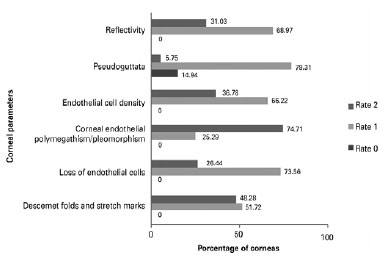
Purpose: Donated corneas are classified as tectonic if there are defects within any layers of the cornea which would prevent a satisfactory visual outcome after transplantation. This study aimed to evaluate whether some tectonic corneas have sufficient endothelial characteristics to allow their use in posterior lamellar keratoplasty, and explored their reclassification for use in this sight-improving procedure.
Methods: A retrospective review of all corneal tissues preserved by the Sorocaba Eye Bank from January to April of 2014 was performed. All donated corneas classified as tectonic were included. Endothelial tissue was defined as healthy and viable for posterior lamellar keratoplasty if endothelial cell density was ≥2000 cells/mm2. Additional parameters analyzed included Descemet folds and stretch marks, loss of endothelial cells, corneal endothelial polymegathism/ pleomorphism, pseudo-guttata, and reflectivity.
Results: During the study period, 2,847 corneas were preserved, of which 423 (14.85%) were classified as tectonic. Of these, 87 (20.56%) were reported as having endothelial viability and were included in the posterior lamellar keratoplasty group. Average corneal endothelial cell density of this group was 2,471 SD ± 256 cells/mm2 (range 2012-2967 cells/mm2).
Conclusion: A significant number of corneas classified as tectonic showed endothelial viability and were included in the posterior lamellar keratoplasty group (20.56%). Despite stromal and/or epithelial alterations, these corneas could have been potentially distributed for posterior lamellar transplantation to improve vision, thus reducing the corneal transplantation waiting period. This study highlights how corneal tissue reclassification could increase the potential amount of corneal tissue available for optical transplantation.
Keywords: Corneal transplantation; Eye banks; Tissue donors; Cornea; Tissue and organ procurement
Arq. Bras. Oftalmol. 201881
| DOI: 10.5935/0004-2749.20180022
Objective: To study the safety parameters associated with intracameral moxifloxacin application five weeks after cataract surgery.
Methods: The study was a prospective case series set in a private hospital in Recife, Pernambuco, Brazil. A consecutive sample of 1,016 cataract surgeries was evaluated. The inclusion criteria were patients with indications for cataract surgery, a minimum of 55 years of age, and no history of allergy to quinolones. Patients were prepared for surgery using a 5% povidone solution diluted as a topical antiseptic agent. The operative technique was phacoemulsification with intraocular lens implantation. A 0.3mL syringe was partially filled with moxifloxacin and 150 µg/0.03 mL of moxifloxacin was administered through the surgical incision at the end of the surgery. Postoperatively, patients were prescribed: (1) 0.5% moxifloxacin eyedrops 5 times daily for 1 week, and (2) 1% prednisolone acetate eyedrops 5 times daily for 1 week, followed by 4 times daily for 1 week and, subsequently, 2 times daily for 3 weeks. The outcomes were incidence of acute endophthalmitis, mean changes from baseline to 5 postoperative weeks in corneal endothelial cell density, corrected distance visual acuity and intraocular pressure.
Results: The mean age was 67 ± 5 years, and 56.2% of the patients were female. There were no cases of endophthalmitis. The mean preoperative corrected distance visual acuity was 58 letters ± 10 (SD), and the mean postoperative corrected distance visual acuity was 80 letters ± 4 (SD). The mean change in corneal endothelial cell density was 249 cells/mm (10.3%). There was almost no difference in intraocular pressure. No studyrelated adverse events were observed.
Conclusion: The results suggest moxifloxacin is a safe option for intracameral use after cataract surgery.
Keywords: Cataract extraction; Endophthalmitis; Antibiotic prophylaxis; Safety; Postoperative complications
Arq. Bras. Oftalmol. 201881
| DOI: 10.5935/0004-2749.20180023
Purpose:To compare the use of topical anesthesia and retrobulbar anesthesia during silicone oil removal with a mixed pars plana technique, through evaluating the pain experience of patients.
Methods: We selected patients according to their behavior during previous vitreoretinal surgery and ophthalmologic examinations and divided them into two anesthesia groups: topical (n=36) and retrobulbar (n=33). We used a mixed technique for the passive removal of silicone oil in both groups. During each step of the surgery, the patients' pain experience and the surgeon's comfort were scored according to a pain scale.
Results: The pain experienced during the application of the anesthesia was significantly greater in the retrobulbar group (p<0.001). The topical group experienced greater pain during trocar insertion (p<0.001). There was no significant difference between the groups regarding the overall pain experience or complications.
Conclusions: The pain experience of the selected patients during silicone oil removal was comparable between the topical and the retrobulbar anesthesia. Topical anesthesia with the mixed pars plana technique is an effective and safe alternative option for silicone oil removal surgery.
Keywords: Pain; Silicone oils; Anesthetics, local; Administration, topical; Vitreoretinal surgery; Patient satisfaction
Arq. Bras. Oftalmol. 201881
| DOI: 10.5935/0004-2749.20180024
Purpose: To evaluate the possible protective effect of breast milk against retinopathy of prematurity by comparing the amount of breast milk received by patients who developed retinopathy of prematurity and those who did not and to determine both the required minimum amount of breast milk and the time of life during which neonates need to receive breast milk for this effect to be significant
Methods: Cohort study of newborns with a birth weight of <1500 g or gestational age of <32 weeks, or both, born between January 2011 and October 2014 and hospitalized within the first 24 h of life in the Hospital Criança Conceição Neonatal Intensive Care Unit in Porto Alegre, RS, Brazil.
Results: The prevalence of retinopathy of prematurity of any degree was 31% (100 of 323 patients) and that of severe retinopathy of prematurity was of 9% (29 of 323 patients). The median amounts of breast milk received daily by patients with and without retinopathy of prematurity were 4.9 mL/kg (interquartile range, 0.3-15.4) and 10.2 mL/kg (1.5-25.5), respectively. The amount of breast milk received in the first 6 weeks of life was inversely associated with the incidence of both retinopathy of prematurity of any degree and severe retinopathy of prematurity in the univariate analyses. However, the statistical significance was maintained only during the sixth week of life in a per-period multivariate analysis controlling for confounding factors.
Conclusions: Small amounts of breast milk are inadequate to prevent retinopathy of prematurity in premature newborns at risk for the disease.
Keywords: Retinopathy of prematurity; Milk, human; Breast feeding; Infant, very low birth weight; Insulin-like growth factor I; Infant, premature
Arq. Bras. Oftalmol. 201881
| DOI: 10.5935/0004-2749.20180025
Purpose: To compare the anterior segment parameters of patients with pseudoexfoliation syndrome, patients with pseudoexfoliation glaucoma, and normal subjects.
Methods: This prospective, controlled, comparative study included 150 eyes of 150 patients. The patients were divided into the pseudoexfoliation syndrome group, the pseudoexfoliation glaucoma group, and the control group (50 patients in each group). Axial length, central corneal thickness, aqueous depth, anterior chamber depth, lens thickness, K1 and K2 keratometry values, and white to white distance measurements were obtained by optical biometry and compared between the groups.
Results: The mean ages of the pseudoexfoliation syndrome, pseudoexfoliation glaucoma, and control patients were 62.18 ± 6.21, 61.80 ± 6.62, and 59.40 ± 6.89 years, respectively. There were no statistically significant differences between the groups in mean age or sex ratio (p>0.05). Mean central corneal thickness was statistically significantly greater, mean aqueous depth and anterior chamber depth were statistically significantly greater, and mean lens thickness was statistically significantly less in the control group than in the pseudoexfoliation syndrome and pseudoexfoliation glaucoma groups (p<0.05). Pairwise comparisons of the pseudoexfoliation syndrome group and the pseudoexfoliation glaucoma group revealed that there were no significant differences between these two groups in central corneal thickness, aqueous depth, anterior chamber depth, and lens thickness (p>0.017).
Conclusions: Patients with pseudoexfoliation glaucoma and pseudoexfoliation syndrome had greater lens thickness, shallower aqueous depth and anterior chamber depth, and less central corneal thickness than normal subjects. None of the anterior segment parameters differed between patients with pseudoexfoliation syndrome and patients with pseudoexfoliation glaucoma.
Keywords: Anterior eye segment; Cornea; Lens; Glaucoma; Exfoliation syndrome
Arq. Bras. Oftalmol. 201881
| DOI: 10.5935/0004-2749.20180026
Purpose: To determine the clinical characteristics and seasonal distribution of patients admitted to the ocular emergency department of a tertiary ophthalmology care center.
Methods: The study cohort includes 27,120 patients who were admitted to ocular emergency room between November 2013 and November 2014. The age, sex, reason for admission, diagnosis, and complete ocular examination reports were recorded for each patient. X-ray and ultrasonographic examinations were performed if necessary.
Results: The mean patient age was 32.83 ± 17.62 years (range, 0-95). The number of males was nearly two times the number of females, with 18,808 (69.4%) males and 8312 (30.6%) females. The diagnoses included viral conjunctivitis (7,859 patients; 29.0%), corneal foreign body (5,286 patients; 19.5%), bacterial conjunctivitis (3,892 patients; 14.4%), corneal abrasions (2,306 patients; 8.5%), and allergic conjunctivitis (1,433 patients; 5.3%) (Table 1). Other frequent diagnoses included subconjunctival hemorrhage, photo keratopathy, chemical eye injury, and penetrating and blunt eye injuries. Allergic conjunctivitis, ocular trauma, and corneal foreign body were more frequent in spring, whereas keratitis and chemical eye injury were more common in winter (chi-square test). The most common reasons for emergency room admission, in order of frequency, were viral conjunctivitis, corneal foreign body, bacterial conjunctivitis, and corneal abrasions.
Conclusion: This study is the first long-term prospective study to evaluate the seasonal distribution and diagnosis of all adult and pediatric patients admitted to the emergency room for ocular conditions. The frequency of ophthalmological conditions seen in the emergency room may vary according to the season.
Keywords: Conjunctivitis; Eye foreign bodies; Hospital emergency service; Seasons; Eye injuries; Corneal injuries
Arq. Bras. Oftalmol. 201881
| DOI: 10.5935/0004-2749.20180027
Purpose:To assess whether the serum levels of mannose-binding lectin of the lectin complement pathway are associated with age-related macular degeneration.
Methods: Patients with age-related macular degeneration and age-matched controls underwent full ophthalmologic examination and optical coherence tomography. Using a time-resolved immunofluorometric assay, blood samples were evaluated to determine the serum mannose-binding lectin levels.
Results: A total of 136 individuals were evaluated, including 68 patients with age-related macular degeneration (34 exudative and 34 nonexudative) and 68 age-matched controls. The median mannose-binding lectin level was 608 ng/mL (range, 30-3,415 ng/mL) in patients with age-related macular degeneration and 739 ng/mL (range, 30-6,039 ng/mL) in controls, with no difference between the groups. Additionally, the median mannose-binding lectin level was 476 ng/mL (range, 30-3,415 ng/mL) in exudative cases and 692 ng/mL (range, 30-2,587 ng/mL) in nonexudative cases.
Conclusions: Serum mannose-binding lectin levels were not associated with age-related macular degeneration or with the exudative and nonexudative forms of the disease.
Keywords: ge-related macular degeneration; Drusen; Immunoregulation; Mannose-binding lectin; Inflammation; Retina
Arq. Bras. Oftalmol. 201881
| DOI: 10.5935/0004-2749.20180028
Purpose: To study a latex biomembrane and conjunctival autograft with regard to the promotion of conjunctival healing in rabbits.
Methods: The study included nine male albino rabbits. In these rabbits, a rectangular area of the conjunctiva was surgically removed from the superonasal quadrant adjacent to the limbus in both eyes. The bare area of the sclerotic coat of the right eye was reconstructed with a latex biomembrane, and the corresponding site of the left eye was reconstructed with a conjunctival autograft. The animals were killed in groups of three at 7, 14, and 21 days after surgery. The tissues from the surgical site, including the cornea, were fixed in formaldehyde, and were then processed in paraffin and stained with hematoxylin and eosin. The nature and intensity of the inflammatory response and the epithelial pattern at the conjunctival surface were evaluated under optical microscopy with longitudinal histological sections through the center of the anatomical specimens.
Results: Until the 14th postoperative day, the inflammatory reaction was greater in the biomembrane group than in the conjunctival autograft group. In the latex biomembrane group, inflammation was less intense and the stroma was thicker on the 14th postoperative day than on the 7th postoperative day. After three weeks, conjunctival healing in both groups showed similar characteristics.
Conclusion: Although healing was slower with a latex biomembrane, tissue reconstitution was almost the same as that with a conjunctival autograft by three weeks. A latex biomembrane is as effective as a conjunctival autograft for the reconstruction of the ocular surface. Owing to the lack of toxicity and allergenicity, a latex biomembrane appears to be a promising therapeutic option for conjunctival reconstruction.
Keywords: Wound healing; Latex; Conjunctiva/transplantation Transplantation, autologous; Histology; Rabbits
Arq. Bras. Oftalmol. 201881
| DOI: 10.5935/0004-2749.20180029
Purpose: We report a simplified Descemet's membrane endothelial keratoplasty (DMEK) technique that involves safe and effective preparation and introduction, correct orientation, and easy unfolding of the donor graft inside the recipient anterior chamber.
Methods: In this retrospective study, we assessed the surgical outcomes of 26 eyes of 23 consecutive patients (mean age, 61.2 ± 11.4 yr; range, 39-82 yr) with Fuchs endothelial corneal dystrophy (n=19) or bullous keratopathy (n=7) who underwent the Samba technique, a simplified DMEK method, at the Sorocaba Ophthalmology Hospital, Sorocaba Eye Bank, Sorocaba, Brazil, between August 2011 and July 2012.
Results: Of the 26 operated eyes, only two (7.7%) experienced partial graft detachment requiring rebubbling, and in those eyes, the graft was reattached successfully with one air bubble. There were no cases of primary graft failure, tissue loss, or pupillary block. All patients with good visual potential achieved a best-corrected visual acuity of 20/30 or better at 6 months, and 82.6% achieved a best-corrected visual acuity of 20/30 or better 1 month postoperatively.
Conclusion: In this retrospective study, the Samba technique, a simplified DMEK procedure, was safe and effective, with an acceptably low rebubbling rate and no incidence of primary graft failure or pupillary block. Moreover, rapid and nearly complete visual recovery was achieved. This simplified DMEK technique can be adopted by corneal surgeons worldwide as a primary treatment for endothelial dysfunction with a less steep learning curve and low rate of postoperative complications.
Keywords: Descemet stripping endothelial keratoplasty; Corneal transplantation; Cornea/surgery
Arq. Bras. Oftalmol. 201881
| DOI: 10.5935/0004-2749.20180030
Purpose: To evaluate microstructural differences between corneas with and without Kayser-Fleischer rings in age-matched subjects with Wilson's disease with neurological symptoms, using confocal laser scanning microscopy.
Methods: The study included 12 subjects with Wilson's disease with neurological symptoms. Twelve corneas presented clinically with classic Kayser-Fleischer rings, visible on slit lamp examination; the other 12 served as controls. The subjects underwent a comprehensive clinical examination. Microstructural analysis using confocal laser scanning microscopy evaluated increased corneal thickness, decreased number of cells, increased debris or specific deposits, and unusual microstructures.
Results: Clinically, the subjects with Kayser-Fleischer rings had similar corneal findings and normal intraocular pressure; two had typical sunflower cataracts and decreased visual acuity. The control eyes all presented normal visual acuity, intraocular pressure, and corneal appearance. The microstructural analysis demonstrated similar findings in all the affected corneas. Compared with the control corneas, there were fewer keratocytes in the anterior stroma (17.380 vs. 22.380/mm3). Round, "hollow" dark areas were observed between the keratocytes; these were universal and similar in appearance in all affected corneas and all cornea layers. In the peripheral posterior stroma, there were dust-like, bright, granular deposits that tended to increase in number and density toward Descemet's membrane, masking the peripheral endothelium. The control corneas presented a normal microstructure apart from dust-like granular deposits in the periphery.
Conclusions: In vivo confocal microscopy is a useful tool for evaluating the corneal microstructure when a Kayser-Fleischer ring is clinically present. The ring consists of granular, bright particles that increase in density toward Descemet's membrane, and is associated with a decreased number of keratocytes and peculiar dark, round areas in all stromal layers, probably a sign of corneal damage. When the ring is not visible in subjects with Wilson's disease, changes to the corneal microstructure are insignificant.
Keywords: Cornea; Hepatolenticular degeneration; Corneal stroma; Descemet membrane; Microscopy, confocal
 ABO is licensed under a Creative Commons Attribution-NonComercial 4.0 Internacional.