
Erika Christina Canarim Martha de Pinho1; Fernando Chahud2; João-José Lachat3; Joaquim José Coutinho-Netto4; Sidney Julio Faria e Sousa1
DOI: 10.5935/0004-2749.20180028
ABSTRACT
Purpose: To study a latex biomembrane and conjunctival autograft with regard to the promotion of conjunctival healing in rabbits.
Methods: The study included nine male albino rabbits. In these rabbits, a rectangular area of the conjunctiva was surgically removed from the superonasal quadrant adjacent to the limbus in both eyes. The bare area of the sclerotic coat of the right eye was reconstructed with a latex biomembrane, and the corresponding site of the left eye was reconstructed with a conjunctival autograft. The animals were killed in groups of three at 7, 14, and 21 days after surgery. The tissues from the surgical site, including the cornea, were fixed in formaldehyde, and were then processed in paraffin and stained with hematoxylin and eosin. The nature and intensity of the inflammatory response and the epithelial pattern at the conjunctival surface were evaluated under optical microscopy with longitudinal histological sections through the center of the anatomical specimens.
Results: Until the 14th postoperative day, the inflammatory reaction was greater in the biomembrane group than in the conjunctival autograft group. In the latex biomembrane group, inflammation was less intense and the stroma was thicker on the 14th postoperative day than on the 7th postoperative day. After three weeks, conjunctival healing in both groups showed similar characteristics.
Conclusion: Although healing was slower with a latex biomembrane, tissue reconstitution was almost the same as that with a conjunctival autograft by three weeks. A latex biomembrane is as effective as a conjunctival autograft for the reconstruction of the ocular surface. Owing to the lack of toxicity and allergenicity, a latex biomembrane appears to be a promising therapeutic option for conjunctival reconstruction.
Keywords: Wound healing; Latex; Conjunctiva/transplantation Transplantation, autologous; Histology; Rabbits
RESUMO
Objetivo: Estudar o uso da biomembrana de látex e o transplante conjuntival autólogo na cicatrização conjuntival em coelhos.
Métodos: Em nove coelhos albinos, neo-zelandeses, machos foram removidas áreas retangulares idênticas, do quadrante supero nasal, adjacente ao limbo, de ambos os olhos. As áreas desnudas da camada esclerótica nos olhos direitos foram recobertas com biomembrana de látex e a dos olhos esquerdos com enxerto conjuntival autólogo. Os animais foram sacrificados em grupos de três, aos 7, 14 e 21 dias após a cirurgia. Os tecidos do local cirúrgico, incluindo a córnea, foram fixados em formaldeído, antes de serem processados em parafina e corados com hematoxilina e eosina. A natureza e a intensidade da resposta inflamatória e o padrão de epitelização da superfície conjuntival foram avaliados sob microscopia óptica, em seções histológicas longitudinais, passando pelo centro dos espécimes anatômicos.
Resultados: Até o décimo quarto dia pós-operatório, o grupo que recebeu a biomembrana apresentou reação inflamatória mais intensa do que o grupo com auto enxerto conjuntival. Aos 14 dias, os olhos com biomembrana apresentavam-se menos inflamados e com estroma mais espesso do que aos 7 dias. Aos 21 dias, a reparação conjuntival de ambos os grupos apresentavam características semelhantes.
Conclusão: Apesar de apresentar uma cicatrização mais lenta, a biomembrana de látex se mostrou tão eficaz quanto o auto enxerto conjuntival na reconstrução da superfície ocular após três semanas de cicatrização pós-operatória. Devido as suas baixas toxicidade e alergenicidade, este material parece ser uma opção terapêutica promissora na reconstrução da conjuntiva.
Descritores: Cicatrização de feridas; Latex; Conjuntiva/transplante; Transplante autólogo; Histologia; Coelhos
INTRODUCTION
A latex biomembrane has been developed at the Neurochemistry Laboratory of the Faculty of Medicine of Ribeirão Preto, University of São Paulo, Brazil. This membrane is prepared using latex extracted from the rubber tree Hevea brasiliensis. The allergenicity and toxicity of this natural latex are almost completely neutralized during the preparation of the membrane. This membrane has important biochemical properties, such as angiogenic activity, cell adhesion promotion, and provisional extracellular matrix formation(1). In animals, it has been shown to enhance healing of the esophagus mucosa and abdominal wall, and it can function as a partial substitute for the pericardium(2). In humans, it has been used successfully for reconstructing the tympanic membrane(3) and healing lower limb ulcers(4,5). Additionally, it has shown excellent biocompatibility(6,7) and low allergenicity(4,6), and it can be sterilized with ethylene oxide(8).
Treatment for diseases of the bulbar conjunctiva, such as symblepharon, frequently requires removal of large areas of adherent tissue, and this generates large areas of bare sclera. Similar outcomes are noted in the treatment of extensive conjunctival tumors. In both cases, it is often required to surgically reconstruct the conjunctiva. Conjunctival autograft or amniotic membrane implantation is currently used for this procedure. The use of a conjunctival autograft is very efficient, but it is limited by the difficulty of harvesting tissue without causing functional impairment at the donor site(9). A natural amniotic membrane has numerous properties that promote healing. However, it is unclear whether these properties are retained after preparation of the membrane for surgical use. Moreover, as it is a human material, there is a risk of disease transmission(10,11). Numerous other conjunctival substitutes have been proposed, including oral mucosa and nasal mucosa, and a recent study has assessed the use of extracellular matrix by tissue engineering for conjunctiva transplantation in rabbits(12).
In a previous study, we found that a latex biomembrane was not only well tolerated but also capable of accelerating the growth of epithelial cells along the ocular surface. When compared with conjunctival healing by secondary intention, it provided better results in a shorter time(13-15). This finding presented the possibility of a new form of conjunctival reconstruction, without the limitations associated with existing methods. Considering that a latex biomembrane has already been compared with secondary intention conjunctival healing, the next logical step before proposing its use in human eyes is to compare its use with conjunctival autograft use, which is the gold standard method of conjunctival reconstruction. Therefore, the aim of this study was to assess the effects of a latex biomembrane and conjunctival auto-graft on wound healing of the rabbit conjunctiva, and to compare the use of a latex biomembrane with the use of a conjunctival autograft.
METHODS
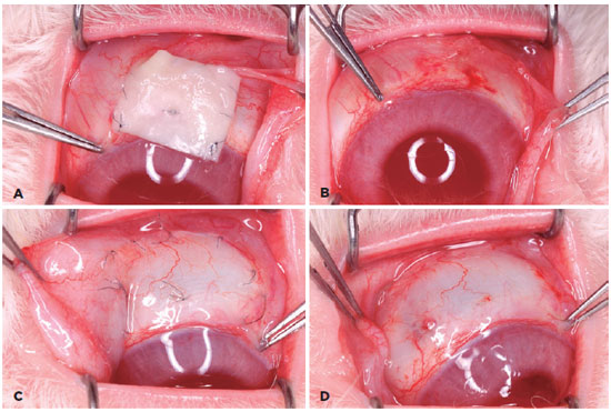
The study included nine male albino rabbits (Oryctolagus cuniculus). These rabbits were three months old and weighed from 2.5 to 2.9 kg. They were anesthetized by intravenous injection of sodium thiopental. In these rabbits, a rectangular area of the conjunctiva (1.5 cm wide and 1.0 cm long) was surgically removed from the superonasal quadrant adjacent to the limbus in both eyes (Figure 1). The bare area of the sclerotic coat of the right eye was then reconstructed with a latex biomembrane (80-µm thick) having dimensions identical to the dimensions of the original wound. The corresponding area of the left eye was reconstructed with conjunctival flaps obtained from the inferior conjunctiva of the same eye. In both cases, the grafts were sutured to the edges of the conjunctival lesion with mononylon 10-0 (Ethicon, 7718, Johnson & Johnson, São Paulo, Brazil). Postoperatively, all animals were treated with neomycin and dexamethasone eye drops (Maxitrol®, Alcon, São Paulo, Brazil), thrice daily, until sacrifice. The rabbits were killed in groups of three at 7, 14, and 21 days after surgery. A lethal dose of anesthetic was used before decapitation. The biomembranes were kept in place in all animals until the 14th postoperative day. At that time, the three animals that had been kept alive for the next seven days were also anesthetized to remove the latex biomembranes and the sutures from both eyes (Figure 1). The tissues from the surgical sites, including those from the adjacent corneas, were fixed in formaldehyde and were then processed in paraffin and stained with hematoxylin and eosin. Longitudinal histological sections were made through the center of the anatomical specimens. The rabbits were handled according to the ARVO Statement for the Use of Animals in Ophthalmic and Vision Research, and the experiment was approved by the ethics research committee of our institution.

The latex was obtained from the rubber tree Hevea brasiliensis (clone RRhim 600). The crude material was obtained from Globbor Inc. Corp. Rubbers LTD (Guapiacu, São Paulo, Brazil). The biomembrane was prepared at the Neurochemistry Laboratory of the Faculty of Medicine of Ribeirão Preto, University of São Paulo, Brazil, as described by Mrué. During preparation, the latex takes the shape of the mold, and it has a controlled thickness, a smooth surface, and an elastic consistency(1).
The essential features with regard to the analysis of tissue repair in this study have been described elsewhere(13-15). Accordingly, the following six items were used to assess tissue repair under light microscopy (ECCMP and FC): (1) inflammatory infiltrate: mild, moderate, or severe; (2) nature of the inflammatory infiltrate: acute (neutrophils and eosinophils), chronic (lymphocytes, plasma cell, and macrophage), or mixed (combination of both); (3) degree of wound epithelization: partial or full; (4) number of epithelial layers; (5) presence of goblet cells: present or absent; and (6) thickness of the conjunctival stroma: partial or full.
RESULTS
The animals were healthy until the end of the experiment, and they showed no local or systemic adverse reactions. The results of histological analyses in both groups are presented in table 1.
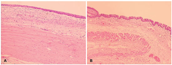
On the 7th postoperative day, the inflammatory reaction was greater and level of tissue repair was lower in the latex biomembrane group than in the conjunctival autograft group. Throughout the 2nd and 3rd weeks after surgery, the inflammation intensity decreased and the cellularity of the process gradually changed from an acute pattern to a chronic pattern, with a slower rate of change in the biomembrane group. For example, on the 14th postoperative day, only eyes that had been treated with autografts showed goblet cells and an abundant connective tissue layer at the healing site (Figure 2).

In the latex biomembrane group, inflammation was less intense and the stroma was thicker on the 14th pos-toperative day than on the 7th postoperative day. Epithelialization was not complete in all cases, but the number of epithelial layers in the areas that were already covered approached normal. From the 21st day, the distinction between the groups with regard to the stage of the healing process became irrelevant.
DISCUSSION
The presence of goblet cells in the conjunctival autograft group in the first postoperative week suggested that this approach tends to resist the temporary deprivation of tissue irrigation and innervation (Figure 2). Goblet cells are important markers of the functionality of the conjunctiva owing to their vulnerability to infection, denervation, and hypoxia. Previous studies have shown that these cells reappear only when the epithelial layer is fully reconstructed with little inflammatory reaction(16,17). The maintenance of stromal thickness during the healing process also supports the conjecture that the graft is resistant to issues with the surgical procedure. These data confirm the belief that the conjunctival autograft approach is an effective procedure for filling the discontinuous areas in the conjunctival coating of the eye.
In the latex biomembrane group, the observation of the greatest inflammatory reaction on the 7th postoperative day was not surprising considering the phlogistic properties of the latex biomembrane, manifested by angiogenesis, increased cell adhesion, and provisional extracellular matrix production(1), which are essential steps in the tissue remodeling process. The amplified level of inflammation observed in the first phase of tissue repair can have a positive effect on the subsequent stages of tissue healing(18).
The absence of goblet cells in the biomembrane group on the 14th postoperative day indicates that the conjunctiva was not fully reconstructed at that time. The biomembrane group required one more week to show goblet cells, and at this point, the findings were similar between the biomembrane and autograft groups. The complexity of these observations could not be assessed with only fluorescein clinical evaluation of the rabbit ocular surface. This study could proportionate more details about Goblet cells return however it would be necessary more animals with daily sacrifices.
In summary, a latex biomembrane requires a minimum of three weeks for reconstruction and complete epithelization of discontinuous areas in the conjunctiva in contrast to two weeks required by a conjunctival autograft. Considering the results of this study and evidence that a latex biomembrane can promote tissue regeneration(19) without toxic and allergic effects(20), a latex biomembrane appears to be a promising therapeutic option for conjunctival reconstruction in humans, particularly in cases with shortage of healthy conjunctiva for autotransplantation.
REFERENCES
1. Mrué, F. Neoformação tecidual induzida por biomembrana de látex natural com polilisina. Aplicabilidade em neoformação esofágica e da parede abdominal. Estudo experimental em cães [tese]. Ribeirão Preto: Universidade de São Paulo; 2000.
2. Sader SL, Coutinho-Netto J, Barbieri-Neto J, Mazzetto AS, Alves-Junior PA, Vanni JC, Sader AA. Substituição parcial do pericárdio de cães por membrana de látex natural. Rev Bras Cir Cardiovasc. 2000; 15(4):338-44.
3. Oliveira JA, Hipólito MA, Coutinho-Netto J. La regeneration du tympan avecl'utilization de matériel biosynthétique nouveau. Annais du Congres Français d'Oto-rhino-laringologie et de Chirurgie de la Face et Ducou, Paris. 1999. p.106:273.
4. Frade MA, Valverde RV, De Assis RV, Coutinho-Netto J, Foss NT. Chronic phlebophatic cutaneous ulcer: a therapeutic proposal. Int J Dermatol. 2001;40(3):237-40.
5. Potério-Filho J, Silveira SF, Potério GM, Mrué F, Coutinho-Netto J. O uso do látex com polilisina 0,1% na cicatrização de úlceras isquêmicas. Rev Soc Bras Ang Cir Vasc. 1999;(Supl):S-156.
6. Mente ED. Desenvolvimento do protótipo de dispositivo para macroencapsulamento de ilhotas pancreáticas a partir de biomembrana natural de látex [tese]. Ribeirão Preto: Universidade de São Paulo; 2002.
7. Lachat J-J, Mrué F, Thomazini JA, Zborowski AC, Ceneviva R, Coutinho-Netto J. Morphological and biochemical studies of the biocompability of a membrane manufactured from the latex of Hevea brasiliensis. Acta Microscopica. 1997;6(Suppl) B):758-9.
8. Mrue F. Substituição do esôfago cervical por prótese biossintética de látex. Estudo experimental em cães [tese]. Ribeirão Preto: Universidade de São Paulo; 1996.
9. Donald TH, Tan MD. Ocular surface transplantation techniques for pterygium surgery. In: Buratto L, Phillips RL, Carito G. Pterygium Surgery. United States of America: Slack; 2000. p. 125-142.
10. Solomon A, Tseng SC. Amniotic transplantation in pterygium surgery. In: Buratto L, Phillips RL, Carito G. Pterygium Surgery. United States of America: Slack, 2000. p. 143-56.
11. Hao YX, DHK Ma, Kim WS, Hwang DG, Zhang F. Identification of anti-neovascularization proteins in human amniotic membrane. Invest Ophthalmol Vis Sci. 1999;40(4):S579.
12. Wen D, Wang H, Liu H. Transplantation of the allogeneic conjunctiva and extracellular matrix. Brarisl Lek Listy. 2014;115(3):136-9.
13. Pinho EC. O uso de uma membrana derivada de látex natural na superfície ocular. Estudo experimental em coelhos [dissertação]. Ribeirão Preto: Universidade de São Paulo; 2003.
14. Pinho EC. O uso da biomembrana de látex natural comparado ao transplante conjuntival autólogo na superfície ocular [tese]. Ribeirão Preto: Universidade de São Paulo; 2011.
15. Pinho EC, Faria e Sousa SJ, Chahud F, Lachat J-J, Coutinho-Netto J. Uso experimental da biomembrana de látex na reconstrução conjuntival. Arq Bras Oftalmol. 2004;67(1):27-32.
16. Tseng SC. Staging of conjunctival squamous metaplasia by impression citology. Ophthalmology. 1985;92(6):728-33.
17. Tseng SC, Hirst LW, Maumenee AE, Kenyon KR, Sun TT, Green WR. Possible mechanisms for the loss of goblet cells in mucin-deficient disorders. Ophthalmology. 1984;91(6):545-52.
18. Andrade TA. Atividade da biomembrana de látex natural da seringueira Hevea brasiliensis na neoformação tecidual em camundongos [dissertação]. Ribeirão Preto: Universidade de São Paulo, 2007.
19. Mendonça RJ, Maurício VB, Teixeira LB, Lachat JJ, Coutinho-Netto J. Increased vascular permeability, angiogenesis and wound healing induced by the serum of natural latex of the rubber tree Hevea brasiliensis. Phytother Res. 2010;24(5):764-8.
20. Frade MA, Coutinho-Netto J, Gomes FG, Mazzucato EL, Andrade TA, Foss NT. Natural biomembrane dressing and hipersensitivity. An Bras Dermatol. 2011;86(5):885-91.
Submitted for publication:
February 9, 2017.
Accepted for publication:
November 15, 2017.
Funding: No specific financial support was available for this study.
Disclosure of potential conflicts of interest: None of the authors have any potential conflict of interest to disclose.
Approved by the following research ethics committee: Faculdade de Medicina, Universidade de São Paulo (#023/2010),