Arq. Bras. Oftalmol. 200265
| DOI: 10.1590/S0004-27492002000400002
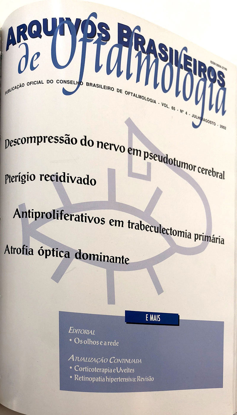
Purpose: To evaluate the efficacy of optic nerve sheath decompression (ONSD) performed in eyes with papilledema and visual loss in patients with pseudotumor cerebri.
Methods: Seventeen patients (24 eyes) submitted to optic nerve sheath decompression from January 1991 until January 1999 were analyzed. All of them had the diagnosis of pseudotumor cerebri and were operated on due to reduction of visual acuity (VA) and/or visual fields (VF). Two patients did not tolerate clinical treatment with diuretics and corticosteroids and the others did not present a satisfactory response to these treatments. Four patients had been previously submitted to lumboperitoneal shunt.
Results: Surgery was unilateral in 10 patients and bilateral in 7. Four patients were submitted to bilateral simultaneous surgery because of the severity of visual loss. Visual acuity and/or visual fields improved in 14 eyes, remained unchanged in 9 and became worse in one of 24 operated eyes. The patient that worsened already had rapidly progressive severe visual loss with a remaining paracentral island of vision and visual acuity of counting finger in the preoperative period. Papilledema resolved in every operated eye, followed by optic atrophy in many of them. Patients who did not improve vision were those who already had severe visual loss at the time of surgery. Moderate intraoperative hemorrhage occurred in two surgeries. Conclusions: Optic nerve sheath decompression can be useful to protect visual function in patients with papilledema from pseudotumor cerebri who do not respond properly to clinical treatment. It can also be helpful as an adjunctive treatment in patients with lumboperitoneal shunt surgery.
Keywords: Papilledema; Pseudotumor cerebri; Pseudotumor cerebri; Pseudotumor cerebri; Surgical decompression; Optic nerve; Visual acuity; Visual fields
Arq. Bras. Oftalmol. 200265
| DOI: 10.1590/S0004-27492002000400003
Purpose: To evaluate and compare long-term outcomes of trabeculectomies with subconjunctival injections of 5-fluorouracil (5-FU) after sugery or use of mitomycin C (MMC) during surgery.
Methods: Retrospective study of 133 eyes who underwent primary trabeculectomy.
Results: Intraocular pressure levels were not statistically significant at any of the postoperative visits when comparing the 5-fluorouracil and mitomycin C groups. In each group, analyzed separately, the intra-ocular pressure levels showed no statistically significant difference before or after surgery. The incidence of postoperative complications was not statistically significant as well, except for a greater incidence of the corneal epithelial defects, more frequent in the 5-fluorouracil group. In both groups, a greater incidence of development/progression of cataract was noted.
Conclusion: Both mitomycin C and 5-fluorouracil promoted an important and stable decrease in intraocular pressure with similar complication rates.
Keywords: Trabeculectomy; Glaucoma; Intraocular pressure; Visual fields; Fluorouracil; Mitomycins; Fluorouracil; Mitomycins; Retrospective studies
Arq. Bras. Oftalmol. 200265
| DOI: 10.1590/S0004-27492002000400004
Purpose: To evaluate the use of conjunctival autograft transplantation in the treatment of recurrent pterygium.
Methods: Retrospective study of 36 patients (36 eyes), with recurrent pterygium, who underwent conjunctival autograft as a treatment option for recurrent pterygium. The minimal follow up was 6 months.
Results: We observed a recurrency rate of 41.6%, 5.5% had loss of graft and 2.7% dehiscence.
Conclusion: Conjunctival autograph is a safe procedure for recurrent pterygium, with few complications, although, the recurrence rate is still high in these patients.
Keywords: Pterygium; Recurrence; Autologous transplantation; Conjunctiva; Mitomycin; Mitomycin; Followup
Arq. Bras. Oftalmol. 200265
| DOI: 10.1590/S0004-27492002000400005
Purpose: Autosomal dominant optic atrophy is a hereditary optic neuropathy characterized by progressive visual loss in childhood, color vision anomalies, visual field defects and temporal pallor of the optic disc. This disease has been mapped to a 1.4 cM interval in chromosome 3q28-29 between markers D3S3669 and D3S3562. One family was mapped to chromosome 18q12.2-12.3. Linkage analysis in three families with autosomal dominant optic atrophy with polymorphic DNA markers for chromosome 3q28-29 and 18q12.2-12.3.
Methods: 57 individuals from three families underwent ophthalmological examination. Genomic DNA was extracted from blood samples. Linkage analysis was performed between the disease and 11 polymorphic markers around 3q28-qter and 18q12.2-12.3. Polymerase chain reaction (PCR) fragments sizes were identified in a scanner gel using a 373 DNA sequencer. These numbers were used as alleles for pedigree analysis. The lod scores were calculated using the MLINK program.
Results: All three families presented optic atrophy with autosomal dominant pattern of inheritance, variable expression and high penetrance. Two families were linked to 3q28-29 markers. A maximal lod score of 3.56, at a recombination fraction of zero, was obtained using the marker D3S3669 in one family. The linkage area was defined in a 2 cM interval by haplotype analysis between markers D3S2418 and D3S1305, because patients III.4 and III.14 showed crossing-overs. The third family was not linked to 3q28-29 neither to 18q12.2-12.3. Conclusions: There is genetic heterogeneity in autosomal dominant optic atrophy, because the third family did not map to any known locus. And a third locus for this disease may exist.
Keywords: Autosomal dominant optic atrophy; Visual acuity; Visual fields; Color vision defects; Linkage (Genetics)
Arq. Bras. Oftalmol. 200265
| DOI: 10.1590/S0004-27492002000400006
Purpose: To verify the effects on optic disc topography of newly diagnosed glaucoma subjects after ingestion of glycerol, a drug that causes major changes in both the intra-ocular pressure (IOP) and cerebrospinal fluid pressure (CSFP).
Methods: We studied 14 patients presenting with intra-ocular pressure over 25 mmHg, without previous use of hypotensive therapy.
Results: Glycerol produced a significant intra-ocular pressure average reduction of 28.2% (95% CI of 20.7 to 32.1%). Despite the obtained intraocular pressure reduction, no significant change was observed in any evaluated topographic parameter (cup area and volume, rim area and volume, maximal and mean cup depth). Conclusions: Overall, no significant change in the topographic parameters, using the "Heildelberg Retina Tomograph", was observed after ingestion of glycerol.
Keywords: Intra-ocular pressure; Optic disc; Optic disc; Glycerol; Glaucoma; Tomography
Arq. Bras. Oftalmol. 200265
| DOI: 10.1590/S0004-27492002000400007
Purpose: To evaluate the initial treatment of infectious corneal ulcers and their laboratory tests.
Methods: Twenty four cases of infectious corneal ulcers seen in the External Corneal Disease Section at the Hospital do Servidor Público Estadual - São Paulo - Brazil, underwent Gram staining and culture for bacterial and fungus. Treatment with fortified cefalotine (50 mg/ml) and gentamicin (14 mg/ml) was started.
Results: Seventeen cultures (70.83%) were positive for bacterial infection and seven (29.17%) showed no growth. In three cases (15.76%) the Gram staining showed correlation with the culture. Three cases did not respond to the initial treatment and had the medications changed according to the results of the cultures. All patients responded to the clinical treatment. Conclusions: Ours results showed that combination of fortified cefalotine and gentamicin eye drops was efficient to treat most of the infectious corneal ulcers.
Keywords: Corneal ulcers; Cephalothin; Gentamicins; Ciprofloxacin; Microbial sensitivity tests; Microbial drug resistance
Arq. Bras. Oftalmol. 200265
| DOI: 10.1590/S0004-27492002000400008
Purpose: To evaluate the complications before and after vitrectomy, their correlation with the time the lens remained in the vitreous cavity, and final visual acuity. The long-term complications were also evaluated.
Methods: Retrospective study of 12 patients who had lens material in the vitreous after cataract surgery. The patients underwent vitreous surgery from November 1994 to June 2000 at the Ophthalmology Service of the Hospital de Clínicas de Porto Alegre (HCPA).
Results: After the displacement of the nucleus into the vitreous cavity, 5 patients had corneal edema, 3 had uveitis, 8 glaucoma, 1 retinal detachment, and all patients had reduced visual acuity. After vitrectomy, 2 patients had hyphema, 6 glaucoma, 2 retinal detachment, 1 phthisis bulbi, 1 cystoid macular edema, 2 perfluorcarbon in the anterior chamber, and 7 corneal edema. Almost all complications resolved on the long-term evaluation (mean 6 months). The length of time the nucleus remained in the vitreous cavity was less than 15 days in 3 patients and more than 15 days in 9 patients.
Conclusion: The results suggest that the fragments retained in the vitreous cavity should be promptly removed to prevent the inflammatory response and to improve the visual prognosis.
Keywords: Crystalline lens; Lens subluxation; Vitrectomy; Preoperative care; Postoperative complications; Visual acuity; Vitreous body; Retrospective studies
Arq. Bras. Oftalmol. 200265
| DOI: 10.1590/S0004-27492002000400009
Purpose: To study the epidemiologic profile of the patients with penetrating ocular trauma (POT) before and after the application of the new traffic code.
Methods: Retrospective study of 253 patients with POT examined at the Ocular Trauma Section (UNIFESP) from January 1997 to April 1999. The patients were divided into 2 groups: Group I, patients with ocular trauma before the new traffic code; Group II, trauma history after the new code. The patients were evaluated regarding different aspects on trauma and ophthalmic evaluation.
Results: The epidemiological findings regarding age, sex and race were similar in both groups. In group I, the patients between 21-50 years presented similar trauma etiology distribution, while in group II, in the same age range, they presented predominance of automotive-related trauma. Regarding seat belt use, 60% and 92% of patients were not using seat belts at the time of the trauma in groups I and II, respectively. 60% of the patients in group II mentioned alcohol consumption against 40% in group I.
Conclusion: Besides the impact measures taken by state to control accidents, ocular trauma damages continue to be related to preventable risk factors like seat belt use and alcohol consumption.
Keywords: Eye injuries; Traffic accidents; Seat belts; Retrospective studies
Arq. Bras. Oftalmol. 200265
| DOI: 10.1590/S0004-27492002000400010
Purpose: To identify improvement in visual performance of low vision students after assessment and management conducted at the Low Vision Service of State University of Campinas (UNICAMP). Method: Fourteen low vision students aged six to 30 years, attended in a room with resources for visual deficiency in Americana and Santa Bárbara d'Oeste -- SP during 1998 received complete ophthalmologic examination, specialized low vision assessment and educational intervention.
Results: The most prevalent cause of vision loss was operated congenital cataract with four cases (28.6%), followed by congenital bilateral toxoplasmic macular scars and eye malformation, both with two cases (14.3%) cases each. Eight students (57.2%) had acuity classified as severe vision loss, four (28.6%) profound, one (7.1%) moderate and one (7.1%) nearly normal vision. Twelve (85.7%) were behind expected school grade. Optical aids were prescribed for 12 (85.8%) students but only 7 (58.3%) acquired the aids thus improving significantly their school performance.
Conclusion: All students improved school performance even considering that 12 (85.7%) had severe to profound vision loss. As a group their performance could even be better if the optical aid prescriptions were acquired by all. This indicates the need of a social work to support such needs. For good results at school and effective student inclusion a partnership between school, family and specialized education is necessary. We recommend to promote the benefits of the resource room.
Keywords: Vision; Therapeutical management; Evaluation studies; Child
Arq. Bras. Oftalmol. 200265
| DOI: 10.1590/S0004-27492002000400011
Purpose: To evaluate morphologic alterations caused by intravitreous injection of bupivacaine, a long-term local anesthetic agent much used in ocular regional blockades, in the retina of albino rabbits.
Methods: The drug was injected at a 0.75% in 0.1ml concentration into the vitreous, close to the retina in one eye, while an equal volume of balanced saline solution was injected into the other eye (control), with indirect ophthalmoscopy performed before, during and immediately after the procedure and at 1hour, 24 hours and 72 hours; both light and electron microscopy were performed at 24 and 72 hours after administration of the anesthetic agent.
Results: Immediately following the injection of bupivacaine, ophthalmoscopy re-vealed the retina to have in all cases a whitened aspect close to the injection site, a phenomenon attributed to the presence of spreading depression, which was also found (at less frequency and intensity), in the control eyes. Further found alterations included: retina edema, 6 (60%); area of vitreous condensation, 5 (50%); and papilla pulse, 2 (20%). Conclusions: Intravitreous injection of bupivacaine at 0.75% concentration (used for retrobulbar, peribulbar local anesthesia or a different technique in extended eye surgeries) triggered no morphological alterations when studied by light microscopy; the injection however did trigger mild edema-suggesting alterations in the horizontal cells of the retina of albino rabbits, studied by electron microscopy within the 24 and 72-hour periods.
Keywords: Retina; Bupivacaine; Bupivacaine; Drug toxicity; Rabbits
Arq. Bras. Oftalmol. 200265
| DOI: 10.1590/S0004-27492002000400012
Purpose: To describe and analyze the clinical manifestation of Usher's syndrome and to study their hereditary standard as well.
Methods: Four patients with Usher's syndrome were studied at Ophthalmologic Clinic of the Getúlio Vargas Hospital, in Teresina-PI, between December, 1997 and March, 1999. The patients were scheduled for tests of visual acuity, refraction, biomicroscopy, direct and indirect ophthalmoscopy, tonometry, retinography, visual field with Goldman perimeter and audiometry. Family's pedigree was built and an anamnesis and indirect binocular ophthalmoscopy of 63 (sixty-three) relatives were performed.
Results: Of the four patients with Usher's syndrome, one was of type I and three of type II. Their visual acuity varied from 20/100 to 20/400 and the dysacusis was from moderate to severe. All patients had retinitis pigmentosa, and tubular visual field.
Conclusion: It is important to make the ophthalmologist aware of Usher's syndrome diagnosis in face of a case of retinitis pigmentosa, calling attention to the autosomal recessive inheritance pattern of the disease as well as to instruct the patients about the clinic manifestations of the disease and the importance of their being treated and attended by an ophthalmogist and otolaryngologist.
Keywords: Retinitis pigmentosa; Retinitis pigmentosa; Eye abnormalities; Hearing loss, sensorineural; Syndrome
Arq. Bras. Oftalmol. 200265
| DOI: 10.1590/S0004-27492002000400013
Purpose: To present how the section of Refractive Surgery of the Federal University of São Paulo assesses the candidates and the reasons to indicate for corneal refractive surgery.
Methods: We examined 1626 patients. Anamnesis, complete ophthalmologic examination and corneal topography were performed in all patients. The patients spontaneously seeked evaluation at the Refractive Surgery Section by telephone without a previous screening. Reasons to refuse patients for refractive surgery were previously established by the Refractive Surgery Section.
Results: Based on current technology and clinical experience, 265 patients (16.29%) were refused for excimer laser corneal refractive surgery. Myopia of patients who had insufficient preoperative corneal pachymetry for the laser treatment was the main cause for refusal (51 patients). Cataract (45 patients), keratoconus (31 patients), amblyopia (21 patients), hyperopia > 5 diopters and mixed astigmatism (19 patients), presbyopia (unaware ness of the need for optical correction after the procedure; 16 patients), pupillary diameter > 5mm (9 patients), single eye (9 patients), progressive myopia (8 patients), postradial keratotomy (7 patients) and low ametropia (7 patients) were among the reasons for the refusal.
Conclusion: Candidates for excimer laser corneal refractive surgery may present risk factors that should be known in order to avoid complications.
Keywords: Refractive errors; Cornea; Myopia; Corneal topography; Keratoconus
Arq. Bras. Oftalmol. 200265
| DOI: 10.1590/S0004-27492002000400014
Purpose: To investigate the level of knowledge among patients with glaucoma about their disease and to compare this group with the number of patients with suspected hypertension or diabetes who know about their disease.
Methods: A descriptive study was conducted during the first stage of the Glaucoma Project in 1999 in the municipality of Piraquara, PR, where the local population older than 40 years was submitted to ophthalmologic examination (tonometry and fundoscopy), measurement of arterial pressure, and blood glucose test.
Results: A total of 922 individuals were examined. Two hundred and six patients were screened as suspected diabetics; of these, 42.72% (n=88) knew about their disease. We detected 625 patients with suspected hypertension, 60.64% of whom (n=379) declared that they were hypertensive. A hundred and fifty pacients were screened as suspected open angle glaucoma; of these, only 3.33% (n=5) knew about their disease and 96.67% (n=145) were not aware of their disease.
Conclusion: The level of knowledge of patients with a diagnosis of glaucoma is extremely low compared to the number of patients with suspected arterial hypertension or diabetes.
Keywords: Hypertension; Diabetes mellitus; Glaucoma; Evaluation studies; Adult
Arq. Bras. Oftalmol. 200265
| DOI: 10.1590/S0004-27492002000400015
Purpose: To describe 5 cases with the diagnosis of canalicu-litis, emphatising the diagnosis of canaliculitis as an important cause of chronic conjunctivitis and blepharitis and also to demonstrate the efficacy of the surgical treatment in these patients.
Methods: Surgical management of 5 cases with diagnosis of canaliculitis.
Results: The gram results were suggestive of Actinomyces israelli and the culture results were negative. The surgical treatment was shown efficacious. Conclusions: The surgical treatment proposed was shown efficacious.
Keywords: Eye infections; bacterial; Actinomycosis; Case report; Male; Female; Adult
Arq. Bras. Oftalmol. 200265
| DOI: 10.1590/S0004-27492002000400016
The authors describe three cases of a rare disease, the diffuse subretinal fibrosis syndrome. They report clinical aspects, fundoscopic features and outcomes after steroid therapy. They also analyze the differential diagnosis between the diseases of white dot syndrome by reviewing the literature. In conclusion, they point out the meaning of early diagnosis and treatment of diffuse subretinal fibrosis syndrome in order to prevent the progression to the fellow eye.
Keywords: Fibrosis; Fibrosis; Choroid diseases; Retinal diseases; Choroiditis; Retina; Syndrome; Adult
Arq. Bras. Oftalmol. 200265
| DOI: 10.1590/S0004-27492002000400017
Purpose: To describe the clinical presentation and diagnostic findings on imaging examination of an unusual case of spontaneous intraconal orbital hematic cyst.
Methods: The patient was submitted to a complete ophthalmic and clinical examination, computerized tomography, orbital ultrasound, magnetic resonance imaging and surgical approach to the lesion.
Results: Clinical examination revealed severe proptosis, congestive signs and visual loss. Computerized tomography revealed homogeneous and well-defined mass lesion in the orbit compressing the globe. On ultrasound the lesion had low internal reflectivity. Magnetic resonance imaging showed in T1 weighted images isointense signal surrounded by hyperintense signal in the periphery and hyperintense signal in T2 weighted images, indicating blood inside the lesion. Surgical approach confirmed the presence of a hematic cyst of the orbit that was drained with excellent functional recovery. Conclusions: Intraconal hematic cyst should be included in the differential diagnosis of well-defined lesion with acute or subacute development in the orbit that present hyperintense signal in both T1 an T2 weighted images on magnetic resonance imaging. Treatment should be done on an emergency basis in cases with disfiguring proptosis and risk of visual loss.
Keywords: Orbital diseases; Cysts; Hemorrhage; Tomography, x-ray computed; Ultrasonography
Arq. Bras. Oftalmol. 200265
| DOI: 10.1590/S0004-27492002000400018
Corticosteroids have been used in clinical practice since 1949, and their potential beneficial effects in Ophthalmology, including inflammatory and autoimmune diseases, are now a fact. Nowadays, these steroids are used, systemically or locally, to treat a constellation of eye diseases. This article considers the most used corticosteroids in Ophthalmology, their indications, their side effects, and routes of administration.
Keywords: Adrenal cortex hormones; Adrenal cortex hormones; Uveitis; Adrenal cortex hormones
Arq. Bras. Oftalmol. 200265
| DOI: 10.1590/S0004-27492002000400019
The present study carries out a review of the theme hypertensive retinopathy. Thus it presents a brief review of the historical data on hypertensive retinopathy. The study reports the classical alterations of hypertensive retinopathy and its classifications, as well as the most recent findings associated with systemic arterial hypertension, the likely patho-physiological mechanisms, and the several methods used for investigation, their advantages and disadvantages; a critical view of the interpretation of signs of the ocular bulb involvement by systemic arterial hypertension; furthermore, based on the diversity of methodologies used in the investigation of retinopathy, comments are made on its reverberation, in the prevalence of hypertensive retinopathy and its implications, as a target organ of systemic arterial hypertension, in an updated context of the metabolic syndrome and of other elements associated with systemic arterial hypertension, such as leptin and endothelin.
Keywords: Hypertension; Retinal diseases; Blood pressure; Ophthalmoscopy


 ABO is licensed under a Creative Commons Attribution-NonComercial 4.0 Internacional.

