
André Luiz Parolin Ribeiro1; Paulo Schor2; Norma Allemann2; Wallace Chamon2; Mauro Silveira de Queiroz Campos2
DOI: 10.1590/S0004-27492002000400013
ABSTRACT
Purpose: To present how the section of Refractive Surgery of the Federal University of São Paulo assesses the candidates and the reasons to indicate for corneal refractive surgery. Methods: We examined 1626 patients. Anamnesis, complete ophthalmologic examination and corneal topography were performed in all patients. The patients spontaneously seeked evaluation at the Refractive Surgery Section by telephone without a previous screening. Reasons to refuse patients for refractive surgery were previously established by the Refractive Surgery Section. Results: Based on current technology and clinical experience, 265 patients (16.29%) were refused for excimer laser corneal refractive surgery. Myopia of patients who had insufficient preoperative corneal pachymetry for the laser treatment was the main cause for refusal (51 patients). Cataract (45 patients), keratoconus (31 patients), amblyopia (21 patients), hyperopia > 5 diopters and mixed astigmatism (19 patients), presbyopia (unaware ness of the need for optical correction after the procedure; 16 patients), pupillary diameter > 5mm (9 patients), single eye (9 patients), progressive myopia (8 patients), postradial keratotomy (7 patients) and low ametropia (7 patients) were among the reasons for the refusal. Conclusion: Candidates for excimer laser corneal refractive surgery may present risk factors that should be known in order to avoid complications.
Keywords: Refractive errors; Cornea; Myopia; Corneal topography; Keratoconus
RESUMO
Objetivo: O objetivo deste estudo é mostrar como o setor de Cirurgia Refrativa da Escola Paulista de Medicina da Universidade Federal de São Paulo avalia seus candidatos e quais as razões para não selecioná-los para cirurgia refrativa. Métodos: Foram examinados 1626 pacientes. Anamnese, avaliação oftalmológica completa e topografia corneana foram realizadas em todos os pacientes. Os pacientes procuraram avaliação no setor de Cirurgia Refrativa espontaneamente sem triagem prévia. Resultados: Não foram selecionados 265 pacientes (16,29%) para cirurgia refrativa na córnea. A principal razão para contra-indicar a cirurgia refrativa corneana por excimer laser foi miopia de pacientes que se apresentaram e que não tinham paquimetria corneana suficiente para o tratamento (51 pacientes). Catarata (45 pacientes), ceratocone (31 pacientes), ambliopia (21 pacientes), hipermetropia > 5 dioptrias (19 pacientes), astigmatismo misto (19 pacientes), presbiopia (pacientes que não sabiam que teriam que usar óculos para leitura após a cirurgia; l6 pacientes), diâmetro pupilar > 5 mm em ambiente iluminado (9 pacientes), olho único (9 pacientes), miopia apresentando progressão (8 pacientes), ceratotomia radial prévia (7 pacientes) e, ametropia baixa (7 pacientes) foram as causas de contra-indicação. Conclusão: Candidatos para cirurgia refrativa corneana podem apresentar fatores de risco que devem ser conhecidos de modo a diminuir os riscos pós-operatórios.
Descritores: Erros de refração; Córnea; Miopia; Topografia da córnea; Ceratocone
INTRODUCTION
Since the 80's, laser light has been studied as a tool for the correction of ametropias. At present, the argon fluoride excimer laser (193 nm) is used for the corneal curvature in an effective, safe and stable way (1). There are two major procedures for laser application to the cornea: photorrefractive keratectomy (PRK) and lamellar photorrefractive keratectomy (LASIK). In PRK, the corneal epithelium is removed and laser is applied to the superficial corneal stroma. In the LASIK technique, a corneal flap is made with a microkeratome and the laser is applied to the underlying stroma. The choice between the techniques depends on the surgeon's experience and the ametropia to be treated.
With the widespread use of excimer laser for correction of ametropias, it is important to select candidates who may benefit from the surgery and, mainly, not to operate in cases where expectancy of results has shown a poor correlation with the clinically obtained results.
Refractive surgery is and should be treated as an elective option instead of spectacles or contact lens wearing, which are clinical methods with less, although present, risks (2).
Regarding safety, complications of the surgical method which lead to loss of lines of vision, such as central islands, optical aberrations, decentration, under- and overcorrection, anomalous wound healing or haze, infections, among others, should be considered (1). In the case of LASIK, the appearance of haze is questioned, but flap - related complications, such as incomplete, irregular flap, folds or flap loss, interface epithelial growth, among others shall be considered (1,3).
The purpose of this study is to show how the Sector of Refractive Surgery of the Federal University of São Paulo assesses the candidates and the reasons for not to operating on corneal refractive surgery to patients.
METHODS
This is a retrospective study of 1626 examined patients, based on a prospective questionnaire, from January to September 1999. The patients spontaneously seeked evaluation at the Refractive Surgery Section.
Anamnesis consisted of demographic data, reason for surgery, occupation, use of contact lenses and use of topical or systemic medication. Visual acuity, refraction under cycloplegia, biomicroscopy, tonometry, retinal examination and corneal topography was performed in all patients, in addition to measurement of the pupillary diameter.
Patients presenting retinopathy were referred to subspecialists for evaluation and treatment before further evaluation.
Ultrasound corneal pachymetry was performed in patients with surgical indication for LASIK.
All patients were advised regarding surgical risks, postoperative care, infection, overcorrection, undercorrection and need for optical correction of presbyopia after the surgery.
The Section uses to indicate PRK for myopia less than or equal to 4 spherical diopters (SD) and LASIK for myopia over 4 SD, astigmatic and hyperopic ametropias. Two types of microkeratomes are utilized: Automated Corneal Shaper (Chiron, USA) and Carriazo-Barraquer (Moria, FR). The excimer laser used in this study was Summit Apex Plus (Summit, USA). Surgeries for myopia were performed with 6.0 - 6.5 mm multizone ablation. In the case of hyperopia, the optical zone was 6.5 - 9.0 mm. For correction of astigmatism, an removable mask with a 5 - 6.5 mm optical zone was used.
Corneal warpage was not listed in this study, since it is considered a temporary contraindication. After regression of the warpage, another ophthalmologic examination was performed, and surgery was indicated, if appropriate.
Preestablished criteria of the Section for surgical contraindication are:
1) Myopia or compound myopic astigmatism:
a) ametropia over 12 diopters at the steepest corneal meridian
b) estimated postoperative keratometry below 32 diopters
c) thickness of the residual stroma (pachymetry - less than 250 micra (insufficient pachymetry).
2) Hyperopia more than 5 SD or with expected final keratometry more than 50 diopters
3) Astigmatism more than 6 diopters
4) Suspected or patent keratoconus, detected by topography
5) Lens opacification, with or without loss of visual acuity
6) Pupillary diameter greater than 5 mm, measured with a rule in a mesopic environment (examination office)
7) Patients who do not accept the probability of using optical correction for presbyopia after the surgery
8) Patients with amblyopia or with only one functional eye
9) Patients who presented progression of myopia and patients under the age of 20 years
10) Situation in which the risk/benefit ratio of the surgery is not satisfactory to the physician or the patient
11) During the period of the study the Sector did not perform surgery for mixed astigmatism
RESULTS
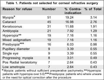
Of the 1626 assessed patients, 265 (16.29%) were not selec-ted for excimer laser corneal refractive surgery. ( Table 1)

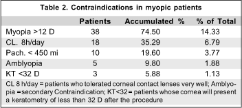
Myopia with insufficient pachymetry was the main factor of contraindication in 51 patients (19.2% of the nonselected patients), thirty-seven of them presenting myopia or compound myopic astigmatism with the most curved meridian more than 12 diopters. None of the contraindicated patients presented sufficient preoperative pachymetry for the residual stromal bed to remain with 250 micra after ablation. ( Table 2)

Lens opacity was the second cause of contraindication for surgery in 45 patients (16.9%). Keratoconus was the third cause of contraindication for surgery (31 patients, 11.6%) followed by amblyopia (21 patients, 7.9%), high hyperopia and mixed astigmatism (19 patients, 7.0% each). Presbyopia (patients who did not accept the high probability of using optical correction for presbyopia after surgery) was the reason for not selecting 16 patients (6.0%) ( Table 3 ). Pupillary diameter greater than 5 mm and single functional eye was the cause for contraindication for surgery in 9 patients each (3.0%). Ametropia progression was the cause for contraindication for surgery in 8 patients (3.0%). In seven patients who had previously undergone radial keratotomy, surgery was contraindicated (2.6%). Low ametropia (which did not cause dependence on optical correction) was the cause for contraindication in 7 patients (2.6%) ( Table 3 ). Other causes of contraindication were: irregular astigmatism, astigmatism more than 5 diopters, glaucoma, Stargardt's disease, patients with psychiatric disorders, rheumatoid arthritis, senile macular degeneration and patients under 20 years old with myopia.

DISCUSSION
Refractive surgery using excimer laser has become a frequent practice among physicians and it is of fundamental importance to known its limitations as well as indications and contraindications in order to correctly advise and select patients.
Both excimer laser corneal refractive surgeries (PRK and LASIK) are considered safe in selected patients. Maghraby et al. (4), compared PRK and LASIK effectiveness, safety and stability for correction of myopia of -2.50 SD to -8.00 SD, and concluded that both presented comparable and satisfactory results. Safety of the procedure seems to be such that Gimbel et al. (5) compared performing simultaneous bilateral LASIK with sequential LASIK and concluded that the former is as safe and effective as the latter.
In our study, insufficient pachymetry for correction in the group of myopic patients was the main cause for contraindication for surgery using excimer laser. In those patients it is important to leave at least 250 micra (6-7) residual stroma to avoid corneal ectasia. Jacobs et al. (8), using a Moria LSK-1 microkeratome, showed that with this type of equipment there is a good reproducibility and precision regarding corneal flap thickness near 160 micra. Ablation depth provided by the excimer laser manufacturer is approximately 0.25 micra per pulse at a radiation exposure of 150 mj/cm 2 (1). With these data it is possible to predict wich patients will remain which less than 250 micra.
Wang et al. (9) showed by the Orbscan system topography of the corneal posterior surface that the risk of post-LASIK corneal ectasia increases when the residual stroma is less than 250 micra. Geggel (10) described a corneal ectasia case after 6.6 SD treatment with the LASIK technique, calling attention to the possibility of occurrence of ectasia even in relatively superficial treatments.
Knorz et al. (11), in a prospective study, showed that predictability significantly decreased in treatment of myopia higher than 15 SD.
For all these reasons, patients with high myopia and/or thin cornea should not be operated on using excimer laser.
Lens opacity was the second cause of contraindication for corneal refractive surgery. Since there is no possibility of determining the time of appearance of clinically significant cataract and, so far, there is no formula able to adequately predict the behavior of residual ametropia after insertion of an intraocular lens in patients submitted to excimer laser corneal refractive surgery, such patients should be contraindicated for surgery. It should be emphasized that surgery for the removal of cataract is performed by inserting an intraocular lens which may compensate the patient's former ametropia. Keratoconus is also an important contraindication for excimer laser surgery. There are no studies proving the long-term efficacy or predictability of refractive correction in corneas with keratoconus. Therefore it is mandatory to perform preoperative corneal topography and to carefully interpret the results.
At present there are some topography softwares for automatic topographic keratoconus detection which use Rabinowitz (12) and Smolek e Klyce (13) criteria, but the localized increase in corneal curvature should raise suspicion of the pathology and postpone the decision about surgery. There are trials in progress to postopone penetrating keratoplasty in these patients, and intracorneal rings or lamellar transplants are future hopes.
Patients with amblyopia and with a single functional eye were not operated on. Even in view of studies, such as that by Gimbel et al. (5), which attest the safety of the procedure, there is a greater risk to the global visual function of these patients; they should be carefully analyzed. In a service with a great number of patients and trainee surgeons, as those of a university service, such cases should not be chosen. Arbelaez et al. (14) showed that in cases of hyperopia LASIK is safe for patients with 1.0 to 5.0 SD. But in cases with more than 5.0 SD, loss of two or more lines of best-corrected visual acuity was significant. In agreement with such results, patients with more than 5.0 diopter hyperopia were not indicated for surgery.
At the time the study was performed, the Sector did not perform surgery for mixed astigmatism.
In this series, the patients not selected because of presbyopia were those who did not accept the fact that they had to use correction for near vision after the surgery.
Holladay et al. (15) published a study showing that large pupil patients suffered from visual disturbance and degradation, the optic of the cornea leading to decrease in contrast sensitivity after LASIK. Halos and false images after PRK occur more often at night in young myopic patients with greater pupillary diameters. These symptoms occur when the effective optical zone of the treatment is smaller than the entrance pupil, a predominat fact in conditions of poor lightening (1). According to such studies, surgery was contraindicated in patients with a pupillary diameter greater than 5 mm in a lightened environment (mesopic condition). Danausory et al. (16) showed that treated eyes with a peripheral transition zone have significantly less haloes and or glare as compared with eyes treated with simple ablation zone. All studied patients were submitted to treatment with an ablation zone greater than or equal to 6 mm.
Corneal warpage due to contact lens use was not listed in this study because it is a temporary contraindication. The need for preoperative examination without contact lens for at least 3 days should be emphasized.
CONCLUSION
In conclusion, candidates for excimer laser corneal refractive surgery may present risk factors that should be known.
Complete preoperative ophthalmologic examination is fundamental for the correct selection of candidates for refractive surgery. Corneal topography has to be performed in all candidates in order to detect subclinical keratoconus. Corneal pachymetry should be carried out in the candidates selected for LASIK. Ablation depth in myopic patients selected for LASIK should be assessed before surgery in order to preserve adequate posterior corneal stroma, avoiding iatrogenic corneal ectasia.
REFERENCES
1. Steinert RF, Bafna S. Surgical correction of moderate myopia: Which method you choose? II. PRK and LASIK are the treatments of choice. Surv Ophthalmol 1998;43:157-79.
2. Lipener C, Parolin Ribeiro AL. Ulcera de córnea bilateral por Pseudomonas em usuário de lente de contato descartável. Arq Bras Oftalmol 1999;62:747-9.
3. Stulting RD, Carr JD, Thompson KP, Waring GO, Wiley WM, Walker JG. Complications of laser in situ keratomileusis for the correction of myopia. Ophthalmology 1999;106:13-20.
4. El-Maghraby A, Salah T, Waring III GO, Klyce S, Ibrahim O. Randomized bilateral comparison of excimer laser in situ keratomileusis and photorrefractive keratectomy for 2.50 to 8.00 diopters of myopia. Ophthalmology 1999;106:447-57.
5. Gimbel HV, Van Westenbrugge JA, Penno EEA, Ferensowicz, Feinerman GA, Chen R. Simultaneous bilateral laser in situ keratomileusis . Ophthalmology 1999;106:1461-9.
6. Probst LE, Machat JJ. Mathematics of laser in situ keratomileusis for high myopia. J Cataract Refract Surg 1998;24:190-5.
7. Seiler T, Koufala K, Richter G. Iatrogenic keratectasia after laser in situ keratomileusis. J Refract Surg 1998;14:312-7.
8. Jacobs BJ, Deutsch TA, Rubenstein JB. Reproducibility of corneal flap thickness in LASIK. Ophthalmic Surg Lasers 1999;30:350-3.
9. Wang Z, Chen J, Yang B. Posterior corneal surface topographic changes after laser in situ keratomileusis are related to residual corneal bed thickness. Ophthalmology 1999;106:406-10; discussion p.409-10.
10. Geggel HS, Talley AR. Delayed onset keractectasia following laser in situ keratomileusis.. J Cataract Refract Surg 1999;25:582-6.
11. Knorz MC, Wiesinger B, Liermann A, Seiberth V, Liesenhoff H (1998). Laser in situ keratomileusis for moderate and high myopia and myopic astigmatism. Ophthalmology 1998;105:932-40.
12. Rabinowitz YS. Keratoconus. Surv Ophthalmol 1998;42:297-319.
13. Smolek MK, Klyce SD. Current keratoconus detection methods compared with a neural network approach. Invest Ophthalmol Vis Sci 1997;38:2290-9.
14. Arbelaez MC, Knorz MC. Laser in situ keratomileusis for hyperopia and hyperopic astigmatism. J Refract Surg 1999;15:406-14.
15. Holladay JT, Dudeja DR, Chang J. Functional vision and corneal changes after laser in situ keratomileusis determined by contrast sensitivity, glare testing, and corneal topography. J Cataract Refract Surg 1999;25:663-9.
16. El Danausory MA. Prospective bilateral study of night glare after laser in situ keratomileusis with single zone and transition zone ablation. J Refract Surg 1998;14:512-6.
From the Refractive Surgery Section - Department of Ophthalmology Universidade Federal de São Paulo (UNIFESP).
1
Orientador do setor de Catarata do Hospital Oftalmológico de Sorocaba e Médico do Setor de Cirurgia Refrativa
do
Departamento
de
Oftalmologia
da
Universidade
Federal
de
São
Paulo
(UNIFESP).
2
Doutor em Oftalmologia pela Universidade Federal de São Paulo e Médico do Setor de Cirurgia Refrativa do Departamento
de
Oftalmologia
da
Universidade
Federal
de
São
Paulo
(UNIFESP).
3
Doutora em Oftalmologia pela Universidade Federal de São Paulo e Médica do Setor de Cirurgia Refrativa do Departamento
de
Oftalmologia
da
Universidade
Federal
de
São
Paulo
(UNIFESP).
4
Livre Docente em Oftalmologia pela Universidade Federal de São Paulo e Médico do Setor de Cirurgia Refrativa
do
Departamento
de
Oftalmologia
da
Universidade
Federal
de
São
Paulo
(UNIFESP).
Endereço para correspondência:
Rua
Benjamin
Constant
681
¾
Bebedouro (SP) CEP 14700-000.
E-mail:
[email protected]
Recebido
para
publicação
em
08.08.2001
Aceito
para
publicação
em
18.03.2002