Arq. Bras. Oftalmol. 199962
| DOI: 10.1590/S0004-27491999000600004
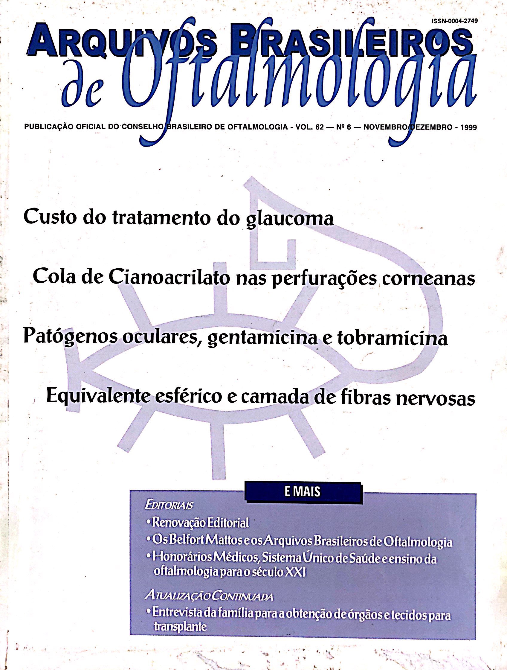
Purpose: To evaluate the cost of glaucoma treatment for an outpatient in a public university hospital. Patients and
Methods: Ninety-four outpatients were ran-domly Glaucoma Clinic of the Ophthalmology Department of the Universidade Federal de São Paulo, submitted to questionnaires at the regarding personal and antiglaucoma medicine expenses. Data collected, tabulated and the sample was divided into two were groups: surgical and nonsurgical.
Results: The median estimated total spent annually on glaucoma treatment was R$ 480.15, equivalent to 13% of the median annual family income of the patients. When the sample was divided into two groups, the figures were quite different. The surgical group had a median annual estimated total expense of R$ 667.94, equivalent to 19% of the median annual family income, whereas the nonsurgical group showed a median annual estimated total expense of R$ 393.54 or 11% of the median annual family income. Sta-tistical difference was found between both groups for total annual cost (p = 0.012).
Conclusion: It was observed that the cost of glaucoma treatment is quite high in respect to the family income, even considering the use of a public hospital.
Keywords: Glaucoma; treatment; cost
Arq. Bras. Oftalmol. 199962
| DOI: 10.1590/S0004-27491999000600005
Purpose: To investigate the indications, outcomes and complications of cyanoacrylate tissue adhesive for stromal thinning and corneal perforations.
Methods: Twenty-six eyes of 26 patients with stromal thinning or corneal perforations (£ 2.5 mm) received methyl 2-cya-noacrylate adhesive and bandage contact lens.
Results: Twelve cases (46%) of corneal perforation were caused by infectious ulcer. For the 26 eyes that received adhesive, 15 (57.7%) needed only one application of the glue and 11 (42.3%) needed two applications. Thirteen eyes (50%) developed for leukoma; 4 (15%) needed surgery and 9 (35%) eyes remained sealed. Visual acuity remained unaltered in 15 eyes (57.7%), improved in 7 (27%) and got worse in 4 (15.3%) eyes.
Conclusion: The cyanoacrylate adhesive is available to all ophthalmologists and is an effective method of temporary or permanent closure of an impending or frank perforation.
Keywords: Cornea; perforations; adhesive
Arq. Bras. Oftalmol. 199962
| DOI: 10.1590/S0004-27491999000600006
Purpose: To evaluate and compare the "in vitro" susceptibility of ocular bacterial isolates to aminoglycoside antibiotics (gentamicin and tobramycin) and to analyze the alteration in susceptibility after 14 years of introduction of tobramycin in Brazilian ophthalmic practice.
Methods: The results of antibiotic susceptibility tests in a period of three years with 887 bacterial strains (Study "A") were retrospectively analyzed and then compared with the results obtained fourteen years ago with 124 microorganisms (Study "B"), when tobramycin was first tested in Brazil for isolated eye strains. In both studies, the data analyzed were from the files of the Ocular Microbiology Laboratory of the Federal University of São Paulo.
Results: The "in vitro" effectiveness of both antibiotics regarding the total number of positive isolates was significantly higher for tobramycin in both studies. An increase in the Staphylococcus aureus strains resistant to tobramycin and only a decreasing trend in the number of microorganisms resistant to gentamicin was observed. With Pseudomonas sp, a strong tendency to the increase of susceptibility to gentamicin and to the appearance of strains resistant to tobramycin occurred. Conclusions: The introduction of tobramycin in the treatment of ocular infections and the possible decrease in number of gentamicin prescriptions during the last fourteen years might be the cause of modifications in the susceptibility observed in the total of positive isolates mainly Staphylococcus aureus and Pseudomonas sp. For the isolated microorganisms from the conjunctiva, the sensitivity to both antibiotics was equivalent.
Keywords: Ocular Infections; Antibiotic susceptibility; Gentamicin; Tobramycin
Arq. Bras. Oftalmol. 199962
| DOI: 10.1590/S0004-27491999000600007
Purpose: To evaluate the effect of spherical equivalent on the acquisition of nerve fiber layer (NFL) thickness with GDX TM Scanning Laser System®.
Methods: Forty-one eyes of 41 volunteers were enrolled in this study. All of them presented with no ocular disease and no visual field defect. The NFL thickness was measured with GDX TM Scanning Laser System® as described in its manual. The values obtained in a group of volunteers with negative spherical equivalent (group I) were compared to those from a group with a positive spherical equivalent (group II) by the Mann-Whitney test.
Results: There was no statistical difference between mea-surements in eyes of group I and those in group II. The NFL thickness measurements were not correlated with the sphe-rical equivalent. Conclusions: In the studied group there was no statistical difference in the GDX TM Scanning Laser System® parameters related to spherical equivalent.
Keywords: Polarimetry; Nerve fiber layer; Spherical equivalent
Arq. Bras. Oftalmol. 199962
| DOI: 10.1590/S0004-27491999000600008
Purpose: To study the incidence of refractive errors in different socioeconomic populational groups and the cost of their correction. Method: 50 patients with refractive errors were divided into 2 groups. Group I was comprised of new outpatients at the Ophthalmic Center of the Hospital Universitário Evangélico de Curitiba (HUEC). Group II consisted of doctors and medical students of HUEC who volunteered for the study. Patients' sex, age, income, educational level, visual acuity, refractive errors and expenses for their correction were recorded. All patients were submitted to ophthalmologic examination.
Results: Patients in group I averaged 48.5 years of age and a monthly income of about 5 times the Brazilian minimum wage (mw). Hyperopia was the most frequent refractive error. The corresponding findings for group II were 24.4 years of age, monthly income over 20 times mw and myopia was the most frequent. Conclusions: Yearly expenses amounted to a minimum of 0.3 mw for glasses, minimum of 1.4 mw for contact lenses and refractive surgery of 5.9 mw. We concluded that refractive surgery is a good indication for both groups in the long run.
Keywords: Refractive errors; Socioeconomic aspects
Arq. Bras. Oftalmol. 199962
| DOI: 10.1590/S0004-27491999000600009
Purpose: To evaluate the ocurrence of ophthalmologic involvement in multibacillary leprosy patients.
Methods: At FIOCRUZ, Rio de Janeiro, 77 multibacillary leprosy patients were evaluated on diagnosis.
Results: On ophthalmologic examination 55.8% of the patients presented ocular involvement and the most fre-quent type was a loss of the sensitivity of the cornea.
Conclusion: Ophthalmologic examination should be a routine examination in leprosy diagnosis in order to pre-vent ophthalmologic lesions in leprosy patients.
Keywords: Eye infections; Bacterial; Mycobacterium leprae
Arq. Bras. Oftalmol. 199962
| DOI: 10.1590/S0004-27491999000600010
Purpose: The prospective study compares the confocal microscopic images of the rabbit and human corneal epithelial layers obtained with two objectives having different numerical apertures (NA).
Methods: Ten rabbit eyes were enucleated and kept in place by a pneumatic support to ensure the best performance of each objective, with no disturbance due to eye movements. Five normal patients were selected after agreement. The rabbit eyes and those of the patients were all examined with slit lamp. Confocal microscopy (Tomey, Erlangen-Tennenlohe, Germany) was performed with Achroplan (Zeiss, Oberkochen, Germany) 40x/NA = 0.75 and 63x/NA = 0.9 objectives. Selected images of the corneal epithelium layers were evaluated qualitatively for the size, shape, light scattering, and reflection of the cells.
Results: The cells in the superficial corneal epithelium of rabbit and human eyes, before desquamation, produced a much brighter reflection than the surrounding cells. This was clearly seen only with the 63x/NA = 0.9 objective. The corneal epithelium subsegments of the rabbit eyes, like the basal cell layer or the intermediate cell layer (wing cells), were visible under the high numerical aperture (63x/NA = 0.9) objective only. These layers, in the human eyes, were more clearly visible using the high numerical aperture objective.
Conclusion: An objective with a high numerical aperture and a high optical resolution provided an improved analysis to study all the layers of the rabbit and human corneal epithelium.
Keywords: Corneal epithelium; Confocal microscopy; Epitélio corneano; Microscopia confocal
Arq. Bras. Oftalmol. 199962
| DOI: 10.1590/S0004-27491999000600011
Introduction: Topographical alterations may occur as a secondary result of the use of hard or soft contact lenses. The expression corneal warpage is used to describe the corneal deformities caused by the lenses. Topographical map of initial keratoconus can be very similar to that of a patient with corneal warpage.
Purpose: To describe a contact lens wearer with keratoconus who developed corneal warpage symptoms that were diag-nosed and studied by topographies and a series of clinical examinations. Case report: A forty-year-old patient with diagnosis of bilateral keratoconus for 12 years was wearing rigid gas permeable contact lenses since the initial diagnosis. On the first exa-mination in 05/95, it was impossible to determine the RE refraction and keratometry was more than 60.00 D. Topogra-phy was carried out being compatible with keratoconus and a Soper RGP lens was fitted with a 20/70 vision. One year later a control topography showed an increase in the central curve and a flattening of the inferior periphery. The use of contact lenses was interrupted and after further six months there was a reversal of the changes, and a new Soper lens was fitted with a better pattern and a 20/40 vision. Discussion: This case shows the occurrence of corneal de-formity in a keratoconus patient using RGP lenses. The au-thor discusses the pathophysiology and clinical and topographical diagnosis of corneal warpage, emphasizing the fact that patients with keratoconus as well as normal patients, can have these alterations which should be differentiated from the keratoconus evolution itself.
Keywords: Keratoconus; Corneal warpage; Contact lens
Arq. Bras. Oftalmol. 199962
| DOI: 10.1590/S0004-27491999000600012
Purpose: Corneal graft rejection suppression after intramus-cular cyclosporine administration at different periods of time was studied on a penetrating keratoplasty rejection model in the rat.
Methods: Inbred Lewis rats were used as recipients and Fischer rats were used as donors in a orthotopic rejection model of corneal transplantation. Intramuscular injection of cyclosporine 10 mg/kg per day was administered up to the 30th postoperative day in 3 groups with different starting periods: at the day of surgery, at the 7th , and at the 9th postoperative day. Grafts were evaluated through clinical and histological examination.
Results: The corneal grafts developed a 100% rejection in about 3 weeks after the penetrating keratoplasty in the control group (n = 5) in which no cyclosporine was adminis-tered. Graft rejection was detected in one case in the groups in which cyclosporine was administered (n = 15), but the process was inhibited during the study period. Histological studies confirmed the clinical evaluations. Conclusions: These data indicate that intramuscular cyclosporine may control the corneal graft rejection, even during the active phase.
Keywords: Cyclosporine A; Keratoplasty; experimental penetrating; Graft rejection
Arq. Bras. Oftalmol. 199962
| DOI: 10.1590/S0004-27491999000600013
Purpose: To show the results of a comparative study between dynamic and normal strategies with Octopus 1-2-3.
Methods: Automatic perimetry using the Octopus 1-2-3 with dynamic and normal strategies was performed on 24 glaucomatous patients (eight males and 16 females) within an average interval of six months between the two exams. All patients had previously submitted to at least one automatic perimetry with the Octopus 1-2-3. The data compared, for both eyes, were: the patient's age, number of questions, mean sensitivity (MS), mean defect (MD), loss variance (LV), short-term fluctuation (SF) and the reliability factor (RF). In the statistical analyses through the paired t test, only the visual fields with RF less than 10 were included. The level of significance was equal to 5% (p < 0.05).
Results: There was no statistically significant difference between the two strategies in relation to age, LV, SF and RF. However, there was statistically significant differences in the duration of the test, number of questions, MS and MD. The dynamic strategy showed a higher diffuse sensitivity and a lower mean defect than the normal strategy. Conclusions: Our results show that the threshold values measured by the dynamic strategy were in close agreement with the values obtained using the normal strategy in patients who have, or are suspected of having, glaucoma and whose visual fields are normal or borderline. They also confirm the claimed reduction in testing time. These results also suggest that, when comparing a visual field exam performed with the dynamic strategy to one performed with a normal strategy, it is necessary to be cautious in regard to MS and MD values.
Keywords: Automated perimetry; Glaucoma; Octopus 1-2-3
Arq. Bras. Oftalmol. 199962
| DOI: 10.1590/S0004-27491999000600014
Samples from the ocular conjunctiva of 23 patients with AIDS, 24 patients infected with human immunodeficiency virus (HIV) and 48 immunocompetent individuals were taken during the months of February, April, July and October. During the year an increase in fungi of the conjunctiva of the AIDS patients, followed by patients infected with HIV and a smaller percentage in healtly people was observed. The most frequently isolated fungus was Penicillium sp followed by Aspergillus sp, Candida sp and Rhodotorula sp.
Arq. Bras. Oftalmol. 199962
| DOI: 10.1590/S0004-27491999000600015
Purpose: To evaluate the prevalence of diabetic retinopathy (DR) in a diabetic association, as well the level of knowledge about the entity among patients.
Methods: This study included 46 patients, at random, from a diabetic association that answered questions about DR and previous ophthalmoscopic examination. Then, they were submitted to indirect ophthalmoscopic evaluation.
Results: Among type 1 diabetic patients, the study found 44.44% of patients with DR and one case of proliferative retinopathy; among type 2, 24.3% of pacients with DR; 65.22% of the total had never been submitted to ophthalmoscopic examination, although 80.43% had already received some information about DR. Conclusions: This study shows that there is low coverage of preventive and detection examinations for DR in this diabetic association, which contrasts with the acceptable level of the patients knowledge, besides being an association directed only to diabetic patients.
Keywords: Diabetes; Retinopathy
Arq. Bras. Oftalmol. 199962
| DOI: 10.1590/S0004-27491999000600016
Purpose: To evaluate the success in reducing intraocular pressure (IOP) using trabeculectomy without antifibro-blastic drugs and trabeculectomy with adjunct 5-fluorou-racil or mitomycin-C as well as to assess if the time of mito-mycin-C exposure modifies this success rate.
Methods: 171 consecutive eyes operated between January 1989 and March 1998 were retrospectively analyzed, 16 eyes having been submitted to simple trabeculectomy, 38 eyes to trabeculectomy and postoperative application of 5-fluorouracil and 117 eyes to trabeculectomy and intra-operative application of mitomycin-C.
Results: There was a significant reduction in IOP with the use of antifibroblastic drugs (p<0.05). Exposure time to mitomycin-C did not present a statistically significant difference (p>0.05). Conclusions: Trabeculectomy associated with antifibro-blastic drugs significantly reduces IOP and promotes short-term stability.
Keywords: Trabeculectomy; 5-Fluoruracil; Mitomycin-C


 ABO is licensed under a Creative Commons Attribution-NonComercial 4.0 Internacional.

