Arq. Bras. Oftalmol. 200265
| DOI: 10.1590/S0004-27492002000200002
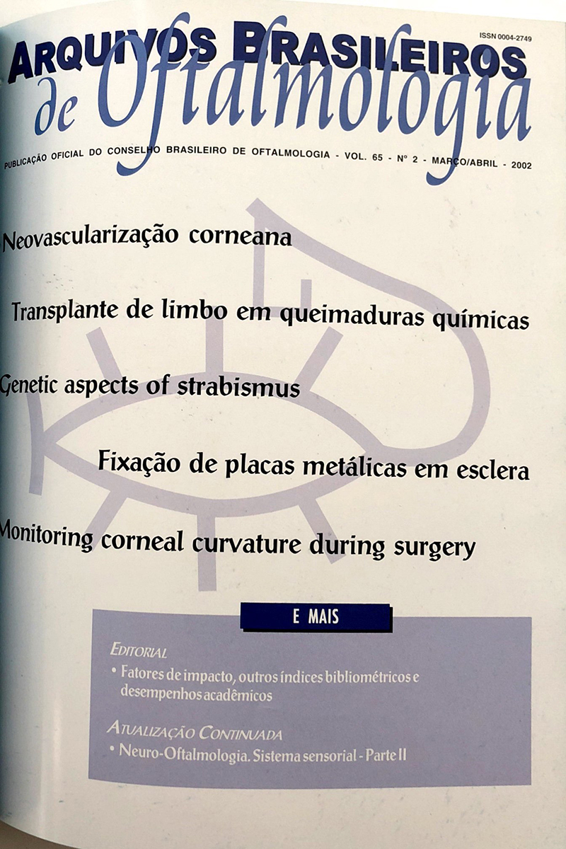
Purpose: To compare the clinical aspects of three experimental models of limbal stem cell (SC) deficiency in rabbits.
Methods: In the present study, 54 rabbits (108 eyes) were divided into 3 experimental groups - (G1), (G2) and (G3). Each group consisted of 18 rabbits. The left eyes were submitted to 3 different experimental techniques - (T1), (T2) and (T3), respectively. A control group was formed with the 54 remaining eyes (RE). In all 3 techniques n-heptanol was used. In T1, the epithelium was removed by applying n-heptanol for 5 minutes. In T2, the n-heptanol application was followed by a 360º conjunctival peritomy at 4 mm beyond the limbus and scraping of the episcleral tissue. In T3, besides the procedure followed in T2, a lamellar dissection towards the limbus was performed, including 1.5 mm of the peripheral cornea and 2 mm of the scleral surface. Six clinical parameters of the corneas were studied: neovascularization, opacity, surface irregularity, epithelial wound healing, epithelial erosion, presence of granuloma and other injuries.
Results: Corneal neovascularization started earlier in G1 and G2. It occurred heterogeneously, ranging from mild to intense, in 100% of the cases. It remained stable from the 28th day up to the end of the experiment (56th day). It was more intense in the upper and lower corneal quadrants and less intense in the nasal and temporal quadrants. Corneal opacity and irregularity of the corneal surface were less intense in G1 than in G2 and G3, which were similar. Reepithelialization started after a 3-day latency period in all the animals submitted to the three experimental techniques and was complete on day 7 in G1 and day 14 in G2 and G3. The rate of occurrence of corneal epithelial erosion and corneal stromal granuloma were similar in the 3 groups. Conclusions: T2 and T3 seemed to be suitable experimental models for total limbal stem cell deficiency and most of the parameters showed similar results. T1 was found to be an appropriate model for the partial-thickness removal of limbal SC. Conjunctivalization and neovascularization occurred in all experimental models.
Keywords: Germ cells; Limbus corneal; Models, animal; Rabbits
Arq. Bras. Oftalmol. 200265
| DOI: 10.1590/S0004-27492002000200003
Purpose: To compare corneal recovery after ocular chemical burn in rabbit eyes treated with conjunctival limbus or keratoconjunctival limbus autografts.
Methods: Thirty-five rabbits underwent a unilateral ocular chemical burn, including the cornea and the limbus, applying a NaOH 1 mol -1 solution. After 30 days, a conjunctival limbus autograft from the contralateral eye was performed in Group 2 (12 eyes); a keratoconjunctival limbus autograft from the contralateral eye was performed in Group 3 (11 eyes) and no surgeries were carried out in the control group (Group 1). Statistical analysis regarding induction and regression of corneal neovascularization, time for corneal re-epithelialization, rate of corneal transparency was performed. The corneal surface phenotype was also analyzed by monoclonal antibodies (AE-5).
Results: After 90 days of follow-up, improvement of corneal transparency, decreased corneal neovascularization and corneal re-ephitelialization were significantly associated with limbal transplantation, carried out in Groups 2 and 3. The control group (Group 1) showed epithelial necrosis in all cases. Corneal surfaces of the operated eyes (Groups 2 and 3) showed a gradual decrease of goblet cells, a gradual increase of corneal epithelial cells, and postoperative stabilization after 30 days. Conclusions: Conjunctival limbus and keratoconjunctival limbus autografts produced the same clinical and epithelial phenotype results in an experimental model of chemical burns in rabbit eyes.
Keywords: Cornea; Chemical burns; Limbal transplantation
Arq. Bras. Oftalmol. 200265
| DOI: 10.1590/S0004-27492002000200004
Purpose: To evaluate the genetic aspects of strabismus.
Methods: Ophthalmic and orthoptic evaluations were performed prospectively on 110 strabismic probands and 478 relatives. We used 3 different criteria in the diagnosis of strabismus: primary diagnosis (dx1) defined as any manifest horizontal or vertical deviation, a secondary diagnosis (dx2) including esophoria (>7 prism diopters) or exophoria (>9 prism diopters), and a tertiary diagnosis (dx3) including abnormal fusional amplitudes, accommodative convergence/accommodation (AC/A) ratio, and/or stereopsis; monofixation syndrome; 4 prism diopters base out; and/or abnormal Maddox test responses. Analyses were carried out within mating types.
Results: Hypotheses of autosomal dominant or recessive inheritance with no sporadics were rejected. Based on the dx1, 25% of the families had more than one individual affected and there was vertical transmission in 13%; adding dx2 there were 36% of the families with more than one affected and 21% had vertical transmission; and adding dx3, there were 73% with more than one affected and 51% with vertical transmission. Conclusions: There is evidence for a pattern consistent with an autosomal dominant form of strabismus in most families.
Keywords: Strabismus; Esotropia; Exotropia; Binocular vision; Orthoptics
Arq. Bras. Oftalmol. 200265
| DOI: 10.1590/S0004-27492002000200005
Purpose: To study two different methods of metal plate fixation: cyanoacrylate adhesive and Mersilene
Ó 6.0 sutures.
Methods: 27 adult rabbits were operated under general anesthesia and the eyes were later examined in order to determine the amount of inflammation. This was done on the 8th, 23rd and 38th day. The adherence of the plate to the sclera was also studied under different perpendicular and tangential traction forces. A comparison was made with a control group.
Results: Clinical signs of inflammatory reaction were found in all eyes in up to the 23rd day and were slightly more intense in the eyes where cyanoacrylate was used. Microscopical signs of inflammation persisted up to 38th day. Different traction forces were applied to the plates and varied from 20 to 80 gf. The latter were unable to detach the plate from the sclera in all eyes at the end of the 38 days. Conclusions: The fixation of metal plates on the scleral surface is a feasible method and can be performed using suture fixation or cyanoacrylate glue.
Keywords: Cyanoacrylates; Sutures; Tensile strength; Rabbits
Arq. Bras. Oftalmol. 200265
| DOI: 10.1590/S0004-27492002000200006
Purpose: To analyze the corneal topography after grade I ocular perforating injuries.
Methods: Twenty-one patients, attended and operated at the Department of Ophthalmology of Santa Casa de Sao Paulo during the year 1998, were included into this controlled clinical transversal study. Computerized corneal topography was performed using the EyeTech CT-2000 Corneal Topographer. Fellow eyes formed the control group.
Results: Fifteen patients (71%) had wounds less than 4 mm. The mean corneal topographic astigmatism was 2.66 ± 2.64 D in the group of perforated eyes, and 0.52 ± 0.25 D in the control group. Statistically significant differences were found comparing the distribution of the patients with topographic corneal astigmatism higher or lower than 2.00 D in the categories of medium corneal wound size (p=0.04) and wound conformation (p=0.02). Localization showed no statistical difference (p=1.00). No statistically significant difference (p=0.98) was found comparing the mean corneal power in the group of perforated eyes with the control group. The final distribution of the topographic corneal pattern showed similarity between the group of perforated eyes and the control group, as well as that of the literature data. Best-corrected visual acuity better or equal to 0.5 was found in 81% of the patients. Conclusions: The topographic astigmatism resulting from corneal laceration was larger than that in the control group, although there was no qualitative change concerning topographic pattern in these eyes, having excluded the cases with irregular topography. Correlation between the final astigmatism and the wound size was found, confirming the critical size of 4 mm. Grade I ocular perforating injuries show good visual outcome, which is not the case with irregular topography or wounds larger than 4 mm.
Keywords: Corneal topography; Eye injuries; Cornea
Arq. Bras. Oftalmol. 200265
| DOI: 10.1590/S0004-27492002000200007
Purpose: Cases of pediatric cataract were assessed at the Department of Cornea and Cataract of the São Geraldo Eye Hospital, Federal University of Minas Gerais, from June 1998 to November 2000.
Methods: Thirty-seven eyes of 31 patients were prospectively analyzed. Thirty-five eyes (94.6%) had congenital cataract and 2 eyes (5.4%) had traumatic cataract. The surgical approach depended on the age of the patients. Patients were classified into the following groups: Group I: (under 1 year of age): manual aspiration of the crystalline lens, with no intraocular lens (IOL) implantation with primary posterior capsulotomy in two eyes; and primary posterior capsulorhexis (PPC) in four eyes and anterior vitrectomy (AV) through limbus in all cases. Group II: (from 1 to 5 years of age): manual aspiration of the crystalline lens with IOL implantation with PPC and AV through limbus. Group III: (from 1 to 5 years of age): manual aspiration of the crystalline lens with IOL implantation with posterior primary capsulotomy and AV through pars-plana. Group IV: (over 5 years of age): manual aspiration of the crystalline lens with IOL implantation without capsulotomy/PPC and AV. In some cases anterior and posterior capsulorhexis were performed utilizing radiofrequency. The primary posterior capsulotomy was performed utilizing the vitreofagus of the Universal -- Alcon phacoemulsificator.
Results: IOL implantation was performed in 31 eyes (83.8%): 28 (90.3%) in-the-bag implantation and 3 (9.7%) in the ciliary sulcus. The follow-up period varied from 2 to 27 months. In group IV, 53.8% (7 eyes), had secondary capsular opacification, which underwent YAG laser capsulotomy.
Conclusion: Use of the radiofrequency for anterior capsulorhexis and PPC as well as the AV improved the surgical outcome of pediatric cataract.
Keywords: Cataract; Cataract extraction; Capsulorhexis; Child
Arq. Bras. Oftalmol. 200265
| DOI: 10.1590/S0004-27492002000200008
Purpose: To determine prognostic factors for local control of uveal melanoma treated by brachytherapy with cobalt 60.
Methods: We evaluated indexes of local tumor control, complications, metastatic disease and global survival in 69 uveal melanoma patients treated with brachytherapy with cobalt 60 from November 1988 to October 1994, at the Hospital do Câncer - A. C. Camargo, São Paulo. We confronted these indexes with different tumoral characteristics and brachytherapy techniques for the determination of the best prognostic factors.
Results: After treatment decrease in height was observed in 55 (79.9%) lesions, in basal diameter in 46 (66.7%), and in visual acuity in 26 (37.2%). 16 (23.2%) eyes were later enucleated due to failure of tumor control in 10 (62.5%) or complications in 6 (37.5%); enucleations were performed on an average 21.1 months after treatment. The mean follow-up time was 44.3 months (10.1-114.3 months). Metastatic disease occurred in 8 (11.6%) patients and the overall five year survival was 89.1%. Complications occurred in 53 (76.8%) patients and eyeball conservation was accomplished in 53 (76.8%). Prognostic factors associated with eyeball conservation and lower complication indexes were: lesion thickness less than 5 mm; basal diameter less than 10 mm; apex doses higher than 12,000 cGy; apex dose tax higher than 45 cGy/h; basal dose lower than 25,000 cGy; basal dose rate lower than 120 cGy/h; and plaque activity lower than 2 mCi.
Conclusion: Tumor size, plaque activity, basal and apex doses and dose rate should be carefully observed in brachytherapy for uveal melanomas in other to optimize its results.
Keywords: Melanoma; Uveal neoplasms; Cobalt; Prognosis
Arq. Bras. Oftalmol. 200265
| DOI: 10.1590/S0004-27492002000200009
Purpose: To investigate the relationship between hypertensive retinopathy (HR) and systemic hypertension (controled or not), gender, age and race.
Methods: 1,954 patients who live in Piraquara, aged over 40 years were examined between 1998 and 2000, during a glaucoma, hypertension and diabetes screening program. Diabetics were excluded. Questionnaire, blood pressure, glycemia and IOP measurement, direct and indirect fundoscopy were performed for all 1,741 patients. Gans classification was used to study the retinal changes.
Results: Considering 1,741 patients, 669 (38.43%) were hypertensive, 645 (37.05%) normotensive and 427 (24.53%) suspected of having hypertension. HR was found in 211 patients (12.12%). 136 (64.46%) were female and 75 (35.54%) male; 135 (63.98%) aged between 40-60 years and 76 (36.02%) aged over 60 years; 75.83% were white and 11.37% black; 154 (73%) were hypertensive, 17 (2.64%) normotensive and 40 (9.37%) hypertension suspects. 12.2% and 25.3% of the hypertensive patients under treatment or not, respectively, had signs of HR.
Conclusion: The prevalence of HR was predominant in hypertensive patients comparing to normotensive and hypertension suspects (p<0.001, OR=5.32). Hypertensive patients without blood pressure control (p<0.01, OR=2.44), aged over 60 years (p<0.001, OR=1.85) and/or black ones (p<0.05 e OR= 1.67) are at increased risk to develop HR.
Keywords: Hypertension; Blood pressure; Retinal diseases; Ocular hypertension; Ophthalmoscopy
Arq. Bras. Oftalmol. 200265
| DOI: 10.1590/S0004-27492002000200010
Purpose: To compare the limits of normality of the electroretinogram (ERG) amplitude and implicit time of two different laboratories.
Methods: Normative ERG data from LKC Technologies, which uses UTAS software were compared to normative data from our laboratory using LKC equipment (EPIC 2000). The former included 110 normal subjects with ages ranging from 22 to 79 years and the latter included 42 normal subjects ranging from 6 to 72 years of age. Single flash ERG using ISCEV (International Society of Clinical Electrophysiology of Vision) recommended routine with ERG-jet contact lens electrodes were performed in both laboratories.
Results: All ERG amplitude parameters, as well as all implicit times were significantly different (p< 0.0001) between both laboratories with the exception of the oscillatory potentials' amplitudes (p = 0.0779) and the b-wave implicit times of the maximal combined response (p = 0.7771). Conclusions: Each electrophysiology laboratory should have its own normal limit standardization, which can be determined either by the 95% confidence interval of its normal data sample, or the 2.5 and 97.5 percentiles. The former is narrower and therefore more sensitive, the latter has a much broader range and therefore is significantly less sensitive.
Keywords: Electroretinography; Electrophysiology; Reference values; Confidence intervals; Comparative study
Arq. Bras. Oftalmol. 200265
| DOI: 10.1590/S0004-27492002000200011
Purpose: Conjunctival discharge is a common complaint among patients treated for retinoblastoma who underwent enucleation and radiotherapy. Lack of data about the microflora in those patients motivated this study.
Methods: One hundred and fourteen orbits of 58 patients were examined. The authors present the results of aerobic bacterial and fungal culture of conjunctival and eyelid margin smears, as well as conjunctival cytology, in 4 different conditions: with and without enucleation, with and without radiation. Microorganisms were identified according to the National Committee for Clinical Laboratory Standards, using Gram and Leishman staining.
Results: Microorganisms were more frequent in irradiated anophthalmic sockets, both in the conjunctiva (54.5%) and eyelid margin (63.6%); however the difference was not statistically significant. On the other hand, in orbits with ocular bulbi, the radiation, in the late phase, represented a limiting factor for the presence of microorganims in the conjunctiva and eyelid margin, but without a statistically significant difference. The coagulase-negative Staphylococcus was the most frequent microorganism in the conjunctiva and eyelid margin in all groups. There was fungus growth, Candida tropicalis, in only one case. Conclusions: Topical antibiotics in patients treated for retinoblastoma who underwent enucleation, with or without radiotherapy, are suggested.
Keywords: Eye; Conjunctiva; Anophthalmia; Retinoblastoma; Bacterial eye infections; Eye enucleation; Staphylococcus; Candida
Arq. Bras. Oftalmol. 200265
| DOI: 10.1590/S0004-27492002000200012
Purpose: The traumatic diseases constitute the great majority of ophthalmic emergencies. Previous knowledge of epidemiologic risk factors makes possible studies on preventive medicine, among others. This study has the aim to evaluate the reasons which lead to look for an ophthalmologic emergency unit.
Methods: The data of patients attended at the "Hospital Governador João Alves Filho", Aracaju, SE -- Brazil, between January 1, 2000 and October 20, 2000, were registered according to their charts, using an exclusive software. 859 patients were selected according to previously established criteria and the following variables were analyzed: sex, age, day of the week, month, diagnosis and chosen medical treatment.
Results: Males represented 72% of all cases; trauma diagnosis was established in 52% of the cases, 59.9% of them being ocular foreign bodies; cases on working days surpassed by 62.5% those on weekends.
Conclusion: Males showed a greater vulnerability to trauma, since the more affected age range was that of the economically active. The most frequent etiology was ocular foreign body and the number of cases on working days was higher than that on weekends.
Keywords: Emergency medical services; Hospital emergency service; Eye foreign bodies; Eye injuries; Occupational accidents
Arq. Bras. Oftalmol. 200265
| DOI: 10.1590/S0004-27492002000200013
Purpose: To study the effects of two drugs (captopril and propranolol) used in the treatment of systemic hypertension, on the intraocular pressure (IOP) of anesthetized dogs.
Methods: 24 dogs, divided into 3 groups of 8 each. In the first group, 1.5 mg/kg IV of captopril (an angiotensin converting enzyme inhibitor) was administered. In the second group, 1.5 mg/kg IV of propranolol (a beta-blocker) was administered. The third group was the control. IOP and blood pressure (BP) were measured by manometry. The perfusion pressure was calculated by the difference between BP and IOP (BP-IOP). The parameters were studied at 6 moments (0, 10, 30, 60, 90 and 120 minutes).
Results: There was significant reduction of IOP (p<0.05) with captopril and propranolol, without difference between the drugs. With captopril the BP and PP decreased markedly at 10 and 30 minutes. With propranolol there was no reduction of BP or PP. Conclusions: Captopril and propranolol reduced IOP. However, the marked reduction of BP, and consequently of PP caused by captopril may be undesirable for irrigation of the optic nerve.
Keywords: Intraocular pressure; Captopril; Propranolol; Hypertension; Dogs
Arq. Bras. Oftalmol. 200265
| DOI: 10.1590/S0004-27492002000200014
Purpose: To examine the effect of patient race on clinical outcomes following excimer laser surgery for myopia and myopic astigmatism.
Methods: A total of 116 eyes from Caucasian patients, 16 eyes from Asian patients and 16 eyes from Hispanic patients who underwent PRK were evaluated retrospectively. PRK procedures were performed by the same surgeon using a 193 nm argon- fluoride excimer laser (VISX) with 160 mJ/cm² fluence and a 6.5 Hz repetition rate at the Doheny Eye Institute. During 6 months of follow-up, changes in the uncorrected visual acuity (UCVA), refraction and spectacle-corrected visual acuity (SCVA) were evaluated. Pairwise comparisons between races were performed for age, sphere and cylinder using independent sample t tests, while UCVA and SCVA were compared using Fisher's exact tests. The accepted level of significance for all tests was alpha = 0.05/3 = 0.0167.
Results: The only differences found were between the Asian versus Caucasian groups related to the spectacle-corrected visual acuity of 20/15 (p=0.01) and in the Asian versus Hispanic groups related to the mean cylinder (p=0.04) at 3 months postoperatively. The comparison of the mean cylinder showed a statistically significant difference between the Asian versus Hispanic groups at 6 months postoperatively (p=0.04). After 6 months, 72.7% of the eyes in the Asian group, 85.7% of the eyes in the Hispanic group and 87.1% of the eyes in the Caucasian group had uncorrected visual acuity of 20/40 or better and the mean sphere and cylinder (±SD) were: --0.55 (± 0.88) and 0.97 (± 0.79); --0.75 (± 1.24) and 0.40 (± 0.45); --1.21 (± 2.55) and 0.75 (± 0.89), respectively.
Conclusion: In this study, there were no statistically significant differences between the three race groups related to the final visual outcome following photorefractive keratectomy. These preliminary results suggest that the clinical outcomes of PRK are not significantly affected by patient race. Larger populations and longer-term studies are needed to definitely determine whether racial differences exist.
Keywords: Myopia; Refraction errors; Keratectomy photorefractive for excimer laser; Racial stocks
Arq. Bras. Oftalmol. 200265
| DOI: 10.1590/S0004-27492002000200015
Purpose: To demonstrate the risk of developing polymegathism in PMMA contact lens users which can be suggestive of endothelial stress.
Methods: Data were obtained from patients who underwent corneal specular microscopy in the last 6 years by the same examiner. Patients were separated in two groups: one of 43 PMMA contact lens users and a control group of 34 patients with a mean age similar to the study group. A contact specular microscope was used and a semiautomated analysis of endothelium mosaic was made.
Results: There were 29 (67.5%) females and 14 (32.5%) males in the study group. The mean age of the study group was 37.86±9.81 and 37.08±6.59 of the control group. The mean time of the lens use was 17±7.5 years. The study group had 53.5% polymegathism and only 11.76% in the control group. The odds ratio found was 8.63 (2.33 < OR < 34.90) and the estimated and uncorrected chi-square was 14.52 with a p<0.01. Among the 23 cases with polymegathism from the study group, 6 (34.78%) had minimal stress, 10 (43.47%) had moderate stress and 7 (21.74%) had marked stress.
Conclusion: A 8 times higher risk was determined for hard contact lens users to develop endothelial stress compared to the control group of the same age.
Keywords: PMMA contact lenses; Corneal endothelium; Polymegathism
Arq. Bras. Oftalmol. 200265
| DOI: 10.1590/S0004-27492002000200016
The author describes a technique for dissection of the nucleus of the cataract into two parts: an external one or external layer, and an internal one or internal nucleus. The anterior portion of the external layer is divided into segmentes like a "pizza" with the phaco tip and a Sinskey hook or a "chopper" and the internal nucleus is dislocated and emulsified. The emulsification of the external layer gives rise to two different surgical models, one for mature and the other for moderately hard cataracts. In the case of mature cataracts we emulsify each segment of the anterior external layer and then the posterior portion, which has already been moved to the anterior chamber. In the case of moderately hard cataracts it is possible to separate a set of layers made of hard anterior and posterior fibers, emulsify them, and leave the superficial layers which continue to protect the posterior capsule. The hard layers are emulsified in the space, which is created by the emulsification of the internal nucleus, which we call the intranuclear space.
Keywords: Crystalline lens; Cataract extraction; Phacoemulsification
Arq. Bras. Oftalmol. 200265
| DOI: 10.1590/S0004-27492002000200017
Purpose: To describe the ophthalmological, dermatological, light microscopy (LM) and scanning electron microscopy (SEM) findings of a new macular dystrophy associated with the loose anagen hair syndrome (LAHS).
Methods: A family of seven patients, four of them affected, was examined. The patients underwent a complete ophthalmological examination, color test (Ishihara and Farnsworth D-15), ecography, angiography, laboratory and dermatological tests, sweat test, light microscopy and scanning electron microscopy of the hair.
Results: In two affected sisters we found on eye fundoscopy pigmentary dispersions in the posterior pole of the retina, with macular staphyloma. In two brothers, the same pigmentary dispersions in the posterior pole was found, with more pigmentation and a yellow coloring in the macular area and without staphyloma. The light microscopy and scanning electron microscopy evaluation of the affected individuals confirmed loose anagen hair syndrome. In a not affected woman and man all examinations were normal, except the light microscopy and scanning electron microscopy that showed some similarities with the affected individuals. As for the way of inheritance, the pedigree is compatible with autosomal recessive inheritance with partial expression of the heterozygote. Conclusions: There is only one report in the international literature of loose anagen hair syndrome association with ocular coloboma. In this study we describe the findings of a new macular dystrophy associated with the loose anagen hair syndrome, a dystrophy whose fundoscopy findings are different between men and women. Since it is the first report in the literature, the described findings strongly suggest that this association can be part of a new nosological entity.
Keywords: Alopecia; Hair; Loose anagen hair syndrome; Hereditary corneal dystrophies; Electron scanning microscopy; Hypotrichosis
Arq. Bras. Oftalmol. 200265
| DOI: 10.1590/S0004-27492002000200018
The authors describe a case of a circumscribed choroidal hemangioma with secondary retinal detachment treated with transpupillary thermotherapy using diode laser. The settings were: 900 mW power, 4.2 mm spot, for 3 minutes, QuadrAspheric
Ò lens (Volk
â) with a magnification factor of 1.97. One month later, there was a significant shrinkage of the hemangioma with full recovery of vision in this eye. The authors discuss some aspects of this method, which seems promising in treatment of this vascular tumors.
Keywords: Induced hyperthermia; Choroid neoplasms; Hemangioma therapy; Retinal detachment; Lasers
Arq. Bras. Oftalmol. 200265
| DOI: 10.1590/S0004-27492002000200019
Purpose: To describe the clinical and pathological aspects, surgical treatment and results of three cases of divided nevus -"kissing nevus"- of the eyelid.
Methods: Review of the clinicopathological data and surgical techniques used to treat these patients.
Results: Cure of the lesions with good esthetic and functional results were obtained in all cases. Conclusions: Surgical excision of the divided nevus of the eyelid in more than one surgical session using flaps from the adjacent skin, and in one case, a free skin graft from the contralateral superior lid, was curative in the studied three cases.
Keywords: Pigmented nevus; Skin transplantation; Eyelids; Reconstructive surgical procedures; Case report
Arq. Bras. Oftalmol. 200265
| DOI: 10.1590/S0004-27492002000200020
This is the second part of a review of papers on the visual afferent system published from 1997 to 1999. In this part the author presents the most important contributions made to areas such as neuroretinitis, optic nerve tumors, idiopathic intracranial hypertension, hereditary optic neuropathies, optic disc drusen, optic nerve hypoplasia, traumatic and toxic optic neuropathy as well as geniculate and retrogeniculate visual disorders. Selected papers are considered in relation to their results and previously established concepts.
Keywords: Neuritis; Geniculate bodies; Afferent neurons; Optic nerve diseases; Visual pathways; Optic nerve; Vision disorders; Optic chiasm; Review


 ABO is licensed under a Creative Commons Attribution-NonComercial 4.0 Internacional.

