Arq. Bras. Oftalmol. 2023;86 (3 )
:1-6
| DOI: 10.5935/0004-2749.20230044
Abstract
Objetivo: O setor nasal do ângulo da câmara anterior pode apresentar maior densidade de canais coletores, o que pode influenciar no resultado de cirurgias angulares. Considerando as diferenças anatômicas no ângulo da câmara anterior, comparamos os resultados das abordagens de trabeculoplastia seletiva a laser nasal e temporal de 180 graus no glaucoma de ângulo aberto.
Métodos: Revisão retrospectiva de prontuários de pacientes com glaucoma de ângulo aberto (primária, pseudoexfoliação e pigmentar), que realizaram pelo menos uma sessão de trabeculoplastia seletiva a laser de 180 graus entre dezembro/2016 e outubro/2018. O setor nasal (N1) ou temporal (T1) foi escolhido a critério do médico. Os pacientes que não apresentaram diminuição da pressão intraocular (PIO) entre 3 e 6 meses foram retratados com trabeculoplastia seletiva a laser de 180 graus no setor de ângulo oposto (T2 e N2). O principal resultado medido foi a diminuição da pressão intraocular no 6º mês de acompanhamento após a última trabeculoplastia seletiva a laser. Uma análise de regressão multivariável avaliou os fatores associados à redução da pressão intraocular após o tratamento.
Resultados: O procedimento foi realizado inicialmente em 45 olhos (N1=25, T1=20 olhos), e repetido no setor ângulo da câmara anterior oposto em 19 olhos (N2 = 11, T2 = 8 olhos). Os testes ANOVA mostraram que apenas a abordagem N1 apresentou diferença significativa na diminuição da pressão intraocular em relação a T1, N2 e T2 (p=0,0014). A pressão intraocular basal (p=0,021) e o setor ângulo da câmara anterior (N1; p=0,044) se correlacionaram com a diminuição da pressão intraocular.
Conclusão: A trabeculoplastia seletiva a laser de 180 graus realizado inicialmente no setor nasal foi associado a uma diminuição mais significativa da pressão intraocular em comparação com a abordagem temporal. Considerando as diferenças setoriais no ângulo da câmara anterior, mais estudos prospectivos são necessários para confirmar nossos achados e fornecer protocolos para trabeculoplastia seletiva a laser mais eficientes.
Keywords: Glaucoma de ângulo aberto; Terapia a laser/métodos; Pressão intraocular; Trabeculoplastia/métodos.
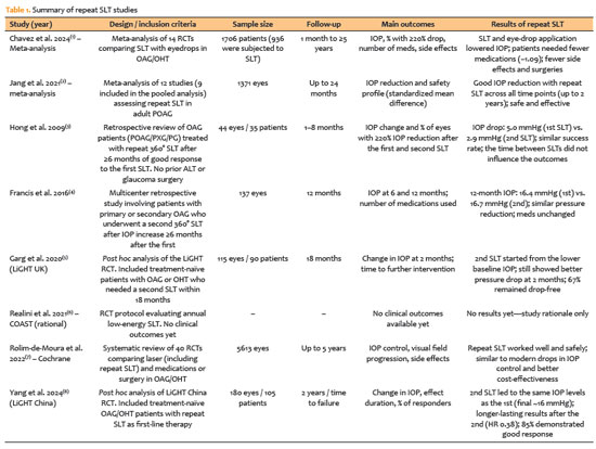
Arq. Bras. Oftalmol. 2025;88 (1 )
:1-8
| DOI: 10.5935/0004-2749.2023-0103
Abstract
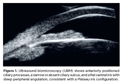
PURPOSE: This study aimed to compare the safety and effectiveness of intraocular pressure reduction between micropulse transscleral cyclophotocoagulation and “slow cook” transscleral cyclophotocoagulation in patients with refractory primary open-angle glaucoma.
METHODS: We included patients with primary open angle glaucoma with at least 12 months of follow-up. We collected and analyzed data on the preoperative characteristics and postoperative outcomes. The primary outcomes were a reduction of ≥20% of the baseline value (criterion A) and/or intraocular pressure between 6 and 21 mmHg (criterion B).
RESULTS: We included 128 eyes with primary open-angle glaucoma. The preoperative mean intraocular pressure was 25.53 ± 6.40 and 35.02 ± 12.57 mmHg in the micropulse- and “slow cook” transscleral cyclophotocoagulation groups, respectively (p<0.001). The mean intraocular pressure was reduced significantly to 14.33 ± 3.40 and 15.37 ± 5.85 mmHg in the micropulse- and “slow cook” transscleral cyclophotocoagulation groups at the last follow-up, respectively (p=0.110). The mean intraocular pressure reduction at 12 months was 11.20 ± 11.46 and 19.65 ± 13.22 mmHg in the micropulse- and “slow cook” transscleral cyclophotocoagulation groups, respectively (p<0.001). The median preoperative logMAR visual acuity was 0.52 ± 0.69 and 1.75 ± 1.04 in the micropulse- and “slow cook” transscleral cyclophotocoagulation groups, respectively (p<0.001). The mean visual acuity variation was -0.10 ± 0.35 and -0.074 ± 0.16 in the micropulse- and “slow cook” transscleral cyclophotocoagulation, respectively (p=0.510). Preoperatively, the mean eye drops were 3.44 ± 1.38 and 2.89 ± 0.68 drugs in the micropulse- and “slow cook” transscleral cyclophotocoagulation groups, respectively (p=0.017), but those were 2.06 ± 1.42 and 1.02 ± 1.46 at the end of the study in the slow cook” and micropulse transscleral cyclophotocoagulation groups, respectively (p<0.001). The success of criterion A was not significant between both groups. Compared with 11 eyes (17.74%) in the slow cook” transscleral cyclophotocoagulation group, 19 eyes (28.78%) in the micropulse transscleral cyclophotocoagulation group showed complete success (p=0.171). For criterion B, 28 (42.42%) and 2 eyes (3.22%) showed complete success after micropulse- and slow cook” transscleral cyclophotocoagulation, respectively (p<0.001).
CONCLUSION: Both techniques reduced intraocular pressure effectively.
Keywords: Sclera/surgery; Glaucoma, open-angle/surgery; Ciliary body/surgery; Intraocular pressure; Laser coagulation/methods; Lasers, semiconductor; Comparative study; Effectiveness
Arq. Bras. Oftalmol. 2026;89 (1 )
:1-8
| DOI: 10.5935/0004-2749.2024-0397
Abstract
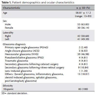
PURPOSE: Glaucoma is one of the leading causes of irreversible blindness worldwide. When topical hypotensive agents or laser trabeculoplasty fail to adequately control the disease, escalation of therapy becomes necessary, with transscleral cyclophotocoagulation being one of the available options. Several variations of transscleral cyclophotocoagulation exist, including traditional continuous wave, MicroPulse, and slow-coagulation techniques. We propose a novel variation – custom slow-coagulation transscleral cyclophotocoagulation – which combines elements of both continuous wave and slow-coagulation approaches. This study aimed to evaluate the outcomes of this technique in patients with refractory glaucoma.
METHODS: This retrospective, interventional study included 104 eyes of 83 patients with refractory glaucoma who underwent custom slow-coagulation transscleral cyclophotocoagulation. Changes in intraocular pressure, visual acuity, the number of glaucoma medications, and postoperative complications were analyzed. A paired t test was used to compare changes in intraocular pressure and visual acuity, while the Wilcoxon signed-rank test was applied to categorical variables. Success rates following custom slow-coagulation transscleral cyclophotocoagulation were estimated using Kaplan–Meier survival analysis.
RESULTS: Mean intraocular pressure decreased significantly from 38.9 ± 15.8 mmHg at baseline to 16.3 ± 9.9 mmHg at Month 12 (p<0.001). The mean number of glaucoma medications also decreased significantly from 3.6 ± 0.6 to 1.8 ± 1.4 (p<0.001). No significant reduction in mean visual acuity was observed during follow-up.
CONCLUSIONS: Custom slow-coagulation transscleral cyclophotocoagulation effectively reduced baseline intraocular pressure and the number of glaucoma medications, with a low rate of complications and no decline in visual acuity over a 12-month follow-up period. This novel technique demonstrated a high safety profile in a Hispanic population and represents a low-cost, minimally invasive procedure with rapid recovery and promising efficacy in intraocular pressure control.
Keywords: Glaucoma/surgery; Sclera; Filtering surgery; Laser coagulation/methods; Lasers, semiconductor/therapeutic use; Intraocular pressure; Blindness/prevention & control; Vision, low/epidemiology; Visual acuity
Arq. Bras. Oftalmol. 2020;83 (6 )
:505-510
| DOI: 10.5935/0004-2749.20200094
Abstract
Objetivo: Avaliar a segurança e o efeito de 12 meses de tratamento com fotocoagulação pelo pattern scanning laser para neoplasia escamosa da superfície ocular em um ambiente com poucos recursos e acesso extremamente limitado a um tratamento cirúrgico.
Métodos: Pacientes adultos com diagnóstico de neoplasia escamosa de superfície ocular foram submetidos a exame oftalmológico completo. Após anestesia tópica e instilação de azul de toluidina 1%, a lesão foi tratada com laser por um tempo de duração que variou de 20 a 100 ms e potência de 600 a 1800 mW. Os pacientes foram examinados semanalmente durante o primeiro mês e foram retratados semanalmente das lesões restantes, conforme necessário. Os pacientes tiveram um seguimento mínimo de 12 meses.
Resultados: Trinta e oito pacientes (38 olhos) foram incluídos no estudo. Todos os pacientes apresentaram neoplasia escamosa da superfície ocular clínica, confirmada por citologia de impressão. A idade dos pacientes variou entre 40 e 83 anos (média de 65.5 anos) e 28 deles eram do sexo masculino (74%). Os pacientes foram divididos em dois grupos: Grupo I (imunocompetente) e grupo II (imunossuprimido). No grupo I, 23 pacientes (74%) apresentaram resposta completa com o controle da lesão após o tratamento com laser. No grupo II, dois dos sete pacientes (28%) apresentaram resposta ao tratamento durante o acompanhamento. A média de aplicações de laser foi de 2,5 (1 a 6 aplicações). Os procedimentos foram bem tolerados.
Conclusões: Os resultados a curto prazo da abordagem de fotocoagulação a laser para o tratamento das lesões conjuntivais de neoplasia escamosa de superfície ocular foram favoráveis, com uma taxa de sucesso de 74% observada em pacientes imunocompetentes. Essa nova estratégia é uma alternativa menos intensiva em recursos que pode demonstrar sua utilidade em ambientes com escassez de salas cirúrgicas e em casos recorrentes. Estudos com acompanhamentos mais longos e amostras maiores são necessários para confirmar nossos achados e avaliar a eficácia do tratamento a laser associado à quimioterapia tópica.
Keywords: Carcinoma de células escamosas/diagnóstico; Neoplasia da túnica conjuntiva/terapia; Terapia a laser; Fotocoagulação
Arq. Bras. Oftalmol. 2021;84 (4 )
:361-366
| DOI: 10.5935/0004-2749.20210052
Abstract
OBJETIVO: Glaucoma é a principal causa de cegueira irreversível no mundo. O pico da pressão intraocular é um dos principais fatores de risco para progressão do glaucoma, e o controle pressórico ainda é o único tratamento efetivo para todas as formas de glaucoma. O objetivo principal deste estudo é comparar a redução basal e do pico da pressão intraocular, obtidas através do Teste de Sobrecarga Hídrica, entre os dois olhos dos mesmos pacientes utilizando latanoprosta 0,005% em um olho e submetidos à aplicação de trabeculoplastia a laser seletiva no olho contralateral.
MÉTODOS: Este é um estudo prospectivo, intervencionista, longitudinal e randomizado. Trinta pacientes consecutivos, glaucomatosos, com pressão intraocular controlada em uso de monoterapia com latanoprosta, foram recrutados de um único centro oftalmológico. Os olhos dos pacientes foram randomizados e um olho foi selecionado para tratamento com trabeculoplastia a laser seletiva e olho contralateral tratado com colírio de latanoprosta 0,005%. Foram avaliados a pressão intraocular basal e pico de pressão intraocular um mês (Teste de Sobrecarga Hídrica 2) e seis meses (Teste de Sobrecarga Hídrica 3) após tratamento.
RESULTADOS: Não houve diferença estatística entre a pressão intraocular pré washout entre os olhos randomizados para trabeculoplastia a laser seletiva e latanoprosta, 13,6 ± 2,1 e 13,3 ± 1,8 mmHg, respectivamente (p=0,182). Em relação à pressão intraocular basal, não houve diferença estatística entre os grupos, tanto no Teste de Sobrecarga Hídrica 2 (p=0,689) e Teste de Sobrecarga Hídrica 3 (p=0,06). Não houve diferença estatística em relação ao pico de pressão intraocular entre os grupos trabeculoplastia a laser seletiva e latanoprosta, no Teste de Sobrecarga Hídrica 2 (p=0,771) e Teste de Sobrecarga Hídrica 3 (p=0,774).
CONCLUSÕES: Em resumo, nosso estudo demonsrou que a eficácia da redução pressórica é similar entre latanoprosta e trabeculoplastia a laser seletiva, e pacientes glaucomatosos que estão com a pressão intraocular clinicamente controlados com latanoprosta e trocam de tratamento para trabeculoplastia a laser seletiva mantém sua pressão intraocular controlada.
Keywords: Glaucoma; Pressão intraocular; Latanoprosta; Lasers
Arq. Bras. Oftalmol. 2025;88 (6 )
:1-5
| DOI: 10.5935/0004-2749.2024-0340
Abstract
PURPOSE: This study aimed to report the surgical outcomes and success predictors of micropulse transscleral cyclophotocoagulation in eyes with refractory glaucoma.
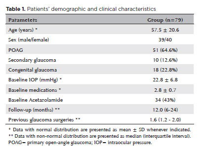
METHODS: This was a noncomparative, interventional case series. Patients with refractory glaucomas, defined as eyes with prior incisional glaucoma surgery failure and uncontrolled intraocular pressure, who underwent micropulse transscleral cyclophotocoagulation between March 2017 and June 2021 were enrolled. A minimum follow-up period of 6 months was required. Preoperative and postoperative intraocular pressure, number of hypotensive medications, surgical complications, and any subsequent related events were recorded. Success criteria were as follows: 1) intraocular pressure reduction ≥20% and intraocular pressure ≤18 mmHg; 2) intraocular pressure reduction ≥30% and intraocular pressure ≤15 mmHg. The need for topical hypotensive medications was not considered a failure.
RESULTS: Seventy-nine (79) eyes (79 patients; mean age, 57.5 ± 20.6 years) were included. Overall, the median follow-up duration was 12.0 (interquartile interval, 6–24) months, and the mean intraocular pressure was reduced from 22.8 ± 6.8 mmHg to 15.5 ± 5.6 mmHg at the last follow-up visit (p<0.001). The mean number of medications was reduced from 2.8 ± 0.7 to 2.0 ± 1.0 (p<0.01). At 12 months postoperatively, the success rates for criteria 1 and 2 were 54.9% and 49.7%, respectively. Aside from one case of corneal ulcer, which fully resolved with clinical treatment, and two cases of persistent hypotony (with no visual acuity loss during follow-up), no other vision-threatening complications were observed during the postoperative period. The magnitude of intraocular pressure reduction at 1 month (adjusted to preoperative intraocular pressure; HR=1.01; p=0.002).
CONCLUSION: Our findings suggest that micropulse transscleral cyclophotocoagulation is a relatively effective alternative for managing refractory glaucomas, with minor postoperative complications. In addition, the initial intraocular pressure reduction was a statistically significant predictor of 1-year success in patients undergoing micropulse transscleral cyclophotocoagulation.
Keywords: Intraocular pressure/physiology; Glaucoma, open-angle/surgery; Trabeculectomy; Laser coagulation/methods; Tonometry, ocular/methods; Postoperative complications; Antihypertensive agents/therapeutic use.
Arq. Bras. Oftalmol. 2026;89 (3 )
:1-8
| DOI: 10.5935/0004-2749.2025-0043
Abstract
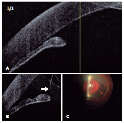
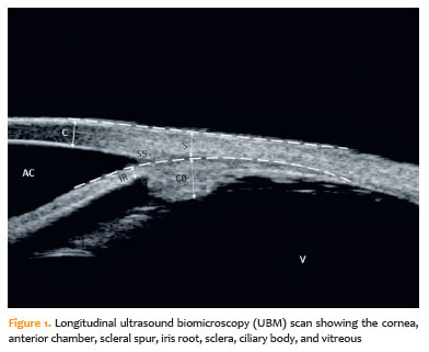
PURPOSE: To evaluate the effect of single-session transscleral diode laser cyclophotocoagulation on intraocular pressure in refractory glaucoma and to determine structural changes using ultrasound biomicroscopy.
METHODS: Forty-three eyes were evaluated. Intraocular pressures at baseline and at the first, third, and sixth months after transscleral diode laser cyclophotocoagulation were compared. Ciliary body thickness, ciliary muscle thickness, ciliary process thickness, iris root thickness, and scleral thickness were assessed at baseline and at the third and sixth months post-treatment.
RESULTS: Reductions in intraocular pressure were significant between baseline and the first month (p=0.018), third month (p<0.001), and sixth month (p<0.001) as well as between the first and third months (p=0.034) and the first and sixth months (p=0.036). Compared with baseline, intraocular pressure reduction rates at the first, third, and sixth months were 34.6%, 56.5%, and 55.3%, respectively, while success rates were 30.2%, 62.8%, and 55.8%, respectively. Decreases in ciliary body thickness, ciliary muscle thickness, and ciliary process thickness were significant between baseline and the third month (p<0.05) and between baseline and the sixth month (p<0.05), whereas changes between the third and sixth months were not significant (p>0.05). Iris root and scleral thicknesses did not change after treatment (p>0.05). At the third and sixth months, significant positive correlations were observed between changes in intraocular pressure and changes in ciliary body thickness and ciliary process thickness (p<0.05).
CONCLUSIONS: To the best of our knowledge, this is one of the few studies comprehensively investigating structural changes after transscleral diode laser cyclophotocoagulation using ultrasound biomicroscopy. Moreover, the relationships between intraocular pressure changes and variations in the ciliary body, ciliary muscle, ciliary process, iris root, and scleral thicknesses were examined in detail. Single-session treatment did not affect iris root or scleral thickness but significantly reduced ciliary body, ciliary muscle, and ciliary process thicknesses. Greater reductions in ciliary body and ciliary process thickness may contribute to more pronounced intraocular pressure reduction.
Keywords: Intraocular pressure; Laser coagulation/methods; Lasers, semiconductor; Microscopy, acoustic; Glaucoma; Ciliary body
Arq. Bras. Oftalmol. 2024;87 (5 )
:0-0
| DOI: 10.5935/0004-2749.2022-0063
Abstract
Objetivo: Comparar os parâmetros de câmara anterior obtidos através da tomografia de coerência óptica de segmento anterior antes e após a iridectomia periférica a laser.
Métodos: Quatorze pacientes com fechamento angular primário e seis com glaucoma primário de ângulo fechado foram prospectivamente avaliados neste estudo. Gonioscopia e tomografia de coerência óptica de segmento anterior com DRI OCT Triton®foram realizadas antes e após a iridectomia periférica a laser. Os seguintes parâmetros de tomografia de coerência óptica de segmento anterior, baseados na localização do esporão escleral, foram avaliados: ângulo de abertura angular a 250 µm, 500 µm e 750 µm, área do espaço entre a íris e o trabeculado a 500 µm, ângulo entre a íris e o trabeculado, extensão do contato entre a íris e o trabeculado e curvatura da íris.
Resultados: A tomografia de coerência óptica de segmento anterior identificou 61% dos indivíduos com dois ou mais quadrantes fechados. A gonioscopia identificou mais quadrantes com ângulo fechado do que tomografia de coerência óptica de segmento anterior antes da iridectomia periférica a laser. Quanto aos parâmetros angulares, apenas ângulo de abertura angular a 250 µm no quadrante nasal não aumentou significativamente após a iridectomia
periférica a laser. A curvatura da íris e a extensão do contato entre a íris e o trabeculado apresentaram redução significativa induzida pelo procedimento a laser. Mesmo nos olhos em que a gonioscopia não identificou aumento da amplitude angular após iridectomia periférica a laser (n=7), ângulo de abertura angular a 750 µm aumentou (nasal: 0,15 ± 0,10 mm para 0,27
± 0,16 mm, p=0,01; temporal: 0,14 ± 0,11 mm para 0,25 ± 0,12 mm, p=0,001), e ICURVE diminuiu (nasal: 0,25 ±
0,04 mm vs. 0,11 ± 0,07 mm, p=0,02; temporal: 0,25 ± 0,07 mm vs. 0,14 ± 0,08 mm, p=0,007).
Conclusão: As alterações na câmara anterior induzidas pelo iridectomia periférica a laser puderam ser avaliadas quantitativamente e documentadas pelo DRI OCT Triton®.
Keywords: Gonioscopia; Tomografia de coerência óptica; Segmento anterior do olho; Glaucoma de ângulo fechado; Iridectomia; Terapia a laser; Lasers
Arq. Bras. Oftalmol. 2025;88 (3 )
:1-6
| DOI: 10.5935/0004-2749.2024-0215
Abstract
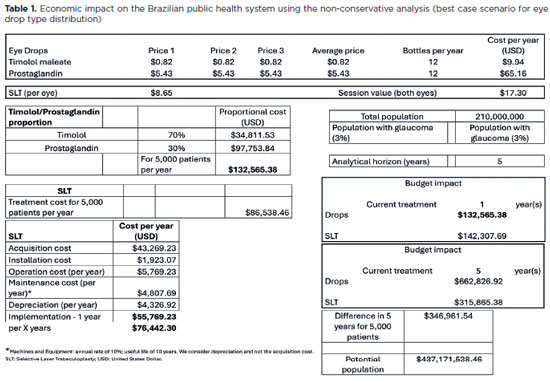
PURPOSE: To evaluate the economic impact of the following initial treatment scenarios for glaucoma on the Brazilian Public Health System (SUS): (1) traditional continuous instillation of hypotensive eye drops and (2) single session of selective laser trabeculoplasty.
METHODS: Economic impact was analyzed in three scenarios, from the least to the most conservative, for a hypothetical cohort of 5,000 individuals with open-angle glaucoma. Thereafter, projections were made on the basis of a glaucoma prevalence of 3% in the 2021 Brazilian population size.
RESULTS: All three scenarios demonstrated that selective laser trabeculoplasty exhibited a significantly lower economic impact than the eye drops on SUS over one and five years. Furthermore, the difference was more than United States Dollar 8 billion at five years when considering 3% of the Brazilian population aged >40 years in 2021.
CONCLUSION: As the initial treatment for primary open-angle glaucoma, selective laser trabeculoplasty exhibited a lower economic impact on SUS than latanoprost and timolol maleate eye drop instillation in all the studied scenarios over one and five-year periods.
Keywords: Glaucoma; Trabeculotomy; Laser therapy; Cost analysis; Health care cost Unified Health System; Brazil
Arq. Bras. Oftalmol. 2024;87 (4 )
:1-5
| DOI: 10.5935/0004-2749.2023-0143
Abstract
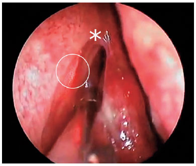
PURPOSE: The purpose of this study is to assess the long-term outcomes of modified transcanalicular diode laser dacryocys torhinostomy in a large cohort of patients affected by primary acquired nasolacrimal duct obstruction.
METHODS: This study, conducted from January 17 to June 2022, encompassed 141 patients (159 procedures) who underwent modified transcanalicular diode laser dacryocystorhinostomy (MT-DCR). The procedure employed an 810-nm diode laser. Patients were monitored for at least a year after the intervention. Anatomical success was determined by ostium patency upon irrigation, while functional success referred to epiphora resolution. Parameters studied included patient demographics, procedure duration, complications, and both anatomical and functional success. Statistical analysis was performed using the Statistical Package for the Social Sciences software, with results considered significant at a 95% confidence interval (p≤0.05).
RESULTS: A total of 159 lacrimal drainage systems (141 patients: 112 women and 29 men) were included in this study. Among them, 18 underwent bilateral procedures. The average patient age was 58 years (range: 34-91 years), and the average surgical duration was 24 minutes (range: 18-35 minutes). One year after the surgery, MT-DCR exhibited anatomical and functional success rates of 84.9% (135/159) and 83% (132/159), respectively.
CONCLUSION: MT-DCR achieved an anatomical success rate of 84.9%, reflecting an excellent outcome. However, further extensive studies with larger sample sizes and longer follow-up periods are necessary to substantiate these findings.
Keywords: Lacrimal duct obstruction; Nasolacrimal duct/surgery; Dacryocystorhinostomy; Lacrimal apparatus diseases; Laser therapy/methods; Lasers, semiconductor/therapeutic use; Regeneration
 ABO is licensed under a Creative Commons Attribution-NonComercial 4.0 Internacional.
13-tab01tb.jpg)
07-fig01.jpg)
09-tab01tb.jpg)
11-fig01tb.jpg)
04-fig01.jpg)