Showing of 1 until 15 from 193 result(s)
Search for: Myopia; Adrenal cortex hormones; Keratomileusis, laser in situ; Intraocular pressure; Glaucoma; Postoperative period; Case reports
Abstract
Objetivo: A refração pós-operatória na cirurgia moderna de catarata por microincisão ganha ainda mais importância em pacientes com cirurgia prévia de ceratomileuse in situ assistida por laser (LASIK). As alterações astigmáticas induzidas cirurgicamente nesses olhos podem diferir não apenas em magnitude, mas também em direção em comparação com córneas virgens. O objetivo deste estudo foi comparar as alterações astigmáticas induzidas cirurgicamente após cirurgia de catarata por microincisão entre córneas pós-LASIK e olhos virgens.
Métodos: Foi revisada uma série de casos de cirurgia de catarata por microincisão em olhos com e sem cirurgia LASIK anterior. Os dados demográficos, o comprimento axial no momento da cirurgia de catarata, a espessura central da córnea, os valores esféricos e cilíndricos, as leituras da ceratometria e o astigmatismo corneano posterior pós-operatório foram avaliados retrospectivamente. O método Alpins modificado foi usado para análise vetorial astigmática e foram avaliados o astigmatismo basal, o astigmatismo induzido cirurgicamente, o vetor de diferença, o efeito de achatamento e o torque.
Resultados: Ao todo, 42 olhos de 24 indivíduos foram avaliados. O Grupo I consistiu em 14 olhos com LASIK prévio; o Grupo II incluiu 28 olhos sem qualquer cirurgia refrativa. A média da espessura corneana central pré-operatória no Grupo I foi significativamente mais fina (p=0,012). Não houve diferença significativa no astigmatismo basal entre os grupos em termos de magnitude e vetores de potência. Após a cirurgia de catarata por microincisão, não houve diferenças significativas nos valores médios esféricos, cilíndricos e leituras médias de ceratometria (todos com p>0,05). No entanto, o astigmatismo induzido cirurgicamente e o vetor de diferença foram significativamente maiores no componente do vetor J45 em olhos pós-LASIK, e o efeito de aumento da inclinação pela cirurgia de catarata por microincisão nas córneas pós-LASIK foi significativo em comparação com olhos virgens (p=0,001, p=0,002 e p=0,018, respectivamente).
Conclusões: A cirurgia de catarata aumentou a inclinação das córneas em ambos os grupos, sendo esse aumento significativamente maior nos olhos pós-LASIK. Certamente, a topografia da córnea antes da cirurgia de catarata é particularmente útil para fornecer interpretações mais precisas do astigmatismo induzido cirurgicamente.
Keywords: Cirurgia de catarata; Ceratomileuse; excimer laser in situ; Cirurgia refrativa; Astigmatismo induzido cirurgicamente; Análise vetorial.
Abstract
Objetivo: Descrever os resultados clínicos do tratamento do crescimento epitelial através da técnica de remoção manual seguido da utilização de um compressor de ar comprimido aquecido após a cirurgia de laser in situ keratomileusis (LASIK).
Métodos: Vinte olhos de 17 pacientes foram incluídos no estudo. Cada paciente havia sido submetido a cirurgia de LASIK com presença de crescimento epitelial e foi submetido a tratamento cirúrgico para sua retirada. O objetivo primário foi identificar a presença de crescimento epitelial recorrente ao final de 3 meses de seguimento. Os objetivos secundários foram as medidas de acuidade visual sem correção, acuidade visual com correção, e complicações pós-operatórias.
Resultados: Dez pacientes (58,8%) eram homens e 7 mulheres. Oito olhos de sete (41,2%) pacientes apresentavam cirurgia de LASIK primária e 12 olhos de 10 pacientes tinham cirurgia de LASIK com retratamento; dezesseis olhos (80%) utilizaram microcerátomo manual e quatro (20%) laser de femtosegundo. A média de idade no momento da cirurgia de remoção do epitélio era de 37,0 anos ± 9,3 (DP) (variando de 24 a 55 anos). Ocorreu recidiva do crescimento epithelial em dois olhos (10%) após 3 meses de seguimento. A acuidade visual sem correção antes da cirurgia era de 0,07 ± 0,09 logMAR, e após a cirurgia passou para 0,02 ± 0,04 logMAR (p=0,06). A chance (odds ration) de aparecimento do crescimento epithelial após uma reoperação de LASIK é 29,41 vezes maior do que no LASIK primário.
Conclusão: A técnica de remoção epitelial manual seguida da utilização de ar comprimido aquecido é segura e efetiva no tratamento do crescimento epitelial após LASIK. Ao final do último acompanhamento, nenhum olho apresentou perda de linhas de visão.
Keywords: Epitélio/crescimento & desenvolvimento; Endotélio corneano; Doenças da córnea; Ceratomileuse assistida por excimer laser in situ; Ceratectomia fotorrefrativa; Procedimentos cirúrgicos refrativos; Acuidade visual
Abstract
Objetivo: O setor nasal do ângulo da câmara anterior pode apresentar maior densidade de canais coletores, o que pode influenciar no resultado de cirurgias angulares. Considerando as diferenças anatômicas no ângulo da câmara anterior, comparamos os resultados das abordagens de trabeculoplastia seletiva a laser nasal e temporal de 180 graus no glaucoma de ângulo aberto.
Métodos: Revisão retrospectiva de prontuários de pacientes com glaucoma de ângulo aberto (primária, pseudoexfoliação e pigmentar), que realizaram pelo menos uma sessão de trabeculoplastia seletiva a laser de 180 graus entre dezembro/2016 e outubro/2018. O setor nasal (N1) ou temporal (T1) foi escolhido a critério do médico. Os pacientes que não apresentaram diminuição da pressão intraocular (PIO) entre 3 e 6 meses foram retratados com trabeculoplastia seletiva a laser de 180 graus no setor de ângulo oposto (T2 e N2). O principal resultado medido foi a diminuição da pressão intraocular no 6º mês de acompanhamento após a última trabeculoplastia seletiva a laser. Uma análise de regressão multivariável avaliou os fatores associados à redução da pressão intraocular após o tratamento.
Resultados: O procedimento foi realizado inicialmente em 45 olhos (N1=25, T1=20 olhos), e repetido no setor ângulo da câmara anterior oposto em 19 olhos (N2 = 11, T2 = 8 olhos). Os testes ANOVA mostraram que apenas a abordagem N1 apresentou diferença significativa na diminuição da pressão intraocular em relação a T1, N2 e T2 (p=0,0014). A pressão intraocular basal (p=0,021) e o setor ângulo da câmara anterior (N1; p=0,044) se correlacionaram com a diminuição da pressão intraocular.
Conclusão: A trabeculoplastia seletiva a laser de 180 graus realizado inicialmente no setor nasal foi associado a uma diminuição mais significativa da pressão intraocular em comparação com a abordagem temporal. Considerando as diferenças setoriais no ângulo da câmara anterior, mais estudos prospectivos são necessários para confirmar nossos achados e fornecer protocolos para trabeculoplastia seletiva a laser mais eficientes.
Keywords: Glaucoma de ângulo aberto; Terapia a laser/métodos; Pressão intraocular; Trabeculoplastia/métodos.
Abstract
Objetivo: Examinar o efeito de infiltrados sub-epiteliais corneanos nas propriedades biomecânicas da córnea após ceratoconjuntivite epidêmica, em comparação com controles saudáveis.
Métodos: Este estudo transversal incluiu pacientes consecutivos com infiltrados sub-epiteliais corneanos bilaterais após ceratoconjuntivite epidêmica e controles saudáveis. Foram medidas a melhor acuidade visual corrigida, uma pontuação do infiltrado sub-epitelial da córnea, a escala de graduação de Fantes e a espessura central da córnea. A histerese da córnea, o fator de resistência da córnea, a pressão intraocular correlacionada à tonometria de Goldmann e a pressão intraocular compensada da córnea foram avaliados com o Ocular Response Analyzer.
Resultados: Este estudo incluiu 66 olhos de 33 pacientes com infiltrados corneanos sub-epiteliais após ceratoconjuntivite epidêmica e selecionou aleatoriamente 37 olhos de 37 voluntários saudáveis. As pontuações médias da escala de Fantes e dos infiltrados sub-epiteliais corneanos nos primeiros olhos acometidos foram respectivamente de 1,8 ± 0,8 e 2,9 ± 1,3. Nos olhos contralaterais, foram respectivamente de 1,3 ± 1,1 e 1,9 ± 1,7 (p=0,009 e p=0,002, respectivamente). O primeiro e o segundo olhos envolvidos tinham córneas significativamente mais finas (respectivamente 526,1 ± 28,1 µm; p=0,005 e 523,4 ± 38,1 µm; p=0,044) em comparação com os controles saudáveis (557,0 ± 38,1 µm). Embora a acuidade visual melhor corrigida tenha mostrado uma correlação positiva com o fator de resistência da córnea (r=0,363, p=0,045) e com a histerese da córnea (r=0,414, p=0,021), a pontuação dos infiltrados sub-epiteliais corneanos mostrou uma correlação negativa com a pressão intraocular correlacionada à tonometria de Goldmann (r=-0,479, p=0,006) e com a pressão intraocular compensada da córnea (r=-0,413, p=0,021).
Conclusão: Os olhos com infiltrados corneanos sub-epiteliais tinham córneas significativamente mais finas em comparação com os controles saudáveis. Ao se medirem os valores de pressão intraocular em pacientes com infiltrados sub-epiteliais corneanos, deve-se levar em consideração tanto as correlações positivas do fator de resistência da córnea e da histerese da córnea com a melhor acuidade visual corrigida quanto as correlações negativas da pressão intraocular correlacionada à tonometria de Goldmann e da pressão intraocular compensada da córnea com a pontuação do infiltrado sub-epitelial da córnea.
Keywords: Ceratoconjuntivite; Pressão intraocular; Epitélio corneano; Corticosteroides; Ciclosporina;Tonometria ocular

Abstract
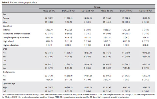
PURPOSE: To evaluate the efficacy of different corticosteroid eye drop formulations (prednisolone acetate 1.0%, dexamethasone 1.0%, and loteprednol etabonate 0.5%) administered for different treatment durations (10 vs. 28 days) in controlling postoperative inflammation following uncomplicated cataract surgery.
METHODS: This randomized, masked clinical trial was conducted at the Instituto Cearense de Oftalmologia. Eligible participants were aged ≥50 yr and scheduled for routine cataract surgery. Exclusion criteria included preexisting ocular disease (elevated intraocular pressure, retinopathy, maculopathy, or uveitis) or concurrent medication use that could confound results. Patients were randomized to receive prednisolone acetate (1.0%), dexamethasone (1.0%), or loteprednol etabonate (0.5%) four times daily for 28 days (with tapering) or for 10 days. Medication bottles, prescriptions, and examiners were masked. Postoperative assessments included ocular symptoms, visual acuity, intraocular pressure, anterior chamber cell count and flare, pachymetry, endothelial cell density, and macular thickness over a 30-day follow-up.
RESULTS: A total of 140 eyes from 140 patients were analyzed (29 prednisolone acetate 1.0%, 18 dexamethasone 1.0%, and 21 loteprednol etabonate 0.5% for 28 days; 28 prednisolone acetate 1.0%, 22 dexamethasone 1.0%, and 22 loteprednol etabonate 0.5% for 10 days). No significant differences were found among the six groups during follow-up. However, eyes treated with dexamethasone (1.0%) showed greater intraocular pressure fluctuations, particularly on Days 7 and 30, and a higher incidence of rebound inflammation in the 28-day regimen. Structural cystoid macular edema without visual impact was observed in 5.9% of eyes in the 28-day groups and 14.2% of eyes in the 10-day groups, as detected by optical coherence tomography at 30 days.
CONCLUSION: Equivalent postoperative inflammation control can be achieved using different corticosteroid eye drop formulations at varying treatment durations following cataract surgery. Brazilian Registry of Clinical Trials (ReBEC): RBR-2frpntv
Keywords: Adrenal cortex hormones; Cataract; Cystoid macular edema; Corticosteroids; Inflammation; Loteprednol etabonate; Ophthalmic solutions; Postoperative period; Intraocular pressure; Visual acuity
Abstract
PURPOSE: This study aimed to report the surgical outcomes and success predictors of micropulse transscleral cyclophotocoagulation in eyes with refractory glaucoma.
METHODS: This was a noncomparative, interventional case series. Patients with refractory glaucomas, defined as eyes with prior incisional glaucoma surgery failure and uncontrolled intraocular pressure, who underwent micropulse transscleral cyclophotocoagulation between March 2017 and June 2021 were enrolled. A minimum follow-up period of 6 months was required. Preoperative and postoperative intraocular pressure, number of hypotensive medications, surgical complications, and any subsequent related events were recorded. Success criteria were as follows: 1) intraocular pressure reduction ≥20% and intraocular pressure ≤18 mmHg; 2) intraocular pressure reduction ≥30% and intraocular pressure ≤15 mmHg. The need for topical hypotensive medications was not considered a failure.
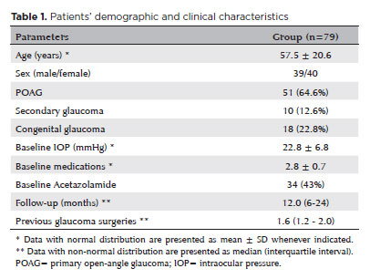
RESULTS: Seventy-nine (79) eyes (79 patients; mean age, 57.5 ± 20.6 years) were included. Overall, the median follow-up duration was 12.0 (interquartile interval, 6–24) months, and the mean intraocular pressure was reduced from 22.8 ± 6.8 mmHg to 15.5 ± 5.6 mmHg at the last follow-up visit (p<0.001). The mean number of medications was reduced from 2.8 ± 0.7 to 2.0 ± 1.0 (p<0.01). At 12 months postoperatively, the success rates for criteria 1 and 2 were 54.9% and 49.7%, respectively. Aside from one case of corneal ulcer, which fully resolved with clinical treatment, and two cases of persistent hypotony (with no visual acuity loss during follow-up), no other vision-threatening complications were observed during the postoperative period. The magnitude of intraocular pressure reduction at 1 month (adjusted to preoperative intraocular pressure; HR=1.01; p=0.002).
CONCLUSION: Our findings suggest that micropulse transscleral cyclophotocoagulation is a relatively effective alternative for managing refractory glaucomas, with minor postoperative complications. In addition, the initial intraocular pressure reduction was a statistically significant predictor of 1-year success in patients undergoing micropulse transscleral cyclophotocoagulation.
Keywords: Intraocular pressure/physiology; Glaucoma, open-angle/surgery; Trabeculectomy; Laser coagulation/methods; Tonometry, ocular/methods; Postoperative complications; Antihypertensive agents/therapeutic use.
Abstract
PURPOSE: This study aimed to evaluate the influence of intrastromal corneal ring segment implants on the intraocular pressure measurements using Goldmann applanation tonometry, rebound tonometry, and noncontact tonometry in keratoconic corneas and analyze the intertonometer agreement.
METHODS: We enrolled 74 eyes in this observational and prospective study. Each participant had a complete eye examination, corneal analysis with Scheimpflug Tomography (Pentacam®), and intraocular pressure evaluation with Goldmann applanation tonometry, rebound tonometry, and noncontact tonometry, before and after intrastromal corneal ring segment implantation (on postoperative days 1, 7, 45, and 90). Intertonometer agreement was assessed using Bland-Altman analysis.
RESULTS: The mean age was 29.9 ± 10.2 years, and 47 (63.5%) eyes had keratoconus grade II. Intraocular pressures were higher for noncontact tonometry preoperatively and on 90 postoperative day (mean ± SD: 12.4 ± and 12.1 ± 2.2 mmHg, respectively), followed by Goldmann applanation tonometry (11.1 ± 3.0 and 11.2 ± 2.7 mmHg, respectively), and were lower for rebound tonometry (9.7 ± and 9.4 ± 3.2 mmHg, respectively). The variation from the Goldmann tonometry on 7 postoperative day to the baseline (p=0.022) and that of noncontact tonometry on 90 postoperative day to the baseline (p=0.021) were statistically significant. The rebound tonometry underestimated intraocular pressure when compared with the Goldmann applanation tonometry by a mean of 1.47 ± 5.19 mmHg. Noncontact tonometry, when compared with Goldmann applanation tonometry, overesti-mated intraocular pressure by a mean of 1.23 ± 4.15 mmHg.
CONCLUSION: Despite statistically significant differences between some postoperative periods, the intraocular pressure measurement differences may not be clinically relevant.
Keywords: Keratoconus; Intraocular pressure; Cornea; Corneal stroma; Postoperative period; Tonometry ocular; Prostheses and implants
Abstract
PURPOSE: Myopia, or nearsightedness, is one of the most common eye conditions worldwide. However, a comparison of the effectiveness of different laser-assisted interventions is lacking. Thus, we aimed to compare the efficacy and safety of LASIK and IntraLASIK in addressing myopia.
METHODS: The study was conducted in two ophthalmology clinics in Beijing, China, in 2022. A total of 84 patients (152 eyes) with different degrees of myopia were examined and underwent LASIK (n=46, 80 eyes) or IntraLASIK (n=38, 72 eyes). Keratometry, corneal topography, pachymetry, visual acuity evaluation, and corneal biomechanical analysis were performed before and after the intervention.
RESULTS: IntraLASIK produced more precise flaps than LASIK, with deviations of <8 mm and 0.1 mm from the intended thickness and diameter, respectively. LASIK resulted in nonuniform flaps, with thickness deviations of 5-86 mm. IntraLASIK demonstrated a superior efficacy for patients with severe myopia and thin corneas, with a mean spherical equivalent of 0.9 D at 6 months compared to the 1.4 D for LASIK. Approximately 91% and 83% of the patients with mild to moderate and severe myopia, respectively, achieved results within ± 0.49 D from the refractive target with IntraLASIK.
CONCLUSIONS: Corneal hysteresis and corneal resistance factor decreased with an increase in laser intensity, and they decreased faster with thinner corneas. Thus, IntraLASIK is more useful than LASIK in patients with thin corneas and severe myopia.
Keywords: Myopia; Lasers; Cornea; Keratomileusis; Laser in situ
Abstract
A ectasia corneana é uma das principais complicações das cirurgias refrativas. Neste caso, descrevemos um caso de ectasia corneana induzida por laser-assisted in situ keratomileusis, que evoluiu com hidrópsia aguda e extravasamento de humor aquoso, necessitando de sutura corneana.
Keywords: Dilatação patológica; Ectasia; Ceratomileuse assistida por excimer laser in situ, Perfuração da córnea; Hidropsia; Ceratocone
Abstract
A paciente era uma mulher de 26 anos com refração manifesta, acuidade visual para longe não corrigida e corrigida de -7,00 × -4,50 a175°, 20/400 e 20/25 no olho direito e -3,25 × -5,25 a 179°, 20/200 e 20/25 no olho esquerdo. A espessura média da córnea foi de 733 e 749 µm, e a espessura epitelial máxima foi de 70 e 68 µm, respectivamente no olho direito e no esquerdo. Inúmeros cistos intraepiteliais foram observados no exame com lâmpada de fenda. Trinta meses após o femto-LASIK, a refração manifesta, a acuidade visual para longe não corrigida e corrigida eram respectivamente de 0,00 × -1,25 a 55°, 20/25 e 20/20 no olho direito e 0,00 × -0,50 a 135°, 20/20 e 20/20 no olho esquerdo. Neste caso de distrofia de Meesmann, a miopia foi tratada com sucesso com femto-LASIK de retalho espesso sem complicações ou ectasia.
Keywords: Ceratomileuse assistida por excimer laser in situ; Miopia; Distrofia corneana epitelial juvenil de Meesmann
Abstract
A ocorrência de ectasia corneana após ceratectomia fotorrefrativa é uma complicação rara, porém grave, em cirurgia refrativa. Os possíveis fatores de risco não são bem avaliados, mas a opinião atual é que a falha na detecção de ceratocone pré-operatório possa ser o principal motivo. Neste relato, descrevemos um caso de ectasia corneana após ceratectomia fotorrefrativa em paciente apresentando padrão tomográfico suspeito no pré-operatório, mas sem alterações degenerativas associadas a ceratocone patológico, conforme revelado por microscopia confocal in vivo da córnea. Além disso, revisamos, na literatura, relatos de casos elegíveis de ectasia pós-ceratectomia fotorrefrativa para encontrar características semelhantes.
Keywords: Córnea/patologia; Dilatação patológica; Ceratoconus; Ceratectomia fotorrefrativa; Ceratomileuse assistida por excimer laser in situ; Microscopia confocal; Humanos; Relato de casos
Abstract
Paciente do sexo masculino, 33 anos, apresentou ceratite infecciosa subaguda unilateral 4 semanas após a cirurgia. A inflamação da córnea foi resistente aos regimes de antibióticos tópicos padrão. A aba da córnea foi derretida e seccionada durante o levantamento e amostragem para diagnóstico. A melhora clínica só foi alcançada após levantamento do retalho, raspagem e diagnóstico microbiológico de micobactérias atípicas e tratamento com amicacina fortificada tópica, claritromicina e claritromicina sistêmica.
Keywords: Córnea/microbiologia; Úlcera da córnea; Infecções oculares bacterianas; Mycobacterium abscessus; Procedimentos cirúrgicos refrativos; Ceratomileuse assistida por excimer laser in situ; Amicacina/uso terapêutico; Claritromicina/uso terapêutico; Humanos; Re
Abstract
We present a case of a patient complaining of monocular diplopia due to a decentered ablation after LASIK. The patient underwent a wavefront-guided retreatment, which resulted in an epithelial ingrowth complication. Additionally, the patient developed cataract, with cataract surgery requiring reliable biometric measurements. Therefore, we opted for corneal treatment and corneal surface regularization. Although we attempted to lift the flap and wash the interface initially, the procedure proved unsuccessful, thereby necessitating immediate flap amputation. Once the corneal surface was regularized in the seventh postoperative month, transepithelial photorefractive keratectomy was successfully performed to homogenize the ocular surface, thereby significantly improving the patient's corrected visual acuity and resolving monocular diplopia. The surface and corneal curvature stabilized by the fifth month after the procedure. Phacoemulsification was then performed along with the implantation of a toric monofocal lens, which was selected using an appropriate formula, resulting in an excellent uncorrected visual acuity.
Keywords: Refractive surgical procedures; Surgical flap/surgery; Keratomileusis laser In situ/methods; Biometry; Corneal topography; Lasers, Excimer/adverse effects; Dipoplia/etiologia; Visual acuity; Humans; Case reports
Abstract
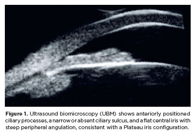
Angle-closure glaucoma is a major cause of visual impairment worldwide, with Plateau iris syndrome presenting management challenges. We present a case report of a 58-year-old woman with advanced, uncontrolled angle-closure glaucoma and Plateau iris. Her history included laser peripheral iridotomy and three glaucoma medications in both eyes. Different treatments were implemented. For the eye with lower intraocular pressure, fewer peripheral anterior synechiae, and milder disease: phacoemulsification with intraocular lens implantation. For the eye with more advanced disease, a two-step approach was used: slow-coagulation transscleral cyclophotocoagulation using the double-arc protocol, followed by phacoemulsification with intraocular lens implantation 2 months later. Both eyes achieved improved visual acuity and intraocular pressure control with fewer medications, without significant complications. This case highlights transscleral cyclophotocoagulation followed by phacoemulsification as an alternative to combined surgeries in uncontrolled angle-closure glaucoma with Plateau iris, offering a simpler technique, more predictable refractive and pressure-control outcomes, and more straightforward postoperative management.
Keywords: Glaucoma, angle-closure/surgery; Iris diseases/surgery; Laser coagulation/methods; Phacoemulsification; Lens implantation, intraocular; Case reports
Abstract
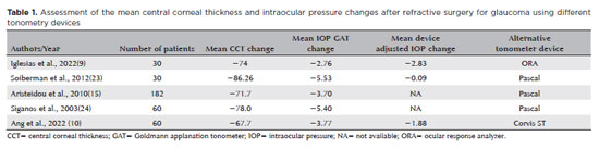
Myopia is a significant risk factor for glaucoma and a growing public health problem worldwide. Detecting glaucomatous changes in highly myopic eyes is diagnostically challenging due to the abnormal appearance of the optic nerve head. These patients also have a greater biomechanical susceptibility to pressure-induced glaucomatous damage. Refractive surgery has become increasingly popular, and many candidates for refractive surgery are myopic. Therefore, we sought to review the aspects of patient evaluation in those who have undergone refractive surgery for myopia concerned with the detection and monitoring of glaucoma development. We identified several important elements of patient evaluation for glaucoma after refractive surgery. These included the need for both structural and functional assessments before and after surgery, and the importance of monitoring for postoperative biomechanical changes in the cornea and their impact on intraocular pressure. We conclude that, in patients who undergo refractive surgery for myopia, it is essential to assess for the presence of glaucoma, to identify staging, and to plan for long-term control of the disease, regardless of IOP.
Keywords: Glaucoma; Intraocular pressure; Myopia; Refractive surgery
ABO is licensed under a Creative Commons Attribution-NonComercial 4.0 Internacional.

About
Issues
Editorial Board
Submission
Official publication of Conselho Brasileiro de Oftalmologia
Rua Casa do Ator, 1.117 - 2º andar - CEP: 04546-004
São Paulo - SP, Brazil
Phone: +55 11 3266-4000