Showing of 1 until 15 from 103 result(s)
Search for: Glaucoma; Artificial intelligence; Large language models; Education, medical; Medical staff, hospital
09-fig01.jpg)
Abstract
Objetivo: Analisar o perfil epidemiológico dos casos de evisceração e enucleação no pronto-socorro oftalmológico de um hospital terciário brasileiro.
Métodos: Análise retrospectiva dos casos tratados no pronto-socorro oftalmológico do Hospital São Paulo (Universidade Federal de São Paulo) entre os anos de 2013 a 2018. Os casos urgentes de evisceração e enucleação foram incluídos e os casos eletivos foram excluídos. A análise dos prontuários médicos foi baseada em: dados demográficos, causas imediatas e associadas ao procedimento, acuidade visual informada, duração dos sintomas antes do atendimento oftalmológico, complicações, distância da residência até o hospital e tempo de hospitalização.
Resultados: 61 enucleações e 121 eviscerações foram incluídas no estudo. Os pacientes tinham uma média de idade de 63,27 ± 18,68 anos; 99 eram do sexo masculino (54,50%) e 83 do sexo feminino (45,60%). As indicações de evisceração e enucleação foram: perfuração corneana com (44,50%) e sem (23,63%) sinais infecciosos, endoftalmite (15,38%), trauma ocular (14,29%), neoplasia (0,55%), queimadura (1,10%) e phthisis bulbi (0,55%). A acuidade visual informada foi de ausência de percepção luminosa (87,36%), percepção luminosa (1.10%), ausência de colaboração (3,30%) e sem dados informados (8,24%). A média de tempo até a busca pelo serviço oftalmológico foi de 18,32 dias. Houve 2 casos de oftalmia simpática após evisceração.
Conclusões: Eviscerações foram predominantemente realizadas em comparação a enucleações em todo o período de estudo. As características demográficas mais comuns foram idade >60 anos e sexo masculino. As principais indicações para procedimentos urgentes de evisceração e enucleação foram perfuração corneana com e sem infecção, endoftalmite e trauma ocular. Este estudo poderia guiar medidas preventivas para evitar procedimentos oculares destrutivos.
Keywords: Evisceração do olho; Enucleação ocular; Úlcera da córnea/epidemiologia; Endoftalmite; Traumatismos oculares; Serviços médicos de emergência; Serviços de saúde ocular.
05-tab01tb.jpg)
Abstract
Objetivos: A pandemia de COVID-19 foi iniciada em março de 2020 e mudou o sistema de saúde. Mudanças na alocação de recursos, sobrecarga de unidades de terapia intensiva, apreensão dos pacientes em procurar atendimento médico não relacionado ao COVID-19 e redução abrupta de todas as consultas e cirurgias não urgentes. Este estudo avalia o impacto em um pronto-socorro oftalmológico após 1 ano de pandemia, avaliando a correlação entre as fases de lockdown, a mortalidade do COVID-19 e as visitas ao pronto-socorro.
Métodos: Estudo observacional retrospectivo que incluiu todos os pacientes admitidos no serviço de emergência oftalmológica do Hospital São Paulo, vinculado a UNIFESP/EPM, entre 1º de janeiro de 2019 e 28 de março de 2021. As visitas foram classificadas e comparadas em um grupo pré-pandemia e pandemia.
Resultados: No período pré-pandemia, o hospital registrou um total de 71.485 atendimentos com média de 194,78 ± 49,74 atendimentos diários, e no grupo pandemia, um total de 41.791 com média de 114,18 ± 43,12 atendimentos diários, redução de 41,4%. Uma diminuição significativa de 16,4% (p<0,001) foi observada na prevalência de conjuntivite aguda e um aumento significativo de 6,4% (p<0,01) na prevalência de corpo estranho da córnea. Foi identificada uma correlação negativa entre a taxa de mortalidade do COVID-19 e as taxas de visita ao pronto-socorro.
Conclusão: Esta análise de um ano mostrou uma redução de 41,4% nas visitas ao pronto-socorro, e uma diminuição significativa nas conjuntivites agudas. A mudança nos hábitos de higiene e o distanciamento social poderiam explicar essa redução, e o aumento da prevalência de traumas corneanos. Achados destacam a necessidade de medidas preventivas e educativas durante os períodos restritivos.
Keywords: SARS-CoV-2; COVID-19; Infecções por coronavírus; Pandemias; Serviços médicos de emergência; Trauma ocular.
08-fig01.jpg)
Abstract
Objetivo: Desenvolver um aplicativo (TopEye) na plataforma iOS para dispositivos móveis que possibilite a captação e interpretação do mapa de cores gerados por qualquer topógrafo corneano através da inteligência artificial (IA).
Métodos: A execução, acompanhamento e avaliação do projeto foi utilizada a metodologia Scrum, processo de desenvolvimento interativo e incremental para gerenciamento de projetos e desenvolvimento ágil de software. O banco de padrões de diagnóstico gerado consiste em 1172 exemplos, divididos em: 275 padrões esféricos, 302 regulares simétricos, 295 regulares assimétricos e 300 irregulares (ceratocone). Para o desenvolvimento da inteligência artificial do aplicativo, foi estabelecido o treinamento da rede com 240 imagens de cada tipo de padrão, totalizando 960 (81,91%) padrões. O restante das imagens, 212 (18,09%), foram utilizadas para testar o aplicativo e usadas para gerar os resultados. O processo é semiautomático, assim a captação da imagem topográfica é realizada com smartphone, o examinador realiza o contorno do relevo corneano manualmente para em seguida a rede neural realizar o diagnóstico.
Resultados: O aplicativo diagnosticou 201 (94,81%) imagens corretamente. De um total de 212 imagens, o algoritmo errou a classificação de apenas 11 (5,19%). A principal ocorrência de erro foi na distinção das classes simétrica e assimétrica. No rastreio do ceratocone o aplicativo alcançou 95,00% de sensibilidade e 98,68% especificidade.
Conclusão: O trabalho resultou na obtenção de um aplicativo eficiente na captura da imagem topográfica pela câmera do smartphone e na interpretação da mesma através da inteligência artificial aplicada.
Keywords: Dispositivos móveis; Inteligência artificial; Topografia corneana; Astigmatismo
Abstract
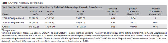
PURPOSE: Natural language models and chatbots, particularly OpenAI’s Generative Pre-Trained Transformer architecture, have transformed human interaction with digital interfaces. The latest versions, including ChatGPT-4o, offer enhanced functionalities compared to their predecessors. This study evaluates the accuracy of ChatGPT-4, ChatGPT-4o, and Claude 3.5 Sonnet in answering questions from the Brazilian Retina and Vitreous Society certification exam.
METHODS: We compiled 200 multiple-choice questions from the Brazilian Retina and Vitreous Society 2018 and 2019 exams. Questions were categorized into three domains: Anatomy and Physiology of the Retina, Retinal Pathology, and Diagnosis and Treatment. Using a standardized prompt developed according to prompt design guidelines, we tested ChatGPT-4, ChatGPT-4o, and Claude 3.5 Sonnet, recording their first responses as final. Three retina specialists performed a qualitative analysis of the answers. Accuracy was determined by comparing responses to the official correct answers. Statistical analysis was conducted using chi-square tests and Cohen’s Kappa.
RESULTS: Claude 3.5 Sonnet achieved the highest overall accuracy (72.5%), followed by ChatGPT-4o (66.0%) and ChatGPT-4 (55.5%). Claude 3.5 Sonnet and ChatGPT-4o significantly outperformed ChatGPT-4 (p<0.01 and p=0.03, respectively), while no significant difference was observed between Claude 3.5 Sonnet and ChatGPT-4o (p=0.16). Model responses agreed 74.5% of the time, with a Cohen’s κ of 0.47. Retinal Pathology was the best-performing domain for all models, whereas Anatomy and Physiology of the Retina and Diagnosis and Treatment were the weakest domains for Claude 3.5 Sonnet and ChatGPT-4, respectively.
CONCLUSIONS: This study is the first to assess Claude 3.5 Sonnet, ChatGPT-4, and ChatGPT-4o in retina specialist certification exams. Claude 3.5 Sonnet and ChatGPT-4o significantly outperformed ChatGPT-4, highlighting their potential as effective tools for studying retina specialist board exams. These findings suggest that the enhanced functionalities of Claude 3.5 Sonnet and ChatGPT-4o offer substantial improvements in medical education contexts.
Keywords: Artificial intelligence; ChatGPT; Retina; Medical education; Ophthalmology, Large language model; Natural language processing
Abstract
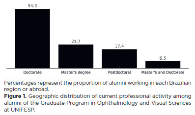
PURPOSE: To evaluate the academic and professional trajectories of graduates from the Graduate Program in Ophthalmology and Visual Sciences at the Escola Paulista de Medicina, Universidade Federal de São Paulo, including geographic distribution, occupational characteristics, and scientific productivity.
METHODS: This descriptive, retrospective, quantitative study included 498 alumni who completed the program between 1979 and 2021. Data were obtained from institutional records and supplemented by public databases (Google Scholar, Lattes Platform, and LinkedIn). The analyzed variables included demographic characteristics, academic background, current professional role, and bibliometric indicators (citation count and h-index). Statistical analyses comprised nonparametric tests and linear regression, with a significance level set at 5%.
RESULTS: Most alumni were Brazilian (96.6%) and physicians (90.7%), predominantly located in the Southeast region (66.9%). Doctoral training was completed by 80.5% of participants. Alumni with current institutional ties to Universidade Federal de São Paulo or Hospital São Paulo demonstrated significantly higher citation counts and h-index values. No significant correlation was observed between time since graduation and citation count (p=0.185). Alumni engaged in academic roles or with postdoctoral training showed greater scientific productivity.
CONCLUSIONS: The findings highlight the strong academic performance and professional integration of alumni from Universidade Federal de São Paulo, particularly within public institutions and the Southeast region of Brazil. Doctoral training and institutional affiliation were associated with higher scientific productivity. Alumni tracking provides valuable insights into the impact of postgraduate programs and informs strategic planning and development.
Keywords: Factual databases; Program evaluation; Medical education; Graduate education; Ophthalmology; Linear models
Abstract
PURPOSE: We developed an artificial intelligence program for calculating intraocular lenses and analyzed its accuracy rate via ultrasonic biometry. This endeavor is aimed at enhancing precision and efficacy in the selection of intraocular lenses, particularly in cases where optical biometry is unavailable.
METHODS: Data was collected from the Hospital de Clínicas de Porto Alegre, which included cases of phacoemulsification with intraocular lens implantation, in which the lens selection was based on ultrasonic biometry. The program, implemented in Python, Java, and PHP, employs the ridge regression method. Two design options were developed: a basic model, which uses only keratometry variables (K1 and K2), axial size and final target refraction in the spherical equivalent, and an advanced model, which incorporates preoperative refraction and the patient's age. The Universal Barrett II formula was used to compare both models.
RESULTS: The sample consisted of 486 eyes from 313 patients, with 350 eyes used for program training and 136 for program validation. The spherical equivalent hit rates, with a variation of ±0.5 D, were 86% and 87.5% for the basic and advanced models, respectively, with no statistically significant difference between them. With the Barret Universal II formula, the success rate was 69%, which was significantly different from the values of the two aforementioned models (p<0.0001). The system was better for medium and long eyes but worse for short eyes (<=22.00 mm).
CONCLUSION: The developed artificial intelligence program was superior to the Barrett formula in terms of performance, in the general context and within the subgroup of patients with longer eyes. This innovation can considerably contribute to the selection of intraocular lenses, particularly in cases where optical biometry is unavailable.
Keywords: Biometry; Intraocular lens; Cataract; Artificial intelligence
Abstract
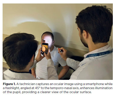
PURPOSE: This pilot study evaluated the diagnostic accuracy of a deep learning model for detecting pterygium in anterior segment photographs taken using smartphones in the Brazilian Amazon. The model’s performance was benchmarked against assessments made by experienced ophthalmologists, considered the clinical gold standard.
METHODS: In this cross-sectional study, 38 participants (76 eyes) from Barcelos, Brazil, were enrolled. Trained nonmedical health workers captured high-resolution anterior segment images using smartphones. These images were analyzed using a deep learning model based on the MobileNet-V2 convolutional neural network. Diagnostic metrics–including sensitivity, specificity, accuracy, positive predictive value, negative predictive value, and area under the receiver operating characteristic curve–were calculated and compared with the ophthalmologists’ evaluations.
RESULTS: The deep learning model achieved a sensitivity of 91.43%, specificity of 90.24%, positive predictive value of 88.46%, negative predictive value of 92.79%, and an area under the curve of 0.91. Logistic regression revealed no statistically significant association between pterygium and demographic variables such as age or gender.
CONCLUSIONS: The deep learning model demonstrated high diagnostic performance in identifying pterygium in a remote Amazonian population. These preliminary findings support the potential use of artificial intelligence–based tools to facilitate early detection and screening in underserved regions, thereby enhancing access to ophthalmic care.
Keywords: Pterygium/diagnostic imaging; Smartphone; Diagnostic techniques, ophthalmological; Deep learning; Telemedicine; Artificial intelligence; Cross-sectional studies; Brazil/epidemiology
Abstract
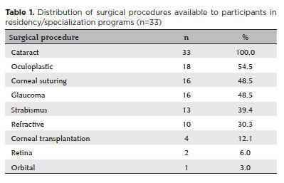
PURPOSE: In Brazil, it has traditionally been standard practice to teach a wide range of surgical techniques to all ophthalmology residents, with the aim of equipping them to manage most ocular conditions. However, with modern developments, access to subspecialists has expanded to nearly the entire country. This raises the question of whether it is still necessary to teach numerous surgical techniques to every resident. This study evaluates the effectiveness of surgical training in Brazilian ophthalmology residency programs to determine if comprehensive surgical training for all residents is truly effective, thereby providing evidence to inform educational policy decisions.
METHODS: A cross-sectional study using a questionnaire distributed to physicians engaged in eye care.
RESULTS: A total of 137 physicians responded to the survey, with 104 (76.0%) having already completed their specialization. The findings indicate that most practicing ophthalmologists received surgical training during residency in cataract, glaucoma, oculoplastic, and strabismus surgeries. Nonetheless, many of these specialists no longer perform most of these surgeries in practice, except for cataract surgery. While 53.8% of those who completed residency reported satisfaction with their training, 35.6% indicated that they wished they had received better surgical preparation.
CONCLUSION: The training of ophthalmology specialists must be made more efficient. Training efficiency is reduced when time and resources are devoted to surgical procedures that many specialists will not perform in their careers.
Keywords: Opthalmologists; Teaching; Education, medical; Ophthalmological surgical procedures; Simulation training; Wet lab; Surveys and questionnaires
Abstract
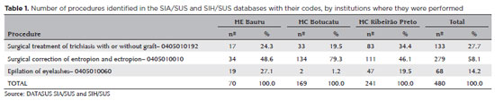
PURPOSE: Trachoma is the major infectious cause of preventable blindness in the world, and its sequelae include the presence of cicatricial entropion and trachomatous trichiasis. Trachoma can be corrected by surgical treatment of the eyelids and, if left untreated, may result in corneal opacification, low vision, and blindness. There are limited data on trachomatous trichiasis in Brazil. This study was conducted to estimate the frequency of entropion and trichiasis surgeries of trachomatous origin based on the records of procedures performed in specialized hospitals that served the Unified Health System (SUS) in the years 2016 and 2017.
METHODS: This was a retrospective study conducted in the oculoplastic sectors of the ophthalmology services of the following three hospitals in the state of São Paulo: Hospital das Clínicas da Faculdade de Medicina de Botucatu (HC Botucatu), Hospital das Clínicas da Faculdade de Medicina de Ribeirão Preto da Universidade de São Paulo (HC Ribeirão Preto), and Hospital Estadual de Bauru (HE Bauru). Medical records corresponding to the codes of interest were evaluated.
RESULTS: In total, 462 medical records were evaluated, including 170 (36.8%) at HC Botucatu, 61 (13.2%) at HE Bauru, and 231 (50.0%) at HC Ribeirão Preto. There were 39 (8.4%) cases of trachomatous trichiasis, ranging from 9 (14.8%) at HE Bauru to 15 (6.5%) at HC Ribeirão Preto.
CONCLUSIONS: The frequency of surgery due to trachoma was low in these oculoplastic services. The state of São Paulo might have reached the goal for trachoma elimination in the surgical component. The questionnaire used for data collection was successfully tested despite some difficulties in collecting data from the medical records. Studies with the same methodology are recommended in other services in the areas of endemic trachoma in the past to understand the frequency of eye lid surgeries performed for treating trachomatous sequelae.
Keywords: Trachoma; Trichiasis; Medical records; Epidemiology; Neglected diseases; Unified Health System; Brazil
Abstract
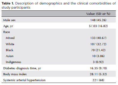
PURPOSE: Diabetic retinopathy screening in low- and middle-income countries is limited by restricted access to specialized care. Portable retinal cameras offer a practical alternative; however, image quality – affected by mydriasis – directly influences the performance of artificial intelligence models. This study evaluated the effect of mydriasis on image gradability and AI-based diabetic retinopathy detection in real-world, resource-limited settings.
METHODS: The proportions of gradable images were compared between mydriatic and non-mydriatic groups. Generalized estimating equations were used to identify factors associated with image gradability, including age, sex, race, diabetes duration, and systemic hypertension. A ResNet-200d model was trained on the mobile Brazilian Ophthalmological dataset and externally validated on both mydriatic and non-mydriatic images. Model performance was evaluated using accuracy, F1 score, area under the curve, and confusion matrix metrics. Sensitivity differences were assessed using the McNemar test, and area under the curves were compared using DeLong's test. The Youden index was used to determine optimal classification thresholds. Agreement between macula- and disc-centered images was analyzed using Cohen's κ.
RESULTS: The mydriatic group demonstrated a higher proportion of gradable images compared with the non-mydriatic group (82.1% vs. 55.6%; p<0.001). In non-mydriatic images, lower gradability was associated with systemic hypertension, older age, male sex, and longer diabetes duration. The AI model achieved better performance in mydriatic images (accuracy, 85.15%; area under the curve, 0.94) than in non-mydriatic images (accuracy, 79.68%; area under the curve, 0.93). The McNemar test showed a significant difference in sensitivity (p=0.0001), whereas DeLong's test revealed no significant difference in area under the curve (p=0.4666). The Youden index indicated that optimal classification thresholds differed based on mydriasis status. Agreement between image fields was moderate to substantial and improved with mydriasis.
CONCLUSION: Mydriasis significantly improves image gradability and enhances AI performance in diabetic retinopathy screening. Nonetheless, in low- and middle-income countries where pharmacologic dilation may be impractical, optimizing model calibration and thresholding for non-mydriatic images is essential to ensure effective AI implementation in real-world clinical environments.
Keywords: Artificial intelligence; Bias; Diabetic retinopathy; Portable camera; Retina
Abstract
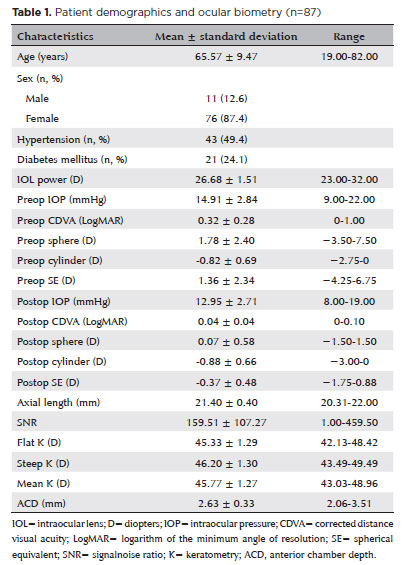
PURPOSE: To compare the refractive prediction error of Hill-radial basis function 3.0 with those of 3 conventional formulas and 11 combination methods in eyes with short axial lengths.
METHODS: The refractive prediction error was calculated using 4 formulas (Hoffer Q, SRK-T, Haigis, and Hill-RBF) and 11 combination methods (average of two or more methods). The absolute error was determined, and the proportion of eyes within 0.25-diopter (D) increments of absolute error was analyzed. Furthermore, the intraclass correlation coefficients of each method were computed to evaluate the agreement between target refractive error and postoperative spherical equivalent.
RESULTS: This study included 87 eyes. Based on the refractive prediction error findings, Hoffer Q formula exhibited the highest myopic errors, followed by SRK-T, Hill-RBF, and Haigis. Among all the methods, the Haigis and Hill-RBF combination yielded a mean refractive prediction error closest to zero. The SRK-T and Hill-RBF combination showed the lowest mean absolute error, whereas the Hoffer Q, SRK-T, and Haigis combination had the lowest median absolute error. Hill-radial basis function exhibited the highest intraclass correlation coefficient, whereas SRK-T showed the lowest. Haigis and Hill-RBF, as well as the combination of both, demonstrated the lowest proportion of refractive surprises (absolute error >1.00 D). Among the individual formulas, Hill-RBF had the highest success rate (absolute error ≤0.50 D). Moreover, among all the methods, the SRK-T and Hill-RBF combination exhibited the highest success rate.
CONCLUSIONS: Hill-radial basis function showed accuracy comparable to or surpassing that of conventional formulas in eyes with short axial lengths. The use and integration of various formulas in cataract surgery for eyes with short axial lengths may help reduce the incidence of refractive surprises.
Keywords: Cataract; Lenses, intraocular; Axial length, eye; Refractive errors; Artificial intelligence
12-tab01.jpg)
Abstract
OBJETIVO: Nos últimos 20 anos, o número de escolas médicas no Brasil aumentou, mas as vagas para especialização em Oftalmologia não acompanharam a demanda crescente. Este estudo quer estimar a demanda por especialização e avaliar a oferta de oportunidades de aprendizado em Oftalmologia.
MÉTODOS: Estudo epidemiológico com pesquisa em banco de dados provenientes do Ministério da Educação e Conselho Brasileiro de Oftalmologia. Estes dados foram checados através de 120 editais publicados pelos serviços de Residência em 2021.
RESULTADOS: De 2002 a 2021, o número de vagas em faculdades de Medicina aumentou 370%, enquanto o número de vagas certificadas de especialização em Oftalmologia aumentou 64%. Houve um desalinhamento de 11.4% entre os dados do Conselho Brasileiro de Oftalmologia e do Ministério da Educação.
CONCLUSÃO: A proporção de graduados em Medicina aumentou muito mais do que a oferta de oportunidades de especialização em Oftalmologia, o impacto disto na busca por vagas de especialização não acreditadas é desconhecido, políticas de monitoramento das vagas de especialização em Oftalmologia devem ser estabelecidas.
Keywords: Oftalmologia; Ensino; Educação médica; Especialização
Abstract
PURPOSE: Standard automated perimetry has been the standard method for measuring visual field changes for several years. It can measure an individual’s ability to detect a light stimulus from a uniformly illuminated background. In the management of glaucoma, the primary objective of perimetry is the identification and quantification of visual field abnormalities. It also serves as a longitudinal evaluation for the detection of disease progression. The development of artificial intelligence-based models capable of interpreting tests could combine technological development with improved access to healthcare.
METHODS: In this observational, cross-sectional, descriptive study, we used an artificial intelligence-based model [Inception V3] to interpret gray-scale crops from standard automated perimetry that were performed in an ophthalmology clinic in the Brazilian Amazon rainforest between January 2018 and December 2022.
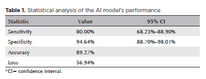
RESULTS: The study included 1,519 standard automated perimetry test results that were performed using Humphrey HFA-II-i-750 (Zeiss Meditech). The Subsequently, 70%, 10%, and 20% of the dataset were used for training, validation, and testing, respectively. The model achieved 80% (68.23%–88.9%) sensitivity and 94.64% (88.8%–98%) specificity for detecting altered perimetry results. Furthermore, the area under the receiver operating characteristic curve was 0.93.
CONCLUSIONS: The integration of artificial intelligence in the diagnosis, screening, and monitoring of pathologies represents a paradigm shift in ophthalmology, enabling significant improvements in safety, efficiency, availability, and accessibility of treatment.
Keywords: Glaucoma; Disease progression; Perimetry; Visual Fields; Visual field tests; Artificial intelligence; Neural networks, computers; Machine learning
07-tab01tb.jpg)
Abstract
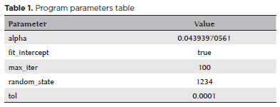
OBJETIVO: Esse estudo tem como objetivo criar um modelo de Machine Learning por um oftalmologista sem experiência em programação utilizando auto Machine Learning predizendo influxo de pacientes em serviço de emergência e casos de trauma.
MÉTODOS: Um dataset de 366,610 visitas em Hospital Universitário da Universidade Federal de São Paulo de 01 de janeiro de 2014 até 31 de dezembro de 2019 foi incluído no treinamento do modelo, incluindo visitas/dia e código internacional de doenças. O treinamento e predição foram realizados com o Amazon Forecast por dois oftalmologistas sem experiência com programação.
RESULTADOS: O período de previsão estimou um volume de 206,37 pacientes/dia em p90, 180,75 em p50, 140,35 em p10 e média de 7,42 casos de trauma/dia em p90, 3,99 em p50 e 0,56 em p10. Janeiro de 2020 teve um total de 6.604 pacientes e média de 206,37 pacientes/dia, 13,5% menos do que a predição em p50. O período teve um total de 199 casos de trauma e média de 6,21 casos/dia, 55,77% mais casos do que a predição em p50.
CONCLUSÃO: O desenvolvimento de modelos era restrito a cientistas de dados com experiencia em programação, porém a transferência de ensino com a tecnologia de auto Machine Learning permite o desenvolvimento de algoritmos por qualquer pessoa sem experiencia em programação. Esse estudo mostra um modelo com valores preditos próximos ao que ocorreram em janeiro de 2020. Fatores que podem ter influenciados no resultado foram feriados e tamanho do banco de dados. Esse é o primeiro estudo que aplicada auto Machine Learning em predição de visitas hospitalares com resultados próximos aos que ocorreram.
Keywords: Aprendizado de máquina; Serviço hospitalar de emergência; Traumatismos oculares; Modelos estatísticos; Algoritmos
Abstract
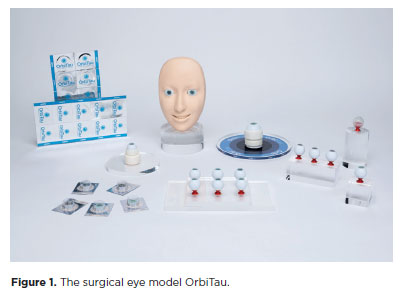
PURPOSE: The OrbiTau surgical simulator is a synthetic eye model developed to enhance cataract surgical training. Herein, we aimed to describe the perspectives of Harvard’s Ophthalmology faculty and residents regarding the effectiveness of OrbiTau.
METHODS: A cross-sectional study was conducted in which 11 surgeons from the Massachusetts Eye and Ear Infirmary, with prior experience utilizing simulated phacoemulsification platforms, conducted cataract surgery with the OrbiTau. Subsequently, they completed a satisfaction questionnaire using the Likert scale.
RESULTS: Regarding the various OrbiTau components, 90.90% of the participants reported that the OrbiTau lens capsule was comparable to that of the human lens during capsulotomy. Furthermore, 72.72% of the participants found that the OrbiTau lens consistency was analogous to that of the human lens nucleus. Approximately 63.63% of the participants reported that the model’s posterior lens capsule resembled the native posterior capsule, and 72.72% of the participants noted that the model’s red reflex was similar to that of the dilated human pupil. Most participants believed that the OrbiTau was easier to use and more realistic than other commercially available simulators.
CONCLUSION: Our single-institution survey of the Orbitau demonstrated that this model realistically replicates ocular structures and may be a viable option for cataract surgery training.
Keywords: Cataract extraction/education; Simulation training/methods; Ophthalmology/education; Phacoemulsification/education; Ophthalmologists/education; Surgeons/education; High fidelity simulation training
 ABO is licensed under a Creative Commons Attribution-NonComercial 4.0 Internacional.
About
Issues
Editorial Board
Submission
Arquivos Brasileiros de Oftalmologia
Official publication of Brazilian Council of Ophthalmology - Conselho Brasileiro de Oftalmologia (CBO)
Rua Casa do Ator, 1.117 - 2nd floor - Zip Code: 04546-004
São Paulo - SP, Brazil
TEL: +55 11 3266-4000
E-mail: [email protected]