Arq. Bras. Oftalmol. 2023;86 (3 )
:1-5
| DOI: 10.5935/0004-2749.20230031
Abstract
Objetivo: Descrever os resultados clínicos do tratamento do crescimento epitelial através da técnica de remoção manual seguido da utilização de um compressor de ar comprimido aquecido após a cirurgia de laser in situ keratomileusis (LASIK).
Métodos: Vinte olhos de 17 pacientes foram incluídos no estudo. Cada paciente havia sido submetido a cirurgia de LASIK com presença de crescimento epitelial e foi submetido a tratamento cirúrgico para sua retirada. O objetivo primário foi identificar a presença de crescimento epitelial recorrente ao final de 3 meses de seguimento. Os objetivos secundários foram as medidas de acuidade visual sem correção, acuidade visual com correção, e complicações pós-operatórias.
Resultados: Dez pacientes (58,8%) eram homens e 7 mulheres. Oito olhos de sete (41,2%) pacientes apresentavam cirurgia de LASIK primária e 12 olhos de 10 pacientes tinham cirurgia de LASIK com retratamento; dezesseis olhos (80%) utilizaram microcerátomo manual e quatro (20%) laser de femtosegundo. A média de idade no momento da cirurgia de remoção do epitélio era de 37,0 anos ± 9,3 (DP) (variando de 24 a 55 anos). Ocorreu recidiva do crescimento epithelial em dois olhos (10%) após 3 meses de seguimento. A acuidade visual sem correção antes da cirurgia era de 0,07 ± 0,09 logMAR, e após a cirurgia passou para 0,02 ± 0,04 logMAR (p=0,06). A chance (odds ration) de aparecimento do crescimento epithelial após uma reoperação de LASIK é 29,41 vezes maior do que no LASIK primário.
Conclusão: A técnica de remoção epitelial manual seguida da utilização de ar comprimido aquecido é segura e efetiva no tratamento do crescimento epitelial após LASIK. Ao final do último acompanhamento, nenhum olho apresentou perda de linhas de visão.
Keywords: Epitélio/crescimento & desenvolvimento; Endotélio corneano; Doenças da córnea; Ceratomileuse assistida por excimer laser in situ; Ceratectomia fotorrefrativa; Procedimentos cirúrgicos refrativos; Acuidade visual
Arq. Bras. Oftalmol. 2025;88 (6 )
:1-6
| DOI: 10.5935/0004-2749.2024-0309
Abstract
PURPOSE: To report the surgical outcomes of patients with primary congenital glaucoma who underwent gonioscopy-assisted transluminal trabeculotomy.
METHODS: This retrospective, noncomparative, interventional study included consecutive patients with primary congenital glaucoma with uncontrolled intraocular pressure undergoing gonioscopy-assisted transluminal trabeculotomy between January 2017 and January 2020. The included participants were followed up for at least 24 months, and only one surgeon performed all the procedures. The number of glaucoma medications, pre- and postoperative intraocular pressure, treatment extension (in quadrants), surgical complications, and any associated events or interventions were documented.
RESULTS: This study included 13 eyes from 10 patients (mean age, 4.5 ± 3.2 years; range, 3 months to 10 years). After a 24-month follow-up, the mean intraocular pressure significantly decreased from 26.1 ± 3.7 to 11.8 ± 2.5 mmHg (p<0.001). The mean number of glaucoma medications was reduced from 3.3 ± 0.5 to 0.85 ± 1.0 (p<0.001). At the end of the follow-up interval, all eyes (13 out of 13) had an intraocular pressure between 7 and 15 mmHg. In 11 of 13 eyes (84.6%), gonioscopy-assisted transluminal trabeculotomy was performed in all quadrants (360º). The most frequent postoperative complication was transitory (self-limited) hyphema (7 out of 13 eyes [53.8%]). No sight-threatening adverse events occurred during the entire follow-up period.
CONCLUSIONS: The 2-year follow-up results indicated gonioscopy-assisted transluminal trabeculotomy as an efficient and safe option for primary congenital glaucoma treatment with minimal postoperative complications.
Keywords: Glaucoma, Open-angle/surgery; Gonioscopy; Trabeculectomy/methods; Intraocular pressure; Antihypertensive agents/therapeutic use.
Arq. Bras. Oftalmol. 2025;88 (1 )
:1-8
| DOI: 10.5935/0004-2749.2023-0103
Abstract
PURPOSE: This study aimed to compare the safety and effectiveness of intraocular pressure reduction between micropulse transscleral cyclophotocoagulation and “slow cook” transscleral cyclophotocoagulation in patients with refractory primary open-angle glaucoma.
METHODS: We included patients with primary open angle glaucoma with at least 12 months of follow-up. We collected and analyzed data on the preoperative characteristics and postoperative outcomes. The primary outcomes were a reduction of ≥20% of the baseline value (criterion A) and/or intraocular pressure between 6 and 21 mmHg (criterion B).
RESULTS: We included 128 eyes with primary open-angle glaucoma. The preoperative mean intraocular pressure was 25.53 ± 6.40 and 35.02 ± 12.57 mmHg in the micropulse- and “slow cook” transscleral cyclophotocoagulation groups, respectively (p<0.001). The mean intraocular pressure was reduced significantly to 14.33 ± 3.40 and 15.37 ± 5.85 mmHg in the micropulse- and “slow cook” transscleral cyclophotocoagulation groups at the last follow-up, respectively (p=0.110). The mean intraocular pressure reduction at 12 months was 11.20 ± 11.46 and 19.65 ± 13.22 mmHg in the micropulse- and “slow cook” transscleral cyclophotocoagulation groups, respectively (p<0.001). The median preoperative logMAR visual acuity was 0.52 ± 0.69 and 1.75 ± 1.04 in the micropulse- and “slow cook” transscleral cyclophotocoagulation groups, respectively (p<0.001). The mean visual acuity variation was -0.10 ± 0.35 and -0.074 ± 0.16 in the micropulse- and “slow cook” transscleral cyclophotocoagulation, respectively (p=0.510). Preoperatively, the mean eye drops were 3.44 ± 1.38 and 2.89 ± 0.68 drugs in the micropulse- and “slow cook” transscleral cyclophotocoagulation groups, respectively (p=0.017), but those were 2.06 ± 1.42 and 1.02 ± 1.46 at the end of the study in the slow cook” and micropulse transscleral cyclophotocoagulation groups, respectively (p<0.001). The success of criterion A was not significant between both groups. Compared with 11 eyes (17.74%) in the slow cook” transscleral cyclophotocoagulation group, 19 eyes (28.78%) in the micropulse transscleral cyclophotocoagulation group showed complete success (p=0.171). For criterion B, 28 (42.42%) and 2 eyes (3.22%) showed complete success after micropulse- and slow cook” transscleral cyclophotocoagulation, respectively (p<0.001).
CONCLUSION: Both techniques reduced intraocular pressure effectively.
Keywords: Sclera/surgery; Glaucoma, open-angle/surgery; Ciliary body/surgery; Intraocular pressure; Laser coagulation/methods; Lasers, semiconductor; Comparative study; Effectiveness
Arq. Bras. Oftalmol. 2021;84 (4 )
:361-366
| DOI: 10.5935/0004-2749.20210052
Abstract
OBJETIVO: Glaucoma é a principal causa de cegueira irreversível no mundo. O pico da pressão intraocular é um dos principais fatores de risco para progressão do glaucoma, e o controle pressórico ainda é o único tratamento efetivo para todas as formas de glaucoma. O objetivo principal deste estudo é comparar a redução basal e do pico da pressão intraocular, obtidas através do Teste de Sobrecarga Hídrica, entre os dois olhos dos mesmos pacientes utilizando latanoprosta 0,005% em um olho e submetidos à aplicação de trabeculoplastia a laser seletiva no olho contralateral.
MÉTODOS: Este é um estudo prospectivo, intervencionista, longitudinal e randomizado. Trinta pacientes consecutivos, glaucomatosos, com pressão intraocular controlada em uso de monoterapia com latanoprosta, foram recrutados de um único centro oftalmológico. Os olhos dos pacientes foram randomizados e um olho foi selecionado para tratamento com trabeculoplastia a laser seletiva e olho contralateral tratado com colírio de latanoprosta 0,005%. Foram avaliados a pressão intraocular basal e pico de pressão intraocular um mês (Teste de Sobrecarga Hídrica 2) e seis meses (Teste de Sobrecarga Hídrica 3) após tratamento.
RESULTADOS: Não houve diferença estatística entre a pressão intraocular pré washout entre os olhos randomizados para trabeculoplastia a laser seletiva e latanoprosta, 13,6 ± 2,1 e 13,3 ± 1,8 mmHg, respectivamente (p=0,182). Em relação à pressão intraocular basal, não houve diferença estatística entre os grupos, tanto no Teste de Sobrecarga Hídrica 2 (p=0,689) e Teste de Sobrecarga Hídrica 3 (p=0,06). Não houve diferença estatística em relação ao pico de pressão intraocular entre os grupos trabeculoplastia a laser seletiva e latanoprosta, no Teste de Sobrecarga Hídrica 2 (p=0,771) e Teste de Sobrecarga Hídrica 3 (p=0,774).
CONCLUSÕES: Em resumo, nosso estudo demonsrou que a eficácia da redução pressórica é similar entre latanoprosta e trabeculoplastia a laser seletiva, e pacientes glaucomatosos que estão com a pressão intraocular clinicamente controlados com latanoprosta e trocam de tratamento para trabeculoplastia a laser seletiva mantém sua pressão intraocular controlada.
Keywords: Glaucoma; Pressão intraocular; Latanoprosta; Lasers
Arq. Bras. Oftalmol. 2024;87 (1 )
:1-7
| DOI: 10.5935/0004-2749.2021-0061
Abstract
Objetivo: Avaliar o efeito do tabagismo nos desfechos da trabeculectomia.
Métodos: Uma revisão retrospectiva do gráfico de pacientes com glaucoma submetidos à trabeculectomia foi realizada por um único cirurgião entre 2007 e 2016. Os gráficos foram examinados para uma história documentada de condição de fumante antes da cirurgia. Variáveis pré-operatórias clínicas e demográficas e clínicas foram registradas. Os pacientes foram divididos em dois grupos de acordo com sua história de tabagismo em fumantes e não fumantes. Quaisquer Intervenções relacionadas à bolha, por exemplo, injeções de 5-fluorouracil + lise de sutura com laser, ou revisão da bolha realizada durante o período pós-operatório foram observadas. O sucesso foi definido como pressão intraocular > 5 mmHg e < 21 mm Hg sem (sucesso completo) ou com (sucesso qualificado) medicamentos hipotensores oculares. A falha foi identificada como violação dos critérios mencionados acima.
Resultados: O estudo incluiu 98 olhos de 83 pacientes com idade média de 70,7 ± 11,09 anos, sendo 53% (44/83) dos pacientes do sexo feminino. O diagnóstico mais comum foi o glaucoma de ângulo aberto primário com 47 casos (47,9%). O Grupo de fumantes incluiu 30 olhos de 30 pacientes. Os fumantes, quando comparados aos não fumantes, apresentaram uma melhor acuidade visual pré-operatória significativamente pior (p=0,038), maior espessura central da córnea (p=0,047) e maior pressão intraocular pré-operatória (p=0,011). A taxa de sucesso de um ano para a cirurgia de trabeculectomia foi de 56,7% no Grupo de fumantes contra 79,4% no Grupo de não fumantes (p=0,020). O tabagismo apresentou razão de chances para falha de 2,95 95% de IC (1,6-7,84).
Conclusão: Os fumantes demonstraram uma taxa de sucesso significativamente menor em um ano após a trabeculectomia em comparação com os não fumantes e uma maior necessidade de intervenções relacionadas à bolha.
Keywords: Glaucoma de ângulo aberto; Trabeculectomia; Pressão intraocular; Tabagismo; Tabaco/efeitos adversos; Acuidade visual
Arq. Bras. Oftalmol. 2025;88 (6 )
:1-5
| DOI: 10.5935/0004-2749.2024-0340
Abstract
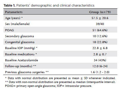
PURPOSE: This study aimed to report the surgical outcomes and success predictors of micropulse transscleral cyclophotocoagulation in eyes with refractory glaucoma.
METHODS: This was a noncomparative, interventional case series. Patients with refractory glaucomas, defined as eyes with prior incisional glaucoma surgery failure and uncontrolled intraocular pressure, who underwent micropulse transscleral cyclophotocoagulation between March 2017 and June 2021 were enrolled. A minimum follow-up period of 6 months was required. Preoperative and postoperative intraocular pressure, number of hypotensive medications, surgical complications, and any subsequent related events were recorded. Success criteria were as follows: 1) intraocular pressure reduction ≥20% and intraocular pressure ≤18 mmHg; 2) intraocular pressure reduction ≥30% and intraocular pressure ≤15 mmHg. The need for topical hypotensive medications was not considered a failure.
RESULTS: Seventy-nine (79) eyes (79 patients; mean age, 57.5 ± 20.6 years) were included. Overall, the median follow-up duration was 12.0 (interquartile interval, 6–24) months, and the mean intraocular pressure was reduced from 22.8 ± 6.8 mmHg to 15.5 ± 5.6 mmHg at the last follow-up visit (p<0.001). The mean number of medications was reduced from 2.8 ± 0.7 to 2.0 ± 1.0 (p<0.01). At 12 months postoperatively, the success rates for criteria 1 and 2 were 54.9% and 49.7%, respectively. Aside from one case of corneal ulcer, which fully resolved with clinical treatment, and two cases of persistent hypotony (with no visual acuity loss during follow-up), no other vision-threatening complications were observed during the postoperative period. The magnitude of intraocular pressure reduction at 1 month (adjusted to preoperative intraocular pressure; HR=1.01; p=0.002).
CONCLUSION: Our findings suggest that micropulse transscleral cyclophotocoagulation is a relatively effective alternative for managing refractory glaucomas, with minor postoperative complications. In addition, the initial intraocular pressure reduction was a statistically significant predictor of 1-year success in patients undergoing micropulse transscleral cyclophotocoagulation.
Keywords: Intraocular pressure/physiology; Glaucoma, open-angle/surgery; Trabeculectomy; Laser coagulation/methods; Tonometry, ocular/methods; Postoperative complications; Antihypertensive agents/therapeutic use.
Arq. Bras. Oftalmol. 2026;89 (3 )
:1-8
| DOI: 10.5935/0004-2749.2025-0043
Abstract
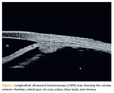
PURPOSE: To evaluate the effect of single-session transscleral diode laser cyclophotocoagulation on intraocular pressure in refractory glaucoma and to determine structural changes using ultrasound biomicroscopy.
METHODS: Forty-three eyes were evaluated. Intraocular pressures at baseline and at the first, third, and sixth months after transscleral diode laser cyclophotocoagulation were compared. Ciliary body thickness, ciliary muscle thickness, ciliary process thickness, iris root thickness, and scleral thickness were assessed at baseline and at the third and sixth months post-treatment.
RESULTS: Reductions in intraocular pressure were significant between baseline and the first month (p=0.018), third month (p<0.001), and sixth month (p<0.001) as well as between the first and third months (p=0.034) and the first and sixth months (p=0.036). Compared with baseline, intraocular pressure reduction rates at the first, third, and sixth months were 34.6%, 56.5%, and 55.3%, respectively, while success rates were 30.2%, 62.8%, and 55.8%, respectively. Decreases in ciliary body thickness, ciliary muscle thickness, and ciliary process thickness were significant between baseline and the third month (p<0.05) and between baseline and the sixth month (p<0.05), whereas changes between the third and sixth months were not significant (p>0.05). Iris root and scleral thicknesses did not change after treatment (p>0.05). At the third and sixth months, significant positive correlations were observed between changes in intraocular pressure and changes in ciliary body thickness and ciliary process thickness (p<0.05).
CONCLUSIONS: To the best of our knowledge, this is one of the few studies comprehensively investigating structural changes after transscleral diode laser cyclophotocoagulation using ultrasound biomicroscopy. Moreover, the relationships between intraocular pressure changes and variations in the ciliary body, ciliary muscle, ciliary process, iris root, and scleral thicknesses were examined in detail. Single-session treatment did not affect iris root or scleral thickness but significantly reduced ciliary body, ciliary muscle, and ciliary process thicknesses. Greater reductions in ciliary body and ciliary process thickness may contribute to more pronounced intraocular pressure reduction.
Keywords: Intraocular pressure; Laser coagulation/methods; Lasers, semiconductor; Microscopy, acoustic; Glaucoma; Ciliary body
Arq. Bras. Oftalmol. 2026;89 (4 )
:1-8
| DOI: 10.5935/0004-2749.2025-0313
Abstract
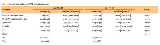
PURPOSE: To compare clinical outcomes associated with different intraoperative mitomycin C exposure times during photorefractive keratectomy for myopia and astigmatism correction.
METHODS: This prospective, comparative, contralateral-eye study included 41 patients (82 eyes), comprising 28 eyes with ablation <60µm and 13 eyes with ablation >60µm, who underwent photorefractive keratectomy with varying mitomycin C application times based on ablation depth. In eyes with ablation <60µm, mitomycin C was applied for 15 s in one eye and 30 s in the fellow eye. In eyes with ablation >60µm, mitomycin C was applied for 30 s in one eye and 60 s in the fellow eye. Outcomes included visual acuity, postoperative pain (visual analog scale), subjective tearing, corneal haze, and refractive results at 3 months.
RESULTS: No statistically significant differences were observed between mitomycin C application times within either group for postoperative pain, tearing, visual acuity, refractive outcomes (spherical, cylindrical, and spherical equivalent), or haze prevalence (p>0.05 for all comparisons). Visual acuity improved in all groups, and no eyes lost ≥2 lines of corrected distance visual acuity.
CONCLUSIONS: Shorter mitomycin C exposure times (15 or 30 s) appear to be as effective and safe as longer durations (30 or 60 s) for haze prevention after photorefractive keratectomy without compromising refractive outcomes or increasing postoperative discomfort at 3-month follow-up.
Keywords: Mitomicin/therapeutic use; Photorefractive keratectomy; Lasers, excimer; Intraoperative period; Miopia/surgery; Astigmatismo/surgery; Corneal opacity; Postoperative pain; Comparative study
Arq. Bras. Oftalmol. 2024;87 (4 )
:1-5
| DOI: 10.5935/0004-2749.2023-0143
Abstract
PURPOSE: The purpose of this study is to assess the long-term outcomes of modified transcanalicular diode laser dacryocys torhinostomy in a large cohort of patients affected by primary acquired nasolacrimal duct obstruction.
METHODS: This study, conducted from January 17 to June 2022, encompassed 141 patients (159 procedures) who underwent modified transcanalicular diode laser dacryocystorhinostomy (MT-DCR). The procedure employed an 810-nm diode laser. Patients were monitored for at least a year after the intervention. Anatomical success was determined by ostium patency upon irrigation, while functional success referred to epiphora resolution. Parameters studied included patient demographics, procedure duration, complications, and both anatomical and functional success. Statistical analysis was performed using the Statistical Package for the Social Sciences software, with results considered significant at a 95% confidence interval (p≤0.05).
RESULTS: A total of 159 lacrimal drainage systems (141 patients: 112 women and 29 men) were included in this study. Among them, 18 underwent bilateral procedures. The average patient age was 58 years (range: 34-91 years), and the average surgical duration was 24 minutes (range: 18-35 minutes). One year after the surgery, MT-DCR exhibited anatomical and functional success rates of 84.9% (135/159) and 83% (132/159), respectively.
CONCLUSION: MT-DCR achieved an anatomical success rate of 84.9%, reflecting an excellent outcome. However, further extensive studies with larger sample sizes and longer follow-up periods are necessary to substantiate these findings.
Keywords: Lacrimal duct obstruction; Nasolacrimal duct/surgery; Dacryocystorhinostomy; Lacrimal apparatus diseases; Laser therapy/methods; Lasers, semiconductor/therapeutic use; Regeneration
Arq. Bras. Oftalmol. 2024;87 (2 )
:1-5
| DOI: 10.5935/0004-2749.2021-0395
Abstract
Objetivos: Avaliar a segurança e eficácia a longo prazo da vitreólise com Nd:YAG laser para moscas volantes sintomáticas, uma vez que permanece como um procedimento controverso devido a falta de evidência científica robusta sobre a manutenção dos resultados e ocorrência de efeitos adversos.
Métodos: Este estudo é uma extensão observacional de um ensaio clínico prospectivo, randomizado, duplo cego, previamente publicado. Oito de treze pacientes que foram submetidos a vitreólise com YAG laser foram acompanhados para uma reavaliação tardia, dezoito meses após o procedimento, para avaliar a eficácia e segurança do procedimento.
Resultados: Todos os pacientes mantiveram a melhora na sintomatologia notada ao final do procedimento original, com 25% dos casos apresentando melhora completa, e uma proporção semelhante (37,5%) demonstrando melhora significativa ou parcial. A melhora objetiva na opacidade foi similar ao achado no seguimento original de 6 meses. O questionário de qualidade de vida NEI-VFQ 25 não demonstrou diferença estatisticamente significativa nas respostas entre o sexto e o décimo oitavo mês de acompanhamento. Nenhum efeito adverso foi notado no exame clínico ou reportado pelos pacientes.
Conclusão: A eficácia da vitreólise observada ao sexto mês do acompanhamento foi mantida até o décimo oitavo mês, com todos os pacientes notando algum grau de melhora quando comparado ao estado pré procedimento. Nenhum efeito adverso tardio foi notado. Um ensaio clínico randomizado maior é necessário para confirmar a segurança do procedimento.
Keywords: Terapia a laser; Lasers de estado sólido; Vitrectomia; Corpo vítreo; Cirurgia vitreorretiniana; Acuidade visual; Doenças oculares; Qualidade de vida; Inquéritos e questionários
Arq. Bras. Oftalmol. 2024;87 (2 )
:1-8
| DOI: 10.5935/0004-2749.2022-0306
Abstract
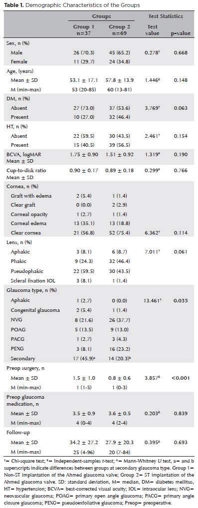
PURPOSE: As superotemporal implantation of the Ahmed glaucoma valve is not always feasible in cases of refractory glaucoma, this study examined the characteristics and surgical outcomes of cases in which the valve was implanted in a nonsuperotemporal quadrant using a modified long scleral tunnel technique.
METHODS: This retrospective case-control study included 37 eyes with nonsuperotemporal quadrant-Ahmed glaucoma valve implantation in Group 1 and 69 eyes with superotemporal Ahmed glaucoma valve implantation in Group 2. The demographic characteristics of these groups, surgical outcomes, including complications, further surgical interventions, and surgical success rates were compared. Surgical success was defined as an intraocular pressure not exceeding 21 mmHg, accompanied by a minimum reduction of 20% in intraocular pressure from the baseline without any additional intraocular pressure-lowering procedures, and the absence of light perception loss or phthisis bulbi.
RESULTS: Group 1 had significantly higher numbers of eyes with secondary glaucoma and preoperative surgical procedures than Group 2 (p<0.05). Both groups had mean preoperative intraocular pressure values, and mean intraocular pressure values at the last visit of 34.2 and 27.9 months, 35.5 ± 1.5 and 35.8 ± 1.2 mmHg, and 14.5 ± 5 and 14.9 mmHg, respectively. Although both groups had 70.2% and 75.8% as their five-year cumulative probability of success, respectively, the rates of complications, revisional surgery, and additional surgical procedures did not differ significantly (p>0.05).
CONCLUSION: The modified long scleral tunnel technique for Ahmed glaucoma valve implantation in nonsuperotemporal quadrants achieves intraocular pressure control and complication rates comparable to superotemporal implantation.
Keywords: Glaucoma/surgery; Sclera/surgery; Glaucoma drainage implant; Intraocular pressure; Tenon capsule
 ABO is licensed under a Creative Commons Attribution-NonComercial 4.0 Internacional.
08-fig01.jpg)

09-tab01tb.jpg)
10-tab01.jpg)
04-fig01.jpg)
02-fig01.jpg)