Arq. Bras. Oftalmol. 2024;87 (1 )
:1-5
| DOI: 10.5935/0004-2749.2021-0037
Abstract
Os autores relatam os achados de eletrorretinograma de campo total e tomografia de coerência óptica (OCT) da toxicidade retiniana ao melfalan intravítreo. Menina de 18 meses com retinoblastoma foi avaliada com fases fotópicas do eletrorretinograma de campo total e tomografia de coerência óptica após o tratamento com melfalan intravítreo. Após a terceira injeção, a criança desenvolveu alterações do epitélio pigmentar da retina próximo ao local da injeção. A resposta fotópica do eletrorretinograma de campo total mostrou diminuição da amplitude das respostas das ondas a e b no olho afetado comparado com o olho sadio. A tomografia de coerência óptica mostrou alterações significativas nas camadas retinianas externas no olho comprometido. A toxicidade do melfalan é dose dependente e, apesar dos benefícios terapêuticos, podem causar alterações retinianas significativas. Este caso demonstra uma avaliação atual e aprofundada da toxicidade retiniana do melfalan intravítreo na retina humana através da tomografia de coerência óptica e sua correlação com as alterações no eletrorretinograma.
Keywords: Retinoblastoma; Efeitos colaterais e reações adversas relacionados a medicamentos; Injeções intravítreas; Melfalan/toxicidade.
Arq. Bras. Oftalmol. 2023;86 (5 )
:1-4
| DOI: 10.5935/0004-2749.20230046
Abstract
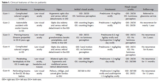
O melanoma iridociliar em anel é um tipo incomum de melanoma uveal. As manifestações clínicas variam desde casos assintomáticos até síndromes mascaradas que mimetizam um glaucoma refratário. As opções de tratamento incluem radioterapia e enucleação. O manejo do melanoma uveal metastático continua desanimador. Novas terapias usando inibidores de checkpoint imunológico estão atualmente em estudo. Apresentamos o caso de uma mulher hispânica de 54 anos com perda progressiva da visão por um melanoma metastático em anel, com semeadura de câmara anterior, tratada com pembrolizumabe.
Keywords: Neoplasia uveal/complicação; Melanoma; Neoplasia da íris/secundário; Corpo ciliar; Anticorpo monoclonal humanizado; Inibidor de checkpoint imunológico; Humanos; Relato de caso.
Arq. Bras. Oftalmol. 2024;87 (3 )
:1-4
| DOI: 10.5935/0004-2749.2021-0239
Abstract
Relatamos um caso de despigmentação aguda bilateral da íris, no qual obtivemos adequado controle da pressão intraocular com o implante do iStent®, após resolução da fase aguda da doença. Paciente feminina, 62 anos, atendida com quadro agudo, bilateral e simultâneo de dor ocular, fotofobia, hipertensão ocular (34 mmHg), pigmentos circulantes na câmara anterior, áreas de despigmentação iriana e sinéquias posteriores. Havia recebido amoxicilina-clavulanato e moxifloxacina orais para pneumonia 2 meses antes. Suspeitando-se de despigmentação aguda bilateral da íris ou de etiologia viral, recebeu acetazolamida, aciclovir e prednisona orais, e colírios prednisolona, betaxolol, brimonidina, dorzolamida e atropina. O quadro se resolveu gradualmente em 4 meses, porém, após 1 ano, desenvolveu catarata bilateral e ainda usava 3 colírios hipotensores (pressão intraocular 16/18 mmHg). A cirurgia combinada de catarata-iStent® foi realizada em ambos os olhos. Um ano depois, a pressão intraocular mantinha-se 11/12 mmHg, sem medicação. O iStent® foi seguro e eficaz no controle deste glaucoma secundário.
Keywords: Doenças da íris; Catarata; Hipertensão ocular; Stents; Gonioscopia
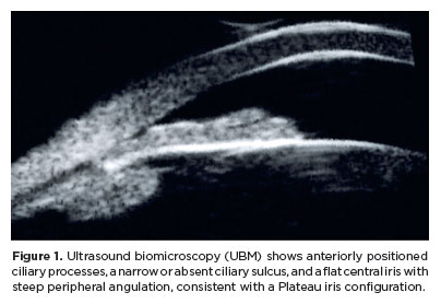
Arq. Bras. Oftalmol. 2025;88 (5 )
:1-4
| DOI: 10.5935/0004-2749.2025-0020
Abstract
Angle-closure glaucoma is a major cause of visual impairment worldwide, with Plateau iris syndrome presenting management challenges. We present a case report of a 58-year-old woman with advanced, uncontrolled angle-closure glaucoma and Plateau iris. Her history included laser peripheral iridotomy and three glaucoma medications in both eyes. Different treatments were implemented. For the eye with lower intraocular pressure, fewer peripheral anterior synechiae, and milder disease: phacoemulsification with intraocular lens implantation. For the eye with more advanced disease, a two-step approach was used: slow-coagulation transscleral cyclophotocoagulation using the double-arc protocol, followed by phacoemulsification with intraocular lens implantation 2 months later. Both eyes achieved improved visual acuity and intraocular pressure control with fewer medications, without significant complications. This case highlights transscleral cyclophotocoagulation followed by phacoemulsification as an alternative to combined surgeries in uncontrolled angle-closure glaucoma with Plateau iris, offering a simpler technique, more predictable refractive and pressure-control outcomes, and more straightforward postoperative management.
Keywords: Glaucoma, angle-closure/surgery; Iris diseases/surgery; Laser coagulation/methods; Phacoemulsification; Lens implantation, intraocular; Case reports
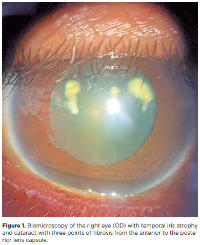
Arq. Bras. Oftalmol. 2025;88 (2 )
:1-4
| DOI: 10.5935/0004-2749.2023-0248
Abstract
Aging and face sagging have many causes, and various techniques are used for treatment, including noninvasive procedures, such as focused ultrasound, which uses the principle of collagen regeneration by coagulative necrosis of the dermis layers using radiofrequency, but this procedure has complications. We reported a case of a 54-year-old female patient who complained of poor visual acuity in her right eye three days after a focused ultrasound facial aesthetic procedure, with the best visual acuity of 20/60. Biomicroscopy of the right eye revealed an acute cataract with three points of fibrosis extending from the posterior to the anterior capsule. The patient underwent phacoemulsification surgery with visual rehabilitation and improved vision of 20/20. We hypothesized that the occurrence of acute cataract was related to the inappropriate use of focused ultrasound.
Keywords: Cosmetic techniques; Skin aging; Rejuvenation; Ultrasonic therapy/adverse effects; High-intensity focused ultrasound ablation/methods; Cataract/etiology; Phacoemulsification; Lens implantation, intraocular; Visual acuity; Humans; Middle age; Female; Case
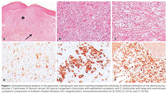
Arq. Bras. Oftalmol. 2025;88 (2 )
:1-4
| DOI: 10.5935/0004-2749.2024-0007
Abstract
Langerhans cell histiocytosis comprises a heterogeneous range of clinical manifestations secondary to clonal proliferation of histiocytes, characterized by the accumulation of these cells in various organs and tissues. The ophthalmological component commonly involved is the orbit. Herein, we report a rare case of Langerhans cell histiocytosis with eyelid involvement, which resulted in severe ocular surface complications, which subsequently significantly impacted the patient's quality of life. This case report highlights the fact that despite being rare, Langerhans cell histiocytosis should be included in the differential diagnosis of eyelid lesions. Furthermore, a multidisciplinary approach with a systemic overview is crucial for managing the ocular complications.
Keywords: Dry eye syndromes; Erdheim-Chester disease/drug therapy; Human; Female; Case reports
 ABO is licensed under a Creative Commons Attribution-NonComercial 4.0 Internacional.
07-fig01.jpg)
11-tab01.jpg)
13-fig01.jpg)
08-fig01.jpg)
10-fig01.jpg)
10-fig01.jpg)
03-fig01.jpg)
-03-fig01.jpg)
01-fig01tb.jpg)
02-fig01.jpg)