Arq. Bras. Oftalmol. 2022;85 (5 )
:450-458
| DOI: 10.5935/0004-2749.20220075
Abstract
Objetivo: Investigar o efeito antiproliferativo de poli (lactídeo-coglicolídeo) com superfície modificada carregada com carboplatina contra células de retinoblastoma.
Métodos: Preparou-se poli (lactídeo-co-glicolídeo) carregado com carboplatina com ou sem alginato de sódio para modifição da superfície, poli com alginato de sódio (lactídeo-co-glicolídeo) e poli (lactídeo-co-glicolídeo). O potencial zeta e o comportamento de liberação de carboplatina foram investigados. A captação celular do fármaco liberado foi observada na linha celular de retinoblastoma Y79. O efeito inibitório das nanopartículas carregadas com carboplatina contra a linha celular Y79 foi avaliado através do ensaio de metiltiazol tetrazólio e Western-blot. Carboplatina nativa e nanopartículas vazias sem carga de carboplatina serviram como controles.
Resultados: O potencial zeta de poli carregado com carboplatina (lactídeo-co-glicolídeo) foi - (26,1 ± 3,1) mV versus - (43,1 ± 8,1) mV em poli com alginato de sódio carregado com carboplatina (lactídeo-co-glicolídeo). A percentagem de libertação de explosão de poli carregado com carboplatina (lactídeo-co-glicolídeo) e poli com alginato de sódio (lactídeo-co-glicolídeo) foram (40,0 ± 8,2)% e (18,9 ± 4,3)% às 24 horas, respectivamente. Uma diferença significativa foi identificada em relação à liberação de fármaco entre poli com alginato de sódio carregado com carboplatina (lactídeo-co-glicolídeo) e poli carregado com carboplatina (lactídeo-co-glicolídeo). A detecção de fluorescência revelou que a carboplatina foi assimilada intensamente no citoplasma da linha celular Y79 que foi exposta ao poli com alginato de sódio carregado com carboplatina (lactídeo-co-glicolídeo). A exposição de poli carregada com carboplatina (lactídeo-co-glicolídeo) ou poli com alginato de sódio (lactídeo-co-glicolídeo) inibiu a expressão de antígeno nuclear de proliferação celular em células Y79 no 3º dia. A extensão da exposição no 5º dia revelou que poli com alginato de sódio (lactídeo-co-glicolídeo) para modificação da superfície foi superior a poli (lactídeo-co-glicolídeo) em termos de inibição do antígeno nuclear de proliferação celular. O teste de viabilidade celular via metiltiazol tetrazólio mostrou um efeito inibitório semelhante. Além disso, as nanopartículas carregadas com carboplatina de concentração mais baixa inibiram a viabilidade celular mais fortemente em comparação com a carboplatina nativa de concentração mais alta no ensaio de metiltiazol tetrazólio.
Conclusões: Poli com alginato de sódio carregado com carboplatina (lactídeo-co-glicolídeo) inibiu a proliferação de células de retinoblastoma com efeito superior em contraste com poli (lactídeo-co-glicolídeo) e carboplatina nativa. O alginato de sódio para modificação da superfície oferece uma estratégia potencial para o sistema de liberação de carboplatina sustentada.
Keywords: Carboplatina; Alginato; Nanopartícula; Retinoblastoma
Arq. Bras. Oftalmol. 2023;86 (5 )
:1-6
| DOI: 10.5935/0004-2749.20230071
Abstract
Objetivo: O melanoma da conjuntiva é um tumor raro e agressivo, com propensão à disseminação metastática regional e distante. Este estudo tem como objetivo analisar os marcadores BRAF e NRAS no melanoma da conjuntiva e sua relação com recidivas tumorais e com o prognóstico do paciente.
Métodos: Este foi um estudo retrospectivo, observacional e unicêntrico de pacientes consecutivos com diagnóstico anatomopatológico de melanoma da conjuntiva feito entre janeiro de 1992 e dezembro de 2019. As mutações BRAF e NRAS foram analisadas com o kit cobas® 4800 (Roche®) em amostras obtidas através de biópsia excisional ou por mapa. Além disso, foi avaliada a presença de lesões pré-cancerosas ou tumorais associadas.
Resultados: Foram incluídos 12 pacientes com amostras histológicas positivas para melanoma da conjuntiva (7 mulheres e 5 homens), com idade média ao diagnóstico de 60 anos e tempo médio de evolução de 6,38 ± 3,4 anos. A mutação BRAF V600E foi encontrada em 3 biópsias (25%), bem como a NRAS Q61X (25%). Ocorreram recidivas em todos os pacientes positivos para mutações de BRAF ou NRAS e 5 desses pacientes desenvolveram disseminação sistêmica (83,33%). Além disso, 4 dos 6 pacientes com BRAF ou NRAS mutante (66,66%) apresentaram achados histopatológicos de lesões tumorais ou pré-cancerosas.
Conclusões: As mutações BRAF e NRAS podem ser fatores de risco para recorrência e menor sobrevida no melanoma da conjuntiva, o que tornaria esses pacientes candidatos a terapias direcionadas e a um acompanhamento mais abrangente e individualizado. Todos esses dados justificam mais estudos prospectivos padronizados.
Keywords: Neoplasias da túnica conjuntiva; Melanoma; Biomarcadores tumorais; Proteínas proto-oncogênicas B-raf; Genes ras.
Arq. Bras. Oftalmol. 2025;88 (1 )
:1-10
| DOI: 10.5935/0004-2749.2023-0073
Abstract
PURPOSE: To describe the epidemiological and clinical profile of hospitalized patients with retinoblastoma in Brazil.
METHODS: Using data from the Hospital Cancer Registry of the , patients with the morphological codes of retinoblastoma who were diagnosed between 2000 to 2018, aged 0–19 years, and followed up in registered hospitals (analytical cases) were selected. The relative and absolute frequencies of demographic, clinical, diagnostic, therapeutic, and outcome variables were described. Hospital performance indicators were calculated and compared between hospitals qualified and not qualified to treat pediatric oncology cases and between hospitals with different case volumes (<20, 20–75, >75 cases).
RESULTS: Of the 2,269 identified analytical cases from 86 institutions, 48% were from the Southeast, 54% were male, and 66% were aged <4 years. The proportion of missing data (NA) was too high for several variables. Approximately 84% of the patients were from the public health system, 40% had a positive family history, and 88% had unilateral involvement. The first treatment included surgery in 58.3% of the patients (NA=2), Approximately 36.6% of these patients achieved complete remission, 10.8% achieved partial remission, and 12.7% died (NA=59%). Hospital performance indicators were within the target in >90% of the patients. The median time between the first appointment and diagnosis (6 days, interquartile range [IQR] 1–14) was significantly lower and the median time to death was longer (343 days, IQR, 212-539) in high-volume hospitals (>75 cases) than in medium- and low-volume hospitals.
CONCLUSIONS: Despite the high proportion of missing data, we found that the delay in diagnosis is due to prehospital factors. Additionally, there is a need for educational programs for healthcare professionals and families that emphasize early identification and referral to specialized centers. Future studies should focus on the impact of Hospital Cancer Registry data completeness on outcomes, causes of delay in diagnosis, regional inequalities, and barriers to accessing specialized services.
Keywords: Retinoblastoma/diagnosis; Retinoblastoma/epidemiology; Patient care; Humans; Children; Adolescents; Brazil.
Arq. Bras. Oftalmol. 2025;88 (4 )
:1-6
| DOI: 10.5935/0004-2749.2024-0278
Abstract
PURPOSE: This study aimed to evaluate the prevalence of orbital conditions in a tertiary ophthalmic outpatient hospital in Sao Paulo, Brazil, with a focus on the main diagnoses and their distribution.
METHODS: A retrospective chart review was conducted involving patients registered and admitted to the orbital disease unit at the Department of Ophthalmology, University of São Paulo Medical School, from January 2004 to March 2018. A total of 838 medical charts were analyzed, of which 37 were excluded due to incomplete data. The remaining charts were categorized into eight diagnostic groups: Graves’ orbitopathy , inflammatory disorders, tumors, vascular lesions, acquired structural abnormalities, congenital structural abnormalities, infectious diseases, and others.
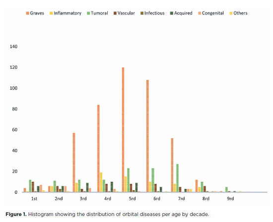
RESULTS: Of the 837,300 ophthalmological appointments, 3,372 (0.4%) were related to orbital diseases. The study included 801 patients, of whom 63.45% were women. The patients’ mean age was 42.86 years. Graves’ orbitopathy was the most common (55%), followed by tumor (17%), inflammatory disorders (9%), vascular lesions (7%), acquired structural abnormalities (5%), congenital structural abnormalities (4%), others (2%), and infectious diseases (1%). The study found significant differences in the incidence and types of orbital diseases, indicating the specialized nature of tertiary care and referral biases.
CONCLUSION: Published data on epidemiological orbital diseases is scarce. Therefore, this study focused on the diverse nature of orbital diseases and their low incidence among ophthalmology appointments. The major trends align with other epidemiological studies, demonstrating a preponderance of Graves’ orbitopathy in middle-aged adults and a bimodal distribution of tumors. These findings are essential in shaping resident training programs and healthcare policies, particularly in tertiary settings. Understanding the epidemiology of orbital diseases can improve diagnostic accuracy, treatment approaches, and patient outcomes as well as support future systemic prospective studies.
Keywords: Orbital diseases; Orbital tumors; Neoplasms; Inflammation; Graves’ ophthalmopathy; Outpatients
Arq. Bras. Oftalmol. 2025;88 (2 )
:1-7
| DOI: 10.5935/0004-2749.2023-0265
Abstract
PURPOSE: Although Brazil has a high prevalence of retinoblastoma, there is a lack of epidemiological data on the disease. Thus, in this study, we aimed to evaluate the epidemiological profile of patients diagnosed with retinoblastoma in the ophthalmology department of a pediatric tertiary referral hospital in Ceara, Brazil.
METHODS: A descriptive and cross-sectional study was conducted by retrospectively analyzing the clinical and socioeconomic data from the medical records of pediatric patients followed-up at the hospital between 2007 and 2021. Retinoblastoma was diagnosed on the basis of a fundoscopic or histopathologic examination.
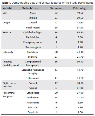
RESULTS: The data of 105 patients were included in the study, and the mean patient age at the time of diagnosis was 1.7 years. Most of the patients were women (50.5%) and hailed from rural areas (57.4%), which was associated with a higher tumor stage. Of the 150 patients, 57.1% initially presented with leukocoria. Ocular hyperemia was associated with more advanced stages of retinoblastoma (p=0.004). Bilateral involvement was observed in 25.7% of the patients and at a significantly younger age (p=0.009). The presence of retinal detachment, vascularized lesions, and vitreous seeds significantly increased the likelihood of requiring enucleation.
DISCUSSION: This study presents an epidemiological description of retinoblastoma in Brazil, which highlights the significance of early detection. Delayed diagnosis is associated with a poorer visual prognosis and higher mortality rate, particularly in patients with unilateral disease. Risk factors for a more severe disease were retinal detachment, vascularized lesions, and vitreous seeds. The correlation between histopathological features and clinical outcomes was limited.
CONCLUSION: Further studies are required to assess the influence of ocular hyperemia, fundoscopic assessment, and histopathologic findings on the prognosis of retinoblastoma. Moreover, it is critical to devise interventions to reduce the time-to-diagnosis in rural areas.
Keywords: Retinoblastoma; Retinal neoplasms; Epidemiology; Prevalence; Risk factors; Delayed diagnosis; Child
Arq. Bras. Oftalmol. 2024;87 (2 )
:1-6
| DOI: 10.5935/0004-2749.2021-0435
Abstract
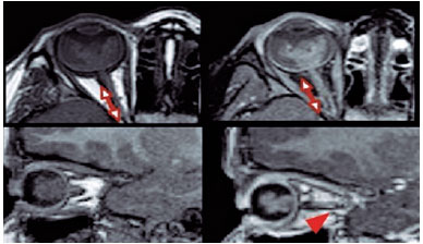
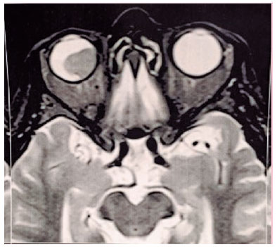
PURPOSE: This study aimed to analyze the association between magnetic resonance imaging apparent diffusion coefficient map value and histopathological differentiation in patients who underwent eye enucleation due to retinoblastomas.
METHODS: An observational chart review study of patients with retinoblastoma that had histopathology of the lesion and orbit magnetic resonance imaging with apparent diffusion coefficient analysis at Hospital de Clínicas de Porto Alegre between November 2013 and November 2016 was performed. The histopathology was reviewed after enucleation. To analyze the difference in apparent diffusion coefficient values between the two major histopathological prognostic groups, Student's t-test was used for the two groups. All statistical analyses were performed using SPSS version 19.0 for Microsoft Windows (SPSS, Inc., Chicago, IL, USA). Our institutional review board approved this retrospective study without obtaining informed consent.
RESULTS: Thirteen children were evaluated, and only eight underwent eye enucleation and were included in the analysis. The others were treated with photocoagulation, embolization, radiotherapy, and chemotherapy and were excluded due to the lack of histopathological results. When compared with histopathology, magnetic resonance imaging demonstrated 100% accuracy in retinoblastoma diagnosis. Optic nerve invasion detection on magnetic resonance imaging showed a 66.6% sensitivity and 80.0% specificity. Positive and negative predictive values were 66.6% and 80.0%, respectively, with an accuracy of 75%. In addition, the mean apparent diffusion coefficient of the eight eyes was 0.615 × 103 mm2/s. The mean apparent diffusion coefficient value of poorly or undifferentiated retinoblastoma and differentiated tumors were 0.520 × 103 mm2/s and 0.774 × 103 mm2/s, respectively.
CONCLUSION: This study revealed that magnetic resonance imaging is useful in the diagnosis of retinoblastoma and detection of optic nerve infiltration, with a sensitivity of 66.6% and specificity of 80%. Our results also showed lower apparent diffusion coefficient values in poorly differentiated retinoblastomas with a mean of 0.520 ×
103 mm2/s, whereas in well and moderately differentiated, the mean was 0.774 × 103 mm2/s.
Keywords: Retinoblastoma; Prognosis; Retinal neoplasms; Orbit; Diffusion magnetic resonance imaging
 ABO is licensed under a Creative Commons Attribution-NonComercial 4.0 Internacional.
02-fig01.jpg)
13-fig01.jpg)

02-fig01.jpg)
-03-fig01.jpg)
13-fig01.jpg)
12-fig01.jpg)