

Showing of 1 until 14 from 230 result(s)
Search for: Eye Banks; Cornea; Tissue donation; Corneal transplantation; COVID-19; Public policy; Brazil.
Abstract
PURPOSE: This study evaluated the proportion of corneas discarded by the Eye Bank of Londrina, Paraná, due to positive serology over a 5-year period and its impact on transplant availability.
METHODS: A cross-sectional study was conducted, analyzing 1,968 corneas from 1,056 donors collected between January 2014 and December 2018 at the Eye Bank of Londrina. Serological tests for hepatitis B (HBsAg and anti-HBc), hepatitis C (anti-HCV), and HIV (anti-HIV 1 and 2) were performed using chemiluminescent microparticle immunoassays. Data were analyzed descriptively and presented in tables and graphs.
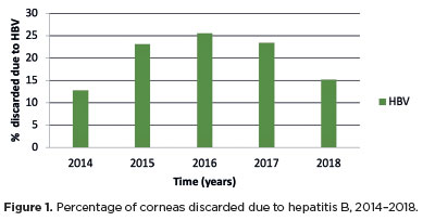
RESULTS: Of the 1,968 corneas processed, 897 (45.57%) were discarded. Among these, 333 (37.12%) tested positive for serological markers. Hepatitis B accounted for 34.67% of positive cases (15% of total donations), hepatitis C for 1.11% (0.50% of total), and HIV for 0.89% (0.4% of total). Hepatitis cases remained stable between 2014 and 2016, with a marked decline in 2017 and 2018. Most discarded corneas were positive for anti-HBc (31.88%) and negative for HBsAg; however, the anti-HBs test was not performed to confirm immunity to the hepatitis B virus.
CONCLUSION: The findings highlight the importance of serological testing to identify and eliminate contaminated corneas, thereby preventing the transmission of infectious diseases to recipients. Positive serology for hepatitis, particularly hepatitis B, was the leading cause of corneal disposal.
Keywords: Cornea; Corneal transplantation; Corneal donation; Eye banks; Hepatitis B virus; Hepatitis C virus; HIV infections; Seropositivity; Serologic tests
Abstract
PURPOSE: To assess the reliability and penetration depth of an automated micropuncture system using a tattoo machine.
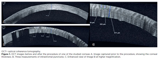
METHODS: Twenty human corneas were obtained and subjected to intrastromal micropuncture using a tattoo machine. Each cornea was divided into two halves: one received pigment, while the other received saline solution as a control. The Cheyenne tattoo machine was operated at 60 Hz, with standardized needle exposure (six passes per application). The machine used cartridges containing five microneedles. The study was registered with Agência Nacional de Vigilância Sanitária ANVISA (numbers 80281110015, 80281110016, and 80281110019). The pigment used was Electric Ink black ink, with a density of 1,271,460 μg/mL. Puncture depth was measured before and after the procedure using both anterior segment optical coherence tomography and histopathological analysis. Puncture depth measurements were analyzed using ImageJ software. Each cornea was measured thrice, and the results were subsequently compared.
RESULTS: No corneal perforations were observed with the use of the tattoo machine, and puncture depth measurements ranged from 107 to 486 µm.
CONCLUSIONS: The use of a tattoo machine represents a viable and accessible approach for keratopigmentation, with potential for both cosmetic and therapeutic applications. Its adaptation for controlled intrastromal drug delivery may enable the targeted treatment of deep infectious keratitis, corneal neovascularization, and stromal inflammatory disorders, representing a promising approach for corneal stromal diseases. Further research is needed to optimize techniques and evaluate long-term safety and efficacy, particularly for the delivery of antimicrobial, anti-inflammatory, and anti-vascular endothelial growth factor agents.
Keywords: Eye banks; Cadaver; Cornea; Corneal stroma; Drug delivery systems; Tissue donors; Tattooing/instrumentation; Punctures
13-tab01.jpg)
Abstract
Objetivo: Avaliar o perfil clínico e epidemiológico dos transplantes de córnea realizados em um centro de referência oftalmológica de Recife no estado de Pernambuco, localizado no nordeste do Brasil.
Métodos: Esse estudo transversal coletou através de prontuários médicos dados clínicos e epidemiológicos de pacientes submetidos a ceratoplastia na Fundação Altino Ventura, de janeiro a dezembro de 2017.
Resultados: Um total de 356 procedimentos foram realizados em 327 pacientes dos quais 165 (50.5%) eram mulheres. A média de idade na cirurgia foi de 50.9 ± 22.6 anos (variação, 10 - 89 anos). A maioria dos pacientes (n=152 [46.5%]) era da capital e região metropolitana. A média de tempo de espera na fila para o transplante de córnea foi de 52.4 ± 58.9 dias (variação, 0 - 460 dias). As principais indicações de transplante foram ceratite infecciosa (n=88 [24.7%]), ceratocone (n=80 [22.5%]) e falência de transplante prévio (n=75 [21.1%]). Transplante penetrante foi a técnica mais realizada (n=213 [59.9%]) e foi mais comum em homens (n=132 [76.7%]), enquanto os transplantes lamelares posteriores (n=143 [41.1%]) foram mais realizados nas mulheres (p<0.001).
Conclusão: Ceratites infecciosas foram a causa mais comum de transplante, com prevalência similar em adultos economicamente ativos de ambos os sexos. Transplante penetrantes foram os prevalentes em homens e os transplantes lamelares em mulheres.
Keywords: Doença da córnea/epidemiologia; Transplante de córnea; Ceratoplastia penetrante; Brasil/epidemiologia

Abstract
PURPOSE: To evaluate the impact of the COVID-19 pandemic and characterize the serological profile of discarded corneal donations in the coverage area of the Banco de Olhos de Londrina, through reverse transcription-polymerase chain reaction testing for COVID-19 and serological screening of cornea donors excluded because of positive test results.
METHODS: This observational retrospective study included 776 cornea donors who’s serological and reverse transcription-polymerase chain reaction test results were processed at the Hospital of Universidade Estadual de Londrina between May 2020 and 2022. The number of corneal donations and tissue utilization rates throughout the years of operation of the Banco de Olhos de Londrina were also analyzed.
RESULTS: The mean donor age was 53.14 years; 332 donors (43%) were female, and 444 (57%) were male. Positive results were identified in 15.76% of donors for hepatitis B core antibody antibodies, 0.65% for hepatitis B surface antigen, 1.03% for hepatitis C antibodies, and 0.52% for human immunodeficiency virus and human T-lymphotropic vírus. Positive reverse transcription-polymerase chain reaction results for SARS-CoV-2 were observed in 2.7% of cases. Older adults were 2.6 times more likely to test positive for SARS-CoV-2 (95% CI, 1.06-6.34) and 3.0 times more likely to test positive for hepatitis B core antibody (95% CI, 1.95-4.41) than younger individuals. A 75.2% reduction in corneal donations was observed in 2020 compared with 2019, accompanied by a 5% increase in tissue utilization, possibly associated with the effectiveness of donor screening during the pandemic.
CONCLUSION: The COVID-19 pandemic had a profound impact on the number of corneal transplants worldwide, in Brazil, and at the Banco de Olhos de Londrina because of the substantial decline in donations during this period. Hepatitis B was the leading cause of corneal tissue discard due to positive serology in both this study and previous reports, highlighting the importance of prevention programs and improved vaccination coverage. Strict legislation, comprehensive serological screening, and appropriate processing of donated tissue remain essential to eliminate potential sources of infection and ensure transplantation safety.
Keywords: Cornea; Corneal transplantation; COVID-19; Eye banks; Serology
Abstract
PURPOSE: To determine the indications and epidemiological characteristics of corneal transplant recipients at a referral center in Amazonas, Brazil, and to evaluate postoperative visual outcomes.
METHODS: This retrospective, descriptive, observational study included adult patients who underwent corneal transplantation performed by a single surgeon at the Instituto de Oftalmologia de Manaus between 2013 and 2023. Demographic characteristics, occupation, comorbidities, surgical indications, graft type, and best-corrected visual acuity at baseline and 12 months postoperatively were analyzed.
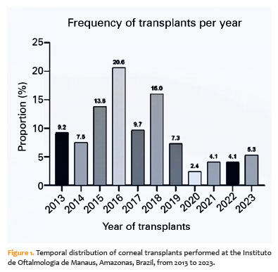
RESULTS: A total of 413 corneal transplantations were performed, predominantly in male patients (64.4%) aged 41–70 years (43.3%). Leucoma was the most common indication (26.2%), followed by perforated corneal ulcers (21.1%), bullous keratopathy (17.9%), and keratoconus (10.7%). Penetrating keratoplasty accounted for 94.9% of all procedures. Optical keratoplasty was the most frequently performed procedure (76.3%), followed by tectonic (22.0%) and therapeutic (1.7%) keratoplasty. At 12 months postoperatively, visual improvement was greatest among patients who underwent optical keratoplasty, particularly those with keratoconus, bullous keratopathy, and leucoma. Functional vision (≤0.3 logMAR) was achieved in 32.6% of eyes overall and in 40.3% of eyes that underwent optical keratoplasty. The number of transplantations declined after 2019, coinciding with the COVID-19 pandemic.
CONCLUSIONS: This study represents the largest retrospective series of corneal transplantations in Amazonas and one of the few such studies from Northern Brazil. The predominance of leucoma and infectious corneal perforation reflects regional socioeconomic and occupational vulnerabilities. Although visual outcomes improved significantly after transplantation, visual recovery was limited by the advanced stage of disease at presentation. These findings underscore the need to expand access to early diagnosis, strengthen eye banking and donor tissue procurement systems, and increase the adoption of lamellar keratoplasty techniques to optimize visual outcomes in the Amazon region.
Keywords: Corneal transplantation; Corneal opacity; Eye infections; Keratoplasty; Epidemiology; Visual acuity; Amazonas; Brazil
Abstract
PURPOSE: Neuro-ophthalmology is a highly specialized subspecialty at the interface of neurology and ophthalmology. Globally, workforce shortages and geographic maldistribution limit access to neuro-ophthalmic care. Although Brazil has a large ophthalmology workforce, regional disparities may restrict access to subspecialty services. The national distribution of neuro-ophthalmologists has not previously been quantified. This study aimed to characterize the geographic distribution of neuro-ophthalmologists in Brazil and identify potential gaps in access to neuro-ophthalmic care.
METHODS: This cross-sectional, population-based study used data from the registry of the Brazilian Neuro-Ophthalmology Association (Associação Brasileira de Neuro-Oftalmologia [ABNO]) to identify practicing neuro-ophthalmologists (data extracted on January 1, 2026). Municipal- and state-level population estimates were obtained from the Brazilian Institute of Geography and Statistics. Specialist density was calculated as the number of neuro-ophthalmologists per million inhabitants. States were classified according to the presence or absence of at least one specialist and were further grouped into Brazil's five macroregions for comparative analyses. Descriptive statistical analyses were performed.
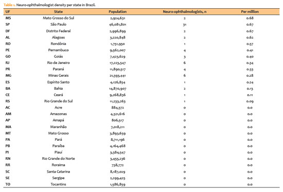
RESULTS: A total of 66 neuro-ophthalmologists were identified across 14 of Brazil's 27 states. Thirteen states had no registered neuro-ophthalmologists, representing approximately 49.4 million people (23.2% of the national population). Five states—São Paulo (n=31), Rio de Janeiro (n=6), Minas Gerais (n=6), Pernambuco (n=4), and Paraná (n=4)—accounted for more than two-thirds of all specialists. Specialist density ranged from 0 to more than 0.7 per million inhabitants, with higher concentrations in the Southeast and no specialists in several states in the North and Northeast. The distribution was highly clustered in metropolitan areas.
CONCLUSION: Brazil exhibits marked geographic maldistribution of neuro-ophthalmologists, with nearly one-quarter of the population lacking local access to specialized care. The concentration of specialists in urban and economically developed regions mirrors international trends and may exacerbate inequities within the public health system. Expanding the workforce and integrating teleophthalmology into neuro-ophthalmic services may help reduce regional disparities and improve access to specialized care.
Keywords: Ophthalmologists; Public health; Health policy; Workforce; Professional practice location; Telemedicine; Teleophthalmology; Brazil
09-tab01.jpg)
Abstract
OBJETIVO: Estudar os dados epidemiológicos, resultados laboratoriais e fatores de risco associados às ceratites infecciosas.
MÉTODOS: Estudo retrospectivo das amostras de cultura de córnea em pacientes com ceratites infecciosas entre Janeiro/2010 a Dezembro/2019. Os resultados foram analisados de acordo com o diagnóstico etiológico de infecção bacteriana, fúngica ou parasitária e correlacionado com os fatores de risco relacionados.
RESULTADOS: Quatro mil, oitocentas e dez amostras corneanas de 4047 pacientes (média de idade de 47,79 ± 20,68 anos; homens em sua maioria (53,7%) foram incluídas.
A prevalência de infecções por bactéria, fungo e Acanthamoeba foram de 69.80%, 7,31%, and 3,51%, respectivamente. A maioria das bactérias mais frequentemente isoladas foram Staphylococcus coagulase-negativo (CoNS) (45,14%), S. aureus (10,02%), Pseudomonas spp. (8,80%), e Corynebacterium spp. (6,21%). Dentre CoNS, o principal agente foi S. epidermidis (n=665). Nas ceratites fúngicas, Fusarium spp. (35,42%) e Candida parapsilosis (16,07%) foram os agentes mais comuns entre os filamentosos e leveduriformes, respectivamente. O uso de lentes de contato foi associado à cultura positiva para Acanthamoeba spp. (OR=19,04; p<0,001) e Pseudomonas spp (OR=3,20; p<0,001). Trauma ocular prévio foi associado a culturas positivas para fungo (OR=1,80; p=0,007), e idade avançada foi associada a culturas positivas para bactéria (OR=1,76; p=0,001).
CONCLUSÕES: Nossos achados demonstraram uma maior positividade para bactérias em amostras de cultura corneana. Dentre estas, CoNS foi mais frequentemente identificado, sendo S. epidermidis o principal agente. Nas ceratites fúngicas, Fusarium spp. Foi o mais comumente isolado. O risco de positividade para Acanthamoeba spp. e Pseudomonas spp. foi maior em usuários de lentes de contato. Trauma ocular aumentou o risco de cultura positiva para fungo, ao passo que idade mais avançada aumentou o risco de infecção bacteriana.
Keywords: Úlcera da córnea/epidemiologia; Úlcera da córnea/microbiologia; Ceratite; Infecções oculares
Abstract
PURPOSE: To evaluate the current scenario of rehabilitation services for people with retinal diseases in Brazil.
METHODS: An exploratory study was conducted between February 2023 and June 2023 using a Google Forms questionnaire that was distributed by patient associations.
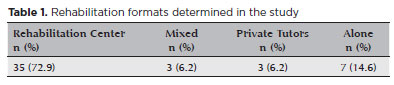
RESULTS: A total of 142 patients, aged 18-80 were included in the study. Forty-eight participants (33.8%) were undergoing rehabilitation, while 94 (66.2%) were not. The main reason for not undergoing rehabilitation was a lack of knowledge about the service (n=41, 43.6%). Healthcare professionals made the most referrals (n=20, 41.7%). Rehabilitation improved the quality of life in 38 (80.9%) participants, and 28 (62.2%) participants were satisfied with the process. There was a statistically significant disparity between patient satisfaction and the locale of rehabilitation implementation. Twenty-three (69.7%) participants who underwent rehabilitation at a specialized center reported satisfaction.
CONCLUSION: The rehabilitation process directly increases the quality of life of individuals with retinal diseases. However, despite the availability of rehabilitation centers in large parts of Brazil, most patients with retinal diseases are not acquainted with the rehabilitation process and do not receive referrals for it. Thus, healthcare providers should increase referrals to rehabilitation centers, and public policies should be formulated to raise awareness among the population regarding the availability of rehabilitation services.
Keywords: Rehabilitation; Quality of life; Retinal diseases; Personal satisfaction; Patient care team; Public policy; Surveys and questionnaires; Brazil
Abstract
PURPOSE: This study aimed to report the use, efficacy, and safety of intracameral voriconazole as an adjuvant treatment for deep fungal keratitis.
METHODS: This was a prospective case series of seven eyes with fungal keratitis with anterior chamber involvement or a corneal ulcer refractory to conventional topical treatment. In addition to topical treatment with 0.15% amphotericin B eye drops, voriconazole 50 μg/ 0.1 mL
was administered to the anterior chamber of each affected eye up to four times within 72 h. The primary outcome measures were healing (fungal eradication) and the need for therapeutic keratoplasty. Best-corrected visual acuity was a secondary outcome measure.
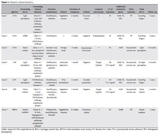
RESULTS: Three cases were confirmed by confocal microscopy, and four were diagnosed from positive culture tests. At presentation, one patient had a best-corrected visual acuity of 20/80, while all others had hand motion or worse. Four cases received one intracameral injection, two cases received three, and one case received four injections. There were no complications after any of the intracameral voriconazole injections. Four patients had imminent corneal perforations and were treated with cyanoacrylate adhesive and bandage contact lenses. Four patients recovered from the infection, and three underwent therapeutic keratoplasty. The final best-corrected visual acuity was improved in two cases but all patients had a final visual acuity of counting fingers or worse.
CONCLUSION: As an adjuvant treatment for deep fungal keratitis, intracameral voriconazole injection is a feasible option. Although fungal eradication was achieved in all patients, three required therapeutic keratoplasty and all patients had unsatisfactory visual acuity outcomes.
Keywords: Antifungal agents; Fungi; Corneal transplantation; Keratitis; Eye infections, fungal; Voriconazole
Abstract
PURPOSES: This study aims to assess and compare the postoperative visual and topographic outcomes, complications, and graft survival rates following deep anterior lamellar keratoplasty and penetrating keratoplasty in patients with macular corneal dystrophy.
METHODS: In this study we enrolled 59 patients (23 male; and 36 female) with macular corneal dystrophy comprising 81 eyes. Out of these, 64 eyes underwent penetrating keratoplasty, while 17 eyes underwent deep anterior lamellar keratoplasty. The two groups were analyzed and compared based on best-corrected visual acuity, corneal tomography parameters, pachymetry, complication rates, and graft survival rates.
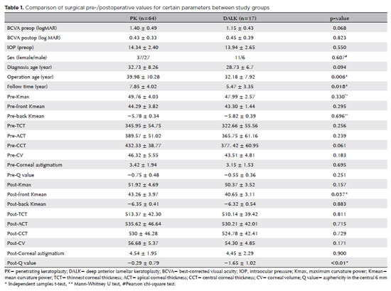
RESULTS: After 12 months, 70.6% of the patients who underwent deep anterior lamellar keratoplasty (DALK) and 75% of those who had penetrating keratoplasty (PK) achieved a best-corrected visual acuity of 20/40 or better (p=0.712). Following surgery, DALK group showed lower front Kmean (p=0.037), and Q values (p<0.01) compared to the PK group. Postoperative interface opacity was observed in seven eyes (41.2%) in the DALK group. Other topography values and other complications (graft rejection, graft failure, cataract, glaucoma, microbial keratitis, optic atrophy) did not show significant differences between the two groups. The need for regrafting was 9.4% and 11.8% in the PK and DALK groups, respectively (p=0.769). Graft survival rates were 87.5% and 88.2% for PK and DALK; respectively (p=0.88 by Log-rank test).
CONCLUSION: Both PK and DALK are equally effective in treating macular corneal dystrophy, showing similar visual, topographic, and survival outcomes. Although interface opacity occurs more frequently after DALK the visual results were comparable in both groups. Therefore, DALK emerges as a viable surgical choice for patients with macular corneal dystrophy without Descemet membrane involvement is absent.
Keywords: Macular corneal dystrophy; Corneal dystrophies; Hereditary; Keratoplasty; Penetrating; Corneal transplantation
Abstract
PURPOSE: To evaluate the clinical results of cryopreserved amniotic membrane transplantation as a treatment option for refractory neurotrophic corneal ulcers.
METHODS: This prospective study included 11 eyes of 11 patients who underwent amniotic membrane transplantation for the treatment of refractory neurotrophic corneal ulcers at Hospital de Clínicas da Universidade Federal do Paraná, in the city of Curitiba, from May 2015 to July 2021. Patients underwent different surgical techniques in which the amniotic membrane was applied with the epithelium facing upward to promote corneal re-epithelialization.
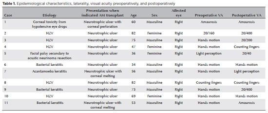
RESULTS: The median age of the patients was 60 years (range, 34-82 years), and 64% were men. The predominant etiology of corneal ulcers was herpes zoster (45% of cases). Approximately one-third of the patients (27%) were chronically using hypotensive eye drops, and more than half (54%) had previously undergone penetrating corneal transplantation. At the time of amniotic membrane transplantation, 18% of the eyes had corneal melting, 9% had corneal perforation, and the others had corneal ulceration without other associated complications (73%). The time between clinical diagnosis and surgical treatment ranged from 9 days to 2 years. The corrected visual acuity was worse than 20/400 in 90% of the patients preoperatively, with improvement in 36% after 3 months of the procedure, worsening in 18% and remaining stable in 36%. Of the patients, 81% complained of preoperative pain, and 66% of them reported total symptom relief after the surgical procedure. In one month, 54.6% of the patients presented a closure of epithelial defect, and half of the total group evolved with corneal thinning. The failure rate was 45.5% of the cases.
CONCLUSION: Cryopreserved amniotic membrane transplantation can be considered a good alternative for treating refractory neurotrophic corneal ulcers, as it resulted in significant improvement in pain (66%) and complete epithelial closure (60%) in many patients at 1 month postoperatively. Notably, the high failure rate highlights the need for further studies to identify patient- and ulcer-related factors that may influence the outcomes of this procedure.
Keywords: Amnion/transplantation; Corneal ulcer; Anterior eye segment; Keratitis
14-fig01.jpg)
Abstract
O presente trabalho traz uma revisão das abordagens terapêuticas para a cegueira da córnea. O estudo detalha as etapas e os elementos envolvidos na cicatrização da córnea. Ele mostra as limitações das estratégias cirúrgicas e farmacológicas usadas para restaurar e manter a transparência da córnea em termos de sobrevida a longo prazo e alcance geográfico. As perspectivas dos agentes anabólicos, incluindo vitamina A, hormônios, fatores de crescimento e novos moduladores pró-mitóticos e anti-inflamatórios para auxiliar a cicatrização da ferida na córnea, são revisadas criticamente. Aqui, apresentamos estudos envolvendo nanotecnologia, terapia gênica e reengenharia de tecidos como possíveis estratégias futuras para atuar de maneira isolada ou combinada com a cirurgia da córnea para prevenir ou reverter a cegueira corneana.
Keywords: Cegueira; Doenças da córnea; Transplante de córnea; Terapia genética; Terapia baseada em transplante de células e tecidos; Células-tronco
Abstract
Os bancos de olhos utilizam procedimentos estéreis na manipulação dos olhos, medidas antissépticas para a descontaminação da superfície ocular e critério rigoroso de seleção do doador. Essa seleção é feita por meio do prontuário médico e de testes sorológicos específicos post mortem. Para orientá-la e uniformizá-la, as associações de bancos de olhos e órgãos governamentais fornecem listas de contraindicações absolutas e relativas de uso do tecido, baseadas nas condições prévias de saúde do doador. Essas listas são as guardiãs do princípio de Hipócrates "primum non nocere" e, como tal, são conservadoras. Entretanto, cada transplante traz o risco de transmissão de agentes potencialmente nocivos ao receptor. O objetivo não é eliminar esse risco, mas limitá-lo a um nível razoável. Existe um equilíbrio entre a segurança e a disponibilidade de córneas. A sabedoria está em manter esse equilíbrio, exercendo a prudência sem rigor exagerado.
Keywords: Bancos de olhos/normas; Transplante de córnea; Seleção do doador; Coleta de tecidos e órgãos
09-fig01.jpg)
Abstract
Em decorrência de complicações, da biocompatibilidade e da escassez de tecido doador para transplantes de córnea natural, foram elaboradas córneas artificiais que são potenciais para reabilitar funções ópticas. Nessa perspectiva, objetivou-se a análise da eficácia da reabilitação visual entre os implantes: Boston tipo I, Boston tipo II, Aurolab, osteo-odonto-ceratoprótese e ceratoprótese de Osso Tibial. De modo geral, a princípio observou-se uma tendência de melhoria da Best-corrected visual acuity em todos os tipos de lentes, mas considerável queda durante acompanhamento a longo prazo. O dispositivo com melhor reabilitação visual em pacientes com superfícies oculares úmidas é a Boston tipo I, seguida pela Aurolab, que é economicamente viável em países emergentes. Ao considerar pacientes com olhos secos, o implante de Boston tipo II apresenta maior reabilitação visual. Por fim, em virtude de não apresentarem dados equiparáveis, as lentes osteo-odonto-ceratoprótese e de osso tibial não puderam ser analisadas.
Keywords: Transplante de córnea; Próteses visuais; Córnea; Reabilitação; Acuidade visual
 ABO is licensed under a Creative Commons Attribution-NonComercial 4.0 Internacional.
About
Issues
Editorial Board
Submission

Arquivos Brasileiros de Oftalmologia
Official publication of Brazilian Council of Ophthalmology - Conselho Brasileiro de Oftalmologia (CBO)
Rua Casa do Ator, 1.117 - 2nd floor - Zip Code: 04546-004
São Paulo - SP, Brazil
TEL: +55 11 3266-4000
E-mail: [email protected]
