Showing of 1 until 14 from 141 result(s)
Search for: Uveitis/epidemiology; Uveitis/etiology; Uveitis/ diagnosis; Toxoplasmosis, ocular; Hospital, university; Brazil/ epidemiology
Abstract
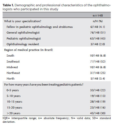
PURPOSE: This study aimed to identify the strategies adopted by Brazilian ophthalmologists to control myopia in clinical practice.
METHODS: This was a prospective cross-sectional study. Data were collected using an online questionnaire.
RESULTS: Responses from 148 participants were collected between March and May 2024. The majority of respondents were general ophthalmologists (51%) and pediatric ophthalmologists (43%). They came from all regions of Brazil, but more than half (52%) were from the Southeast region. Most participants (30%) had over 20 years of clinical practice experience. A significant proportion (89.2%) treated progressive myopia. The most requested complementary exams were optical biometry (83.78%) and corneal topography or tomography (69.59%). Behavioral measures were considered the most effective myopia treatment strategies by 41.2% of the respondents, followed by optical (33.8%) and pharmacological interventions (25%). Most recommended spending more time outdoors (94.59%) and reducing screen time (93.92%). Spectacle lenses for myopia (83.11%) and 0.025% atropine eye drops (54.73%) were the most prescribed treatments after the recommendation of environmental and behavioral changes.
CONCLUSION: This study presents a novel analysis of the clinical strategies for myopia control among Brazilian ophthalmologists. Understanding current clinical practices and identifying possible improvements are essential steps toward developing evidence-based guidelines and professional education aimed at improving patient care.
Keywords: Myopia/epidemiology; Refractive errors; Contact lenses; Myopia/drug therapy; Atropine/therapeutic use; Ophthalmologists; Practice patterns, physicians’; Surveys and questionnaires; Brazil/epidemiology
11-car01.jpg)
Abstract
Objetivo: A alta prevalência e gravidade da toxoplasmose congênita no Brasil, com muitos indivíduos afetados desenvolvendo baixa visão, reforça a importância da avaliação da sua qualidade de vida. Este estudo tem como objetivo adaptar o Children’s Visual Function Questionnaire (CVFQ) para a realidade sociocultural de crianças brasileiras e investigar suas propriedades psicométricas para avaliação da qualidade de vida relacionada à visão nesses indivíduos.
Métodos: Estudo epidemiológico transversal aninhado de coorte de 142 crianças pré-escolares acompanhadas prospectivamente em hospital universitário de referência em Belo Horizonte, Brasil. Todas foram submetidos a exame oftalmológico completo, incluindo medida da acuidade visual e oftalmoscopia binocular indireta. Questionários foram aplicados aos pais e cuidadores, para avaliar a percepção da qualidade de vida, bem como o nível sócio-econômico das famílias. Análise estatística multivariada foi realizada para avaliar as propriedades psicométricas da escala de qualidade de vida.
Resultados: Adaptações na versão brasileira do Children’s Visual Function Questionnaire-7 originaram o Children’s Visual Function Questionnaire-7-BR-toxo, um questionário para avaliar a percepção de pais/cuidadores sobre a qualidade de vida relacionada à visão de crianças pré-escolares com toxoplasmose congênita. Pela descrição, estrutura de variabilidade, e interpretação do agrupamento dos itens do questionário adaptado, identificaram-se seis subescalas: saúde geral, capacidade visual, desempenho visual/visão funcional, comportamento social e pessoal, impacto na família e tratamento. Crianças com baixa visão associada a toxoplasmose congênita tiveram escores mais baixos nas seguintes subescalas: acuidade visual (p=0,004), desempenho visual/visão funcional (p=0,008), impacto na família (p=0,001) e saúde geral (p=0,001).
Conclusão: As propriedades psicométricas foram adequadas no tocante à validade do construto. O Children’s Visual Function Questionnaire-7-BR-toxo foi capaz de registrar o impacto da deficiência visual nas famílias de crianças com toxoplasmose congênita.
Keywords: Qualidade de vida; Baixa visão; Uveíte; Toxoplasmose congênita; Criança
05-tab01.jpg)
Abstract
Objetivo: Caracterizar a população com suspeita de glaucoma encaminhada a um centro público terciário no sul do Brasil e avaliar diferenças no dano dos parâmetros funcionais e estruturais entre os pacientes diagnosticados com diferentes tipos de glaucoma e aqueles classificados como normais e aqueles mantidos como suspeitos de glaucoma.
Métodos: Esta é uma coorte dos pacientes encaminhados para o setor de glaucoma suspeito do Hospital Nossa Senhora da Conceição, Porto Alegre - BR, no período de março de 2016 a dezembro de 2018. Os pacientes foram acompanhados até obterem exames confiáveis (exame oftalmológico completo, campimetria visual, tomografia de coerência óptica) para serem classificados como: normal, glaucoma suspeito, glaucoma com pressão intraocular elevada, glaucoma de pressão normal ou hipertenso ocular.
Resultados: Um total de 135 pacientes foram incluídos neste estudo, sendo que destes, 117 pacientes completaram todos os exames e foram incluídos neste estudo. A maioria dos pacientes foi considerada normal (36,8%), seguido por glaucoma suspeito (25,64%), glaucoma de pressão normal (18,8%), glaucoma com pressão intraocular elevada (12%) e hipertensão ocular (6%). A principal razão para encaminhamento foi escavação do nervo óptico aumentada. Pacientes com glaucoma de pressão normal eram em média mais velhos que os demais (p=0,03). Esses também apresentavam índice de campo visual e desvio médio da campimetria visual piores que sujeitos normal, com suspeita de glaucoma e hipertensos oculares, e tinham a camada de fibra nervosa medida pela tomografia de coerência óptica mais fina que normais e suspeitos de glaucoma (p<0,002). Os pacientes com glaucoma de pressão elevada não diferiram significativamente dos outros grupos.
Conclusão: Pacientes com glaucoma de pressão normal tendem a ser diagnosticados mais tardiamente devido ao fato da pressão intraocular não estar elevada, logo a escavação do disco óptico deve ser maior para gerar a suspeita de glaucoma. Neste estudo, paciente com glaucoma de pressão normal apresentaram doença mais avançada no momento do diagnóstico em comparação com os outros grupos.
Keywords: Glaucoma/diagnóstico; Hipertensão ocular; Glaucoma de ângulo aberto/diagnóstico; Glaucoma de ângulo fechado/diagnóstico; Atenção terciária à saúde; Padrões de prática médica

Abstract
PURPOSE: To describe the epidemiological and clinical profile of hospitalized patients with retinoblastoma in Brazil.
METHODS: Using data from the Hospital Cancer Registry of the , patients with the morphological codes of retinoblastoma who were diagnosed between 2000 to 2018, aged 0–19 years, and followed up in registered hospitals (analytical cases) were selected. The relative and absolute frequencies of demographic, clinical, diagnostic, therapeutic, and outcome variables were described. Hospital performance indicators were calculated and compared between hospitals qualified and not qualified to treat pediatric oncology cases and between hospitals with different case volumes (<20, 20–75, >75 cases).
RESULTS: Of the 2,269 identified analytical cases from 86 institutions, 48% were from the Southeast, 54% were male, and 66% were aged <4 years. The proportion of missing data (NA) was too high for several variables. Approximately 84% of the patients were from the public health system, 40% had a positive family history, and 88% had unilateral involvement. The first treatment included surgery in 58.3% of the patients (NA=2), Approximately 36.6% of these patients achieved complete remission, 10.8% achieved partial remission, and 12.7% died (NA=59%). Hospital performance indicators were within the target in >90% of the patients. The median time between the first appointment and diagnosis (6 days, interquartile range [IQR] 1–14) was significantly lower and the median time to death was longer (343 days, IQR, 212-539) in high-volume hospitals (>75 cases) than in medium- and low-volume hospitals.
CONCLUSIONS: Despite the high proportion of missing data, we found that the delay in diagnosis is due to prehospital factors. Additionally, there is a need for educational programs for healthcare professionals and families that emphasize early identification and referral to specialized centers. Future studies should focus on the impact of Hospital Cancer Registry data completeness on outcomes, causes of delay in diagnosis, regional inequalities, and barriers to accessing specialized services.
Keywords: Retinoblastoma/diagnosis; Retinoblastoma/epidemiology; Patient care; Humans; Children; Adolescents; Brazil.
Abstract
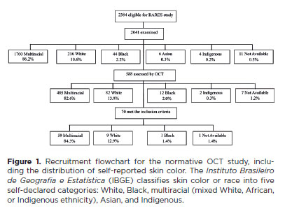
PURPOSE: This study evaluated macular thickness using spectral-domain optical coherence tomography in healthy participants from a population-based eye survey.
METHODS: The Brazilian Amazon Region Eye Survey was a population-based study assessing the prevalence and causes of visual impairment, blindness, and ocular diseases in adults aged ≥45 years from urban and rural areas of Parintins. A subgroup was selected based on inclusion criteria for both eyes: best-corrected visual acuity ≥20/32, normal eye examination results, and no prior ocular surgery. Scans were performed using the iVue optical coherence tomography device. Measurements were taken from the nine subfields defined by the Early Treatment Diabetic Retinopathy Study, examining the full retina as well as the inner and outer retinal layers. Associations of retinal thickness with age and sex were also analyzed. Statistical significance was set at p≤0.05.
RESULTS: In total, 70 healthy participants (25 males), aged 45–65 years (mean=52 ± 5), were included. Mean central foveal thickness was 248.71 ± 18.73 μm. A significant age-related reduction in macular thickness was observed, particularly in the inner superior parafovea (p=0.036), nasal perifovea (p=0.001), superior perifovea (p=0.028), outer layer of inferior parafovea (p=0.049), and the inferior perifovea of the full retina (p=0.029). Males showed significantly greater thickness in the outer layer, especially in the outer parafovea (p=0.004) and perifovea (p<0.0001).
CONCLUSIONS: This study established normative macular thickness values for healthy older adults in the Brazilian Amazon region using spectral-domain optical coherence tomography. Age and sex were found to significantly influence macular thickness and should be considered when interpreting measurements. These data will support future studies of retinal diseases in this population.
Keywords: Retinal diseases/diagnosis; Macula lutea/pathology; Macular degeneration/diagnosis; Diabetic retinopathy/diagnosis; Vision, low; Vision tests; Tomography, optical coherence/methods; Young adult; Cross-sectional studies; Brazil/epidemiology
09-tab01tb.jpg)
Abstract
Objetivos: O objetivo do estudo é avaliar o perfil das visitas ao Pronto-Socorro de Oftalmologia (PS) do Hospital São Paulo, serviço público de atendimento terciário aberto 24 horas em São Paulo - Brasil, pertencente à Universidade Federal de São Paulo, nos últimos 11 anos.
Métodos: Foi realizado um estudo transversal retrospectivo, com base em todos os pacientes (n=634.726) admitidos no pronto-socorro de oftalmologia do Hospital São Paulo entre janeiro de 2009 e dezembro de 2019.
Resultados: De 2009 a 2019, houve um aumento no influxo de 39,2% com importante variação nos atendimentos ao longo dos anos, a mediana de idade foi de 38 ± 20,4 anos, o sexo masculino representou 53,3% e os pacientes únicos representaram 53,1%. Verificou-se que 79,5% das visitas ocorreram das 7h às 17h e 80,8% nos dias da semana. Os diagnósticos mais frequentes foram conjuntivite aguda seguida de blefarite, ceratite, hordéolo / calázio e corpo estranho corneano.
Conclusão: Ao longo do período de análise do estudo, houve importante aumento nas apresentações, com predominância de atendimentos não urgentes e baixa proporção de pacientes com uma única visita. Nossos resultados evidenciam o perfil das consultas oftalmológicas, podendo gerar mudanças no sistema público de saúde visando a melhoria da qualidade do atendimento e acesso às emergências oftalmológicas na cidade de São Paulo.
Keywords: Serviço hospitalar de emergência; Epidemiologia; Traumatismos oculares; Oftalmopatias.
13-tab01.jpg)
Abstract
Objetivo: Avaliar o perfil clínico e epidemiológico dos transplantes de córnea realizados em um centro de referência oftalmológica de Recife no estado de Pernambuco, localizado no nordeste do Brasil.
Métodos: Esse estudo transversal coletou através de prontuários médicos dados clínicos e epidemiológicos de pacientes submetidos a ceratoplastia na Fundação Altino Ventura, de janeiro a dezembro de 2017.
Resultados: Um total de 356 procedimentos foram realizados em 327 pacientes dos quais 165 (50.5%) eram mulheres. A média de idade na cirurgia foi de 50.9 ± 22.6 anos (variação, 10 - 89 anos). A maioria dos pacientes (n=152 [46.5%]) era da capital e região metropolitana. A média de tempo de espera na fila para o transplante de córnea foi de 52.4 ± 58.9 dias (variação, 0 - 460 dias). As principais indicações de transplante foram ceratite infecciosa (n=88 [24.7%]), ceratocone (n=80 [22.5%]) e falência de transplante prévio (n=75 [21.1%]). Transplante penetrante foi a técnica mais realizada (n=213 [59.9%]) e foi mais comum em homens (n=132 [76.7%]), enquanto os transplantes lamelares posteriores (n=143 [41.1%]) foram mais realizados nas mulheres (p<0.001).
Conclusão: Ceratites infecciosas foram a causa mais comum de transplante, com prevalência similar em adultos economicamente ativos de ambos os sexos. Transplante penetrantes foram os prevalentes em homens e os transplantes lamelares em mulheres.
Keywords: Doença da córnea/epidemiologia; Transplante de córnea; Ceratoplastia penetrante; Brasil/epidemiologia
Abstract
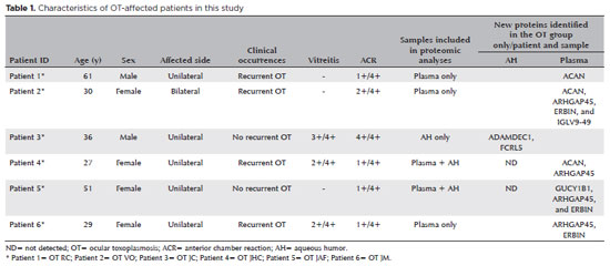
PURPOSE: To characterize the extracellular vesicle protein cargo in the aqueous humor and plasma of patients with ocular toxoplasmosis.
METHODS: Aqueous humor and plasma were collected from six patients with active ocular toxoplasmosis and six patients with cataract. Extracellular vesicles were isolated, and western blotting and mass spectrometry were performed for protein analysis.
RESULTS: All plasma samples from patients with ocular toxoplasmosis and cataract were positive for the tetraspanins CD63 and TSG101. However, the aqueous humor from patients with ocular toxoplasmosis was positive only for CD63. Sixty-seven new unreported proteins were identified in the aqueous humor and plasma of patients with the ocular toxoplasmosis and cataract. Of the 67 proteins, 10 and 7 were found only in the cataract and ocular toxoplasmosis groups, respectively. In general, these proteins were involved in immune system activation and retina homeostasis and were related to infections and retina-associated diseases. Conclusion: The distinct protein signatures between ocular toxoplasmosis and cataract may be helpful in the differential diagnosis of ocular toxoplasmosis. However, more studies are needed to better understand the role of these proteins in the pathogenesis of ocular toxoplasmosis.
Keywords: Extracellular vesicles; Proteomics; Toxoplasma gondii; Ocular toxoplasmosis, Aqueous humor; Plasma; Liquid biopsy
Abstract
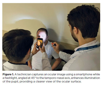
PURPOSE: This pilot study evaluated the diagnostic accuracy of a deep learning model for detecting pterygium in anterior segment photographs taken using smartphones in the Brazilian Amazon. The model’s performance was benchmarked against assessments made by experienced ophthalmologists, considered the clinical gold standard.
METHODS: In this cross-sectional study, 38 participants (76 eyes) from Barcelos, Brazil, were enrolled. Trained nonmedical health workers captured high-resolution anterior segment images using smartphones. These images were analyzed using a deep learning model based on the MobileNet-V2 convolutional neural network. Diagnostic metrics–including sensitivity, specificity, accuracy, positive predictive value, negative predictive value, and area under the receiver operating characteristic curve–were calculated and compared with the ophthalmologists’ evaluations.
RESULTS: The deep learning model achieved a sensitivity of 91.43%, specificity of 90.24%, positive predictive value of 88.46%, negative predictive value of 92.79%, and an area under the curve of 0.91. Logistic regression revealed no statistically significant association between pterygium and demographic variables such as age or gender.
CONCLUSIONS: The deep learning model demonstrated high diagnostic performance in identifying pterygium in a remote Amazonian population. These preliminary findings support the potential use of artificial intelligence–based tools to facilitate early detection and screening in underserved regions, thereby enhancing access to ophthalmic care.
Keywords: Pterygium/diagnostic imaging; Smartphone; Diagnostic techniques, ophthalmological; Deep learning; Telemedicine; Artificial intelligence; Cross-sectional studies; Brazil/epidemiology
Abstract
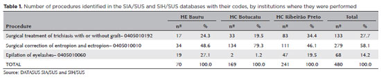
PURPOSE: Trachoma is the major infectious cause of preventable blindness in the world, and its sequelae include the presence of cicatricial entropion and trachomatous trichiasis. Trachoma can be corrected by surgical treatment of the eyelids and, if left untreated, may result in corneal opacification, low vision, and blindness. There are limited data on trachomatous trichiasis in Brazil. This study was conducted to estimate the frequency of entropion and trichiasis surgeries of trachomatous origin based on the records of procedures performed in specialized hospitals that served the Unified Health System (SUS) in the years 2016 and 2017.
METHODS: This was a retrospective study conducted in the oculoplastic sectors of the ophthalmology services of the following three hospitals in the state of São Paulo: Hospital das Clínicas da Faculdade de Medicina de Botucatu (HC Botucatu), Hospital das Clínicas da Faculdade de Medicina de Ribeirão Preto da Universidade de São Paulo (HC Ribeirão Preto), and Hospital Estadual de Bauru (HE Bauru). Medical records corresponding to the codes of interest were evaluated.
RESULTS: In total, 462 medical records were evaluated, including 170 (36.8%) at HC Botucatu, 61 (13.2%) at HE Bauru, and 231 (50.0%) at HC Ribeirão Preto. There were 39 (8.4%) cases of trachomatous trichiasis, ranging from 9 (14.8%) at HE Bauru to 15 (6.5%) at HC Ribeirão Preto.
CONCLUSIONS: The frequency of surgery due to trachoma was low in these oculoplastic services. The state of São Paulo might have reached the goal for trachoma elimination in the surgical component. The questionnaire used for data collection was successfully tested despite some difficulties in collecting data from the medical records. Studies with the same methodology are recommended in other services in the areas of endemic trachoma in the past to understand the frequency of eye lid surgeries performed for treating trachomatous sequelae.
Keywords: Trachoma; Trichiasis; Medical records; Epidemiology; Neglected diseases; Unified Health System; Brazil
Abstract
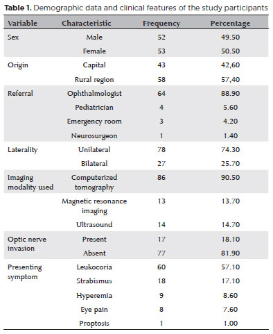
PURPOSE: Although Brazil has a high prevalence of retinoblastoma, there is a lack of epidemiological data on the disease. Thus, in this study, we aimed to evaluate the epidemiological profile of patients diagnosed with retinoblastoma in the ophthalmology department of a pediatric tertiary referral hospital in Ceara, Brazil.
METHODS: A descriptive and cross-sectional study was conducted by retrospectively analyzing the clinical and socioeconomic data from the medical records of pediatric patients followed-up at the hospital between 2007 and 2021. Retinoblastoma was diagnosed on the basis of a fundoscopic or histopathologic examination.
RESULTS: The data of 105 patients were included in the study, and the mean patient age at the time of diagnosis was 1.7 years. Most of the patients were women (50.5%) and hailed from rural areas (57.4%), which was associated with a higher tumor stage. Of the 150 patients, 57.1% initially presented with leukocoria. Ocular hyperemia was associated with more advanced stages of retinoblastoma (p=0.004). Bilateral involvement was observed in 25.7% of the patients and at a significantly younger age (p=0.009). The presence of retinal detachment, vascularized lesions, and vitreous seeds significantly increased the likelihood of requiring enucleation.
DISCUSSION: This study presents an epidemiological description of retinoblastoma in Brazil, which highlights the significance of early detection. Delayed diagnosis is associated with a poorer visual prognosis and higher mortality rate, particularly in patients with unilateral disease. Risk factors for a more severe disease were retinal detachment, vascularized lesions, and vitreous seeds. The correlation between histopathological features and clinical outcomes was limited.
CONCLUSION: Further studies are required to assess the influence of ocular hyperemia, fundoscopic assessment, and histopathologic findings on the prognosis of retinoblastoma. Moreover, it is critical to devise interventions to reduce the time-to-diagnosis in rural areas.
Keywords: Retinoblastoma; Retinal neoplasms; Epidemiology; Prevalence; Risk factors; Delayed diagnosis; Child
Abstract
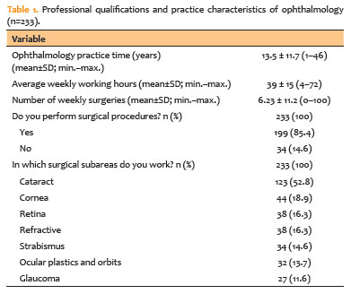
PURPOSE: To assess musculoskeletal symptoms, identify the most affected body areas, and investigate factors associated with the development of musculoskeletal disorders among ophthalmologists in Brazil.
METHODS: A survey was conducted using an online questionnaire and snowball sampling. Statistical analyses were performed using Jamovi version 2.3.28, and graphs were generated using RStudio version 2023.06.2 + 561.
RESULTS: A total of 233 participants (42 ophthalmology residents and 191 ophthalmologists) were included, with a mean age of 40.4 years (standard deviation 11.3; range 25–73 years). Musculoskeletal symptoms were reported by 83% of participants. The cervical region (57.1%), upper back (54.5%), and lumbar region (53.6%) were the most frequently reported sites of pain. A high body mass index was identified in 54.9% of the sample, and 50.2% of participants reported using painkillers in the previous year for musculoskeletal symptoms. The mean duration of professional activity in ophthalmology was 13.5 years, and the mean weekly workload was 39 hours. A significant association was observed between weekly workload and the presence of musculoskeletal disorders (p=0.045).
CONCLUSION: This study demonstrated a high prevalence of musculoskeletal disorders among ophthalmologists in Brazil, particularly involving the cervical, lumbar, and upper back regions, consistent with findings reported in international studies. Important contributing factors include long working hours, a high patient volume, and repetitive or awkward postures during examinations and procedures. Preventive strategies and improvements in working conditions are needed to protect the health and well-being of ophthalmologists.
Keywords: Musculoskeletal Diseases/epidemiology; Back pain; Lumbar Vertebrae; Occupational diseases/epidemiology; Ergonomics; Ophthalmic practice; Ophthalmologists/statistics & numerical data; Brazil/epidemiology
Abstract
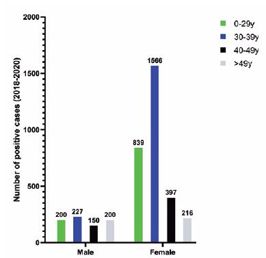
PURPOSE: To describe a 2019 acute toxoplasmosis outbreak in the city of São Paulo, Brazil, and to evaluate the laboratory serological profile for toxoplasmosis for three consecutive years. The ophthalmological manifestations of the patients involved in the outbreak were also studied.
METHODS: A cross-sectional descriptive study of a toxoplasmosis outbreak in São Paulo, Brazil, between February and May 2019. Epidemiological data were described, as were the observed ocular manifestations. As part of this study the number of patients with positive IgM toxoplasmosis serology was obtained from a large laboratory network (DASA) for three consecutive years, including the year of the outbreak (2018, 2019, 2020).
RESULTS: Eighty-three individuals were identified in the outbreak and two clusters were studied. The clinical picture of at least 77% of the patients, the epidemiological analysis, and the short incubation period (5-8 days) suggested contamination by oocysts. Serological laboratory data analysis revealed an increase of positive toxoplasmosis IgM in 2019 of 73% compared to the previous year. Ophthalmological examination revealed that at least 4.8% of the patients developed toxoplasmic retinochoroiditis, none of whom had been treated during the acute systemic disease.
CONCLUSION: Our findings indicate vegetable contamination as the possible source of this outbreak, a high prevalence of toxoplasmosis in São Paulo during the outbreak period, and a drop in the number of tests during the COVID-19 pandemic. Retinochoroiditis was observed in at least 4.8% of the cases. We confirm the need to implement effective means for the prevention, diagnosis, and treatment of the disease. This may involve raising awareness among the population of the importance of vegetable hygiene, and improved quality control of food and water.
Keywords: Toxoplasmosis/etiology; Food parasitology; Water/parasitology; Uveitis, posterior/parasitology; Chorioretinitis/parasitology; Visual acuity; Disease outbreaks; Eye manifestations; Humans.
04-fig01.jpg)
Abstract
A infecção pelo Toxoplasma gondii pode causar manifestações oculares tanto após a sua forma congênita quanto a sua forma adquirida. Reportamos aqui dois casos de toxoplasmose congênita sintomática com envolvimento ocular em irmãos não gêmeos, com intervalo de 2 anos entre gestações. A transmissão vertical da toxoplasmose em gestações sucessivas, outrora considerada impossível, é um evento plausível mesmo em indivíduos imunocompetentes.
Keywords: Toxoplasmose ocular/congênita; Toxoplasmose ocular/ genética; Toxoplasmose congênita; Toxoplasma gondii; Uveite
 ABO is licensed under a Creative Commons Attribution-NonComercial 4.0 Internacional.
About
Issues
Editorial Board
Submission
Arquivos Brasileiros de Oftalmologia
Official publication of Brazilian Council of Ophthalmology - Conselho Brasileiro de Oftalmologia (CBO)
Rua Casa do Ator, 1.117 - 2nd floor - Zip Code: 04546-004
São Paulo - SP, Brazil
TEL: +55 11 3266-4000
E-mail: [email protected]