Arq. Bras. Oftalmol. 2025;88 (1 )
:1-8
| DOI: 10.5935/0004-2749.2023-0103
Abstract
PURPOSE: This study aimed to compare the safety and effectiveness of intraocular pressure reduction between micropulse transscleral cyclophotocoagulation and “slow cook” transscleral cyclophotocoagulation in patients with refractory primary open-angle glaucoma.
METHODS: We included patients with primary open angle glaucoma with at least 12 months of follow-up. We collected and analyzed data on the preoperative characteristics and postoperative outcomes. The primary outcomes were a reduction of ≥20% of the baseline value (criterion A) and/or intraocular pressure between 6 and 21 mmHg (criterion B).
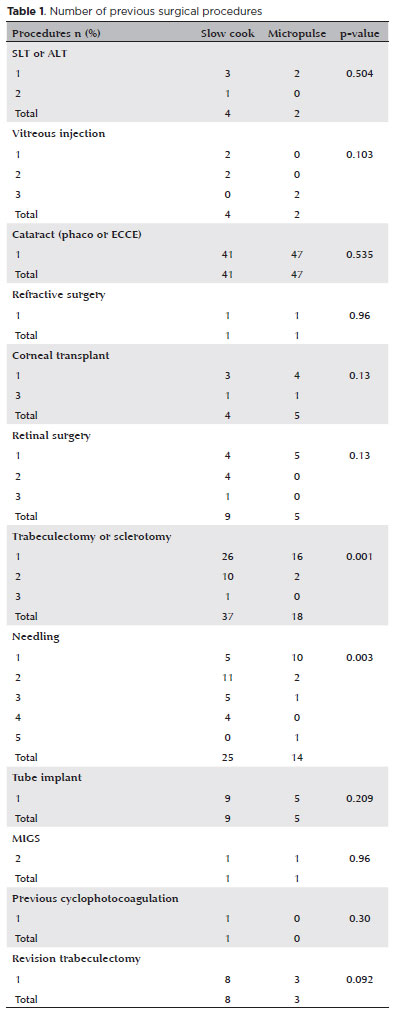
RESULTS: We included 128 eyes with primary open-angle glaucoma. The preoperative mean intraocular pressure was 25.53 ± 6.40 and 35.02 ± 12.57 mmHg in the micropulse- and “slow cook” transscleral cyclophotocoagulation groups, respectively (p<0.001). The mean intraocular pressure was reduced significantly to 14.33 ± 3.40 and 15.37 ± 5.85 mmHg in the micropulse- and “slow cook” transscleral cyclophotocoagulation groups at the last follow-up, respectively (p=0.110). The mean intraocular pressure reduction at 12 months was 11.20 ± 11.46 and 19.65 ± 13.22 mmHg in the micropulse- and “slow cook” transscleral cyclophotocoagulation groups, respectively (p<0.001). The median preoperative logMAR visual acuity was 0.52 ± 0.69 and 1.75 ± 1.04 in the micropulse- and “slow cook” transscleral cyclophotocoagulation groups, respectively (p<0.001). The mean visual acuity variation was -0.10 ± 0.35 and -0.074 ± 0.16 in the micropulse- and “slow cook” transscleral cyclophotocoagulation, respectively (p=0.510). Preoperatively, the mean eye drops were 3.44 ± 1.38 and 2.89 ± 0.68 drugs in the micropulse- and “slow cook” transscleral cyclophotocoagulation groups, respectively (p=0.017), but those were 2.06 ± 1.42 and 1.02 ± 1.46 at the end of the study in the slow cook” and micropulse transscleral cyclophotocoagulation groups, respectively (p<0.001). The success of criterion A was not significant between both groups. Compared with 11 eyes (17.74%) in the slow cook” transscleral cyclophotocoagulation group, 19 eyes (28.78%) in the micropulse transscleral cyclophotocoagulation group showed complete success (p=0.171). For criterion B, 28 (42.42%) and 2 eyes (3.22%) showed complete success after micropulse- and slow cook” transscleral cyclophotocoagulation, respectively (p<0.001).
CONCLUSION: Both techniques reduced intraocular pressure effectively.
Keywords: Sclera/surgery; Glaucoma, open-angle/surgery; Ciliary body/surgery; Intraocular pressure; Laser coagulation/methods; Lasers, semiconductor; Comparative study; Effectiveness
Arq. Bras. Oftalmol. 2023;86 (3 )
:1-7
| DOI: 10.5935/0004-2749.20230029
Abstract
Objetivo: Caracterizar a população com suspeita de glaucoma encaminhada a um centro público terciário no sul do Brasil e avaliar diferenças no dano dos parâmetros funcionais e estruturais entre os pacientes diagnosticados com diferentes tipos de glaucoma e aqueles classificados como normais e aqueles mantidos como suspeitos de glaucoma.
Métodos: Esta é uma coorte dos pacientes encaminhados para o setor de glaucoma suspeito do Hospital Nossa Senhora da Conceição, Porto Alegre - BR, no período de março de 2016 a dezembro de 2018. Os pacientes foram acompanhados até obterem exames confiáveis (exame oftalmológico completo, campimetria visual, tomografia de coerência óptica) para serem classificados como: normal, glaucoma suspeito, glaucoma com pressão intraocular elevada, glaucoma de pressão normal ou hipertenso ocular.
Resultados: Um total de 135 pacientes foram incluídos neste estudo, sendo que destes, 117 pacientes completaram todos os exames e foram incluídos neste estudo. A maioria dos pacientes foi considerada normal (36,8%), seguido por glaucoma suspeito (25,64%), glaucoma de pressão normal (18,8%), glaucoma com pressão intraocular elevada (12%) e hipertensão ocular (6%). A principal razão para encaminhamento foi escavação do nervo óptico aumentada. Pacientes com glaucoma de pressão normal eram em média mais velhos que os demais (p=0,03). Esses também apresentavam índice de campo visual e desvio médio da campimetria visual piores que sujeitos normal, com suspeita de glaucoma e hipertensos oculares, e tinham a camada de fibra nervosa medida pela tomografia de coerência óptica mais fina que normais e suspeitos de glaucoma (p<0,002). Os pacientes com glaucoma de pressão elevada não diferiram significativamente dos outros grupos.
Conclusão: Pacientes com glaucoma de pressão normal tendem a ser diagnosticados mais tardiamente devido ao fato da pressão intraocular não estar elevada, logo a escavação do disco óptico deve ser maior para gerar a suspeita de glaucoma. Neste estudo, paciente com glaucoma de pressão normal apresentaram doença mais avançada no momento do diagnóstico em comparação com os outros grupos.
Keywords: Glaucoma/diagnóstico; Hipertensão ocular; Glaucoma de ângulo aberto/diagnóstico; Glaucoma de ângulo fechado/diagnóstico; Atenção terciária à saúde; Padrões de prática médica
Arq. Bras. Oftalmol. 2022;85 (2 )
:166-173
| DOI: 10.5935/0004-2749.20220034
Abstract
Objetivos: Mensurar a perfusão do complexo retina/coróide com ressonância magnética em olhos com fechamento angular primário agudo (FAPA).
Métodos: Três sequências de ressonância magnética, duas anatômicas e uma de perfusão com gadolínio, foram adquiridas em pacientes com fechamento angular primário agudo. Regiões de interesse foram desenhadas na sequência de perfusão e sobrepostas à sequência anatômica. O volume de sangue relativo nos 2 primeiros segundos foi considerado como referência, e sua variação nos 28 segundos subsequentes foi analisada.
Resultados: Cinco olhos de 5 pacientes com fechamento angular primário agudo foram incluídos (3 com crise unilateral e 2 com crise bilateral). Três olhos contralaterais e 2 olhos de 2 pacientes saudáveis, pareados por sexo e idade, foram incluídos no grupo controle. Pacientes com fechamento angular primário agudo incluíam 4 (80%) mulheres, com idade média de 65,8 ± 12,37 anos, pressão intraocular média de 56,2 ± 14,67 mmHg, pressão arterial média de 113,4 ± 8,17 mmHg e pressão de perfusão ocular de 57,2 ± 13,46mmHg. No grupo controle, pressão intraocular média foi de 15,6 ± 2,61 mmHg (p=0,0625), pressão arterial média de 107,4 ± 6,57 mmHg (p=1,00) e pressão de perfusão ocular de 91,8 ± 6,72 mmHg (p=0,0625). O volume de sangue relativo do complexo retina/coróide foi de -0,127 ± 0,048 nos olhos em fechamento angular primário agudo e -0,213 ± 0,116 nos olhos controles (p=0,3125).
Conclusões: A sequência de ressonância magnética com gadolínio não demonstrou diferença na perfusão de retina/coroide em olhos com fechamento angular primário agudo.
Keywords: Glaucoma de ângulo fechado; Imagem por ressonância magnética; Gadolínio; Retina; Perfusão
Arq. Bras. Oftalmol. 2026;89 (1 )
:1-6
| DOI: 10.5935/0004-2749.2025-0052
Abstract
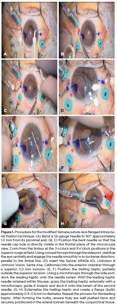
PURPOSE: To evaluate whether two simplified modifications of flanged intrascleral fixation techniques (Yamane and Canabrava) provide comparable refractive outcomes and complication rates while reducing surgical complexity in trocar-assisted vitrectomy.
METHODS: This retrospective observational study included 88 patients who underwent flanged fixation surgery with vitrectomy. In the modified Yamane technique, a single-path sclerotomy with bilateral symmetry was performed instead of an angled sclerotomy. In the modified Canabrava technique, the intraocular lens was inserted first, followed by the creation of a circular polypropylene loop with 2-mm flange spacing. Postoperative refractive parameters, including intraocular lens astigmatism, and complications such as intraocular lens iris capture were analyzed.
RESULTS: Of the 88 patients, 70 underwent the modified Yamane technique, and 18 underwent the modified Canabrava technique. No significant differences were observed between the two techniques regarding refractive outcomes or postoperative complications, except for surgical duration, which was significantly shorter (p<0.001) in one technique. Mean intraocular lens astigmatism was −0.675 D for Yamane and −0.666 D for Canabrava.
CONCLUSION: Optimizing needle engagement for symmetry in the Yamane technique and narrowing flange spacing while ensuring a circular polypropylene configuration in the Canabrava technique may reduce surgical complexity and improve postoperative outcomes.
Keywords: Polypropylenes; Yamane technique; Vitrectomy; Astigmatism; Lenses, intraocular; Postoperative complications; Suture techniques; Iris.
Arq. Bras. Oftalmol. 2026;89 (3 )
:1-8
| DOI: 10.5935/0004-2749.2025-0043
Abstract
PURPOSE: To evaluate the effect of single-session transscleral diode laser cyclophotocoagulation on intraocular pressure in refractory glaucoma and to determine structural changes using ultrasound biomicroscopy.
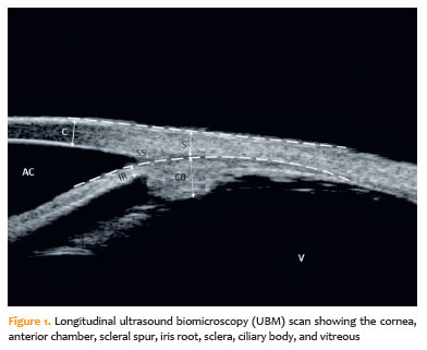
METHODS: Forty-three eyes were evaluated. Intraocular pressures at baseline and at the first, third, and sixth months after transscleral diode laser cyclophotocoagulation were compared. Ciliary body thickness, ciliary muscle thickness, ciliary process thickness, iris root thickness, and scleral thickness were assessed at baseline and at the third and sixth months post-treatment.
RESULTS: Reductions in intraocular pressure were significant between baseline and the first month (p=0.018), third month (p<0.001), and sixth month (p<0.001) as well as between the first and third months (p=0.034) and the first and sixth months (p=0.036). Compared with baseline, intraocular pressure reduction rates at the first, third, and sixth months were 34.6%, 56.5%, and 55.3%, respectively, while success rates were 30.2%, 62.8%, and 55.8%, respectively. Decreases in ciliary body thickness, ciliary muscle thickness, and ciliary process thickness were significant between baseline and the third month (p<0.05) and between baseline and the sixth month (p<0.05), whereas changes between the third and sixth months were not significant (p>0.05). Iris root and scleral thicknesses did not change after treatment (p>0.05). At the third and sixth months, significant positive correlations were observed between changes in intraocular pressure and changes in ciliary body thickness and ciliary process thickness (p<0.05).
CONCLUSIONS: To the best of our knowledge, this is one of the few studies comprehensively investigating structural changes after transscleral diode laser cyclophotocoagulation using ultrasound biomicroscopy. Moreover, the relationships between intraocular pressure changes and variations in the ciliary body, ciliary muscle, ciliary process, iris root, and scleral thicknesses were examined in detail. Single-session treatment did not affect iris root or scleral thickness but significantly reduced ciliary body, ciliary muscle, and ciliary process thicknesses. Greater reductions in ciliary body and ciliary process thickness may contribute to more pronounced intraocular pressure reduction.
Keywords: Intraocular pressure; Laser coagulation/methods; Lasers, semiconductor; Microscopy, acoustic; Glaucoma; Ciliary body
 ABO is licensed under a Creative Commons Attribution-NonComercial 4.0 Internacional.
05-tab01.jpg)
05-fig01.jpg)
04-fig01.jpg)
15-fig01.jpg)
01-fig01tb.jpg)
03-fig01.jpg)
02-fig01.jpg)