Arq. Bras. Oftalmol. 2020;83 (4 )
:283-288
| DOI: 10.5935/0004-2749.20200040
Abstract
Objetivo: Comparar as alterações nos parâmetros do segmento anterior após a cirurgia ExPRESS Mini Glaucoma Shunt vs. trabeculectomia usando a câmera Scheimpflug Pentacam rotativa.
Métodos: Neste estudo comparativo prospectivo, 27 pacientes com glaucoma tratados no Centro Médico Rabin de 2009 a 2013 foram incluídos neste estudo comparativo prospectivo: 19 participantes (19 olhos) foram submetidos ao implante de derivação ExPRESS e 12 (13 olhos) foram submetidos à trabeculectomia. Alterações nos parâmetros da câmara anterior no dia 1 e em 3 meses de pós-operatório foram avaliadas pelas imagens de Scheimpflug.
Resultados: A pressão intraocular diminuiu significativamente em relação aos valores iniciais nos dois grupos. A diminuição nos dois grupos foi semelhante no 3º mês pós-operatório (p=0,82). A cirurgia com ExPRESS causou um aumento temporário do astigmatismo posterior da córnea (p=0,008) e uma diminuição temporária da profundidade da câmara anterior (p=0,016) e do volume (p=0,006) no primeiro dia do pós-operatório. Ao final de três meses, esses parâmetros não foram mais estatisticamente significativos (p=0,065, p=0,51 e p=0,57, respectivamente). A trabeculectomia causou um aumento temporário do astigmatismo anterior e posterior da córnea no primeiro dia do pós-operatório (p=0,003 e p=0,005, respectivamente), mas isso não foi observado ao final de 3 meses (p=1,0 e p=1,0, respectivamente). Após 3 meses, tanto o EXPRESS quanto a trabeculectomia mostraram alterações semelhantes nos parâmetros da câmara anterior.
Conclusões: O implante ExPRESS Mini para glaucoma e a trabeculectomia diminuíram significativamente a pressão intraocular e tiveram efeitos temporários nos parâmetros do segmento anterior, com pequenas diferenças entre os métodos.
Keywords: Glaucoma/cirurgia; Implantes para drenagem de glaucoma; Trabeculectomia/métodos; Pressão intraocular
Arq. Bras. Oftalmol. 2023;86 (3 )
:1-6
| DOI: 10.5935/0004-2749.20230044
Abstract
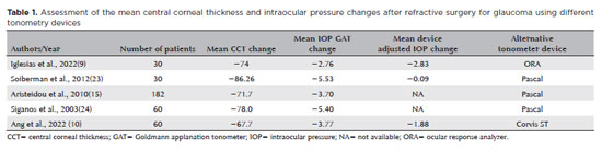
Objetivo: O setor nasal do ângulo da câmara anterior pode apresentar maior densidade de canais coletores, o que pode influenciar no resultado de cirurgias angulares. Considerando as diferenças anatômicas no ângulo da câmara anterior, comparamos os resultados das abordagens de trabeculoplastia seletiva a laser nasal e temporal de 180 graus no glaucoma de ângulo aberto.
Métodos: Revisão retrospectiva de prontuários de pacientes com glaucoma de ângulo aberto (primária, pseudoexfoliação e pigmentar), que realizaram pelo menos uma sessão de trabeculoplastia seletiva a laser de 180 graus entre dezembro/2016 e outubro/2018. O setor nasal (N1) ou temporal (T1) foi escolhido a critério do médico. Os pacientes que não apresentaram diminuição da pressão intraocular (PIO) entre 3 e 6 meses foram retratados com trabeculoplastia seletiva a laser de 180 graus no setor de ângulo oposto (T2 e N2). O principal resultado medido foi a diminuição da pressão intraocular no 6º mês de acompanhamento após a última trabeculoplastia seletiva a laser. Uma análise de regressão multivariável avaliou os fatores associados à redução da pressão intraocular após o tratamento.
Resultados: O procedimento foi realizado inicialmente em 45 olhos (N1=25, T1=20 olhos), e repetido no setor ângulo da câmara anterior oposto em 19 olhos (N2 = 11, T2 = 8 olhos). Os testes ANOVA mostraram que apenas a abordagem N1 apresentou diferença significativa na diminuição da pressão intraocular em relação a T1, N2 e T2 (p=0,0014). A pressão intraocular basal (p=0,021) e o setor ângulo da câmara anterior (N1; p=0,044) se correlacionaram com a diminuição da pressão intraocular.
Conclusão: A trabeculoplastia seletiva a laser de 180 graus realizado inicialmente no setor nasal foi associado a uma diminuição mais significativa da pressão intraocular em comparação com a abordagem temporal. Considerando as diferenças setoriais no ângulo da câmara anterior, mais estudos prospectivos são necessários para confirmar nossos achados e fornecer protocolos para trabeculoplastia seletiva a laser mais eficientes.
Keywords: Glaucoma de ângulo aberto; Terapia a laser/métodos; Pressão intraocular; Trabeculoplastia/métodos.
Arq. Bras. Oftalmol. 2025;88 (1 )
:1-8
| DOI: 10.5935/0004-2749.2023-0103
Abstract
PURPOSE: This study aimed to compare the safety and effectiveness of intraocular pressure reduction between micropulse transscleral cyclophotocoagulation and “slow cook” transscleral cyclophotocoagulation in patients with refractory primary open-angle glaucoma.
METHODS: We included patients with primary open angle glaucoma with at least 12 months of follow-up. We collected and analyzed data on the preoperative characteristics and postoperative outcomes. The primary outcomes were a reduction of ≥20% of the baseline value (criterion A) and/or intraocular pressure between 6 and 21 mmHg (criterion B).
RESULTS: We included 128 eyes with primary open-angle glaucoma. The preoperative mean intraocular pressure was 25.53 ± 6.40 and 35.02 ± 12.57 mmHg in the micropulse- and “slow cook” transscleral cyclophotocoagulation groups, respectively (p<0.001). The mean intraocular pressure was reduced significantly to 14.33 ± 3.40 and 15.37 ± 5.85 mmHg in the micropulse- and “slow cook” transscleral cyclophotocoagulation groups at the last follow-up, respectively (p=0.110). The mean intraocular pressure reduction at 12 months was 11.20 ± 11.46 and 19.65 ± 13.22 mmHg in the micropulse- and “slow cook” transscleral cyclophotocoagulation groups, respectively (p<0.001). The median preoperative logMAR visual acuity was 0.52 ± 0.69 and 1.75 ± 1.04 in the micropulse- and “slow cook” transscleral cyclophotocoagulation groups, respectively (p<0.001). The mean visual acuity variation was -0.10 ± 0.35 and -0.074 ± 0.16 in the micropulse- and “slow cook” transscleral cyclophotocoagulation, respectively (p=0.510). Preoperatively, the mean eye drops were 3.44 ± 1.38 and 2.89 ± 0.68 drugs in the micropulse- and “slow cook” transscleral cyclophotocoagulation groups, respectively (p=0.017), but those were 2.06 ± 1.42 and 1.02 ± 1.46 at the end of the study in the slow cook” and micropulse transscleral cyclophotocoagulation groups, respectively (p<0.001). The success of criterion A was not significant between both groups. Compared with 11 eyes (17.74%) in the slow cook” transscleral cyclophotocoagulation group, 19 eyes (28.78%) in the micropulse transscleral cyclophotocoagulation group showed complete success (p=0.171). For criterion B, 28 (42.42%) and 2 eyes (3.22%) showed complete success after micropulse- and slow cook” transscleral cyclophotocoagulation, respectively (p<0.001).
CONCLUSION: Both techniques reduced intraocular pressure effectively.
Keywords: Sclera/surgery; Glaucoma, open-angle/surgery; Ciliary body/surgery; Intraocular pressure; Laser coagulation/methods; Lasers, semiconductor; Comparative study; Effectiveness
Arq. Bras. Oftalmol. 2026;89 (1 )
:1-8
| DOI: 10.5935/0004-2749.2024-0397
Abstract
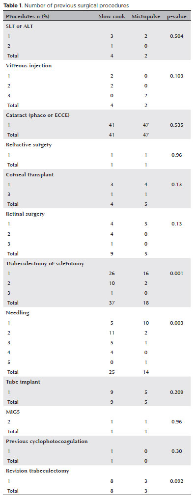
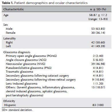
PURPOSE: Glaucoma is one of the leading causes of irreversible blindness worldwide. When topical hypotensive agents or laser trabeculoplasty fail to adequately control the disease, escalation of therapy becomes necessary, with transscleral cyclophotocoagulation being one of the available options. Several variations of transscleral cyclophotocoagulation exist, including traditional continuous wave, MicroPulse, and slow-coagulation techniques. We propose a novel variation – custom slow-coagulation transscleral cyclophotocoagulation – which combines elements of both continuous wave and slow-coagulation approaches. This study aimed to evaluate the outcomes of this technique in patients with refractory glaucoma.
METHODS: This retrospective, interventional study included 104 eyes of 83 patients with refractory glaucoma who underwent custom slow-coagulation transscleral cyclophotocoagulation. Changes in intraocular pressure, visual acuity, the number of glaucoma medications, and postoperative complications were analyzed. A paired t test was used to compare changes in intraocular pressure and visual acuity, while the Wilcoxon signed-rank test was applied to categorical variables. Success rates following custom slow-coagulation transscleral cyclophotocoagulation were estimated using Kaplan–Meier survival analysis.
RESULTS: Mean intraocular pressure decreased significantly from 38.9 ± 15.8 mmHg at baseline to 16.3 ± 9.9 mmHg at Month 12 (p<0.001). The mean number of glaucoma medications also decreased significantly from 3.6 ± 0.6 to 1.8 ± 1.4 (p<0.001). No significant reduction in mean visual acuity was observed during follow-up.
CONCLUSIONS: Custom slow-coagulation transscleral cyclophotocoagulation effectively reduced baseline intraocular pressure and the number of glaucoma medications, with a low rate of complications and no decline in visual acuity over a 12-month follow-up period. This novel technique demonstrated a high safety profile in a Hispanic population and represents a low-cost, minimally invasive procedure with rapid recovery and promising efficacy in intraocular pressure control.
Keywords: Glaucoma/surgery; Sclera; Filtering surgery; Laser coagulation/methods; Lasers, semiconductor/therapeutic use; Intraocular pressure; Blindness/prevention & control; Vision, low/epidemiology; Visual acuity
Arq. Bras. Oftalmol. 2021;84 (4 )
:361-366
| DOI: 10.5935/0004-2749.20210052
Abstract
OBJETIVO: Glaucoma é a principal causa de cegueira irreversível no mundo. O pico da pressão intraocular é um dos principais fatores de risco para progressão do glaucoma, e o controle pressórico ainda é o único tratamento efetivo para todas as formas de glaucoma. O objetivo principal deste estudo é comparar a redução basal e do pico da pressão intraocular, obtidas através do Teste de Sobrecarga Hídrica, entre os dois olhos dos mesmos pacientes utilizando latanoprosta 0,005% em um olho e submetidos à aplicação de trabeculoplastia a laser seletiva no olho contralateral.
MÉTODOS: Este é um estudo prospectivo, intervencionista, longitudinal e randomizado. Trinta pacientes consecutivos, glaucomatosos, com pressão intraocular controlada em uso de monoterapia com latanoprosta, foram recrutados de um único centro oftalmológico. Os olhos dos pacientes foram randomizados e um olho foi selecionado para tratamento com trabeculoplastia a laser seletiva e olho contralateral tratado com colírio de latanoprosta 0,005%. Foram avaliados a pressão intraocular basal e pico de pressão intraocular um mês (Teste de Sobrecarga Hídrica 2) e seis meses (Teste de Sobrecarga Hídrica 3) após tratamento.
RESULTADOS: Não houve diferença estatística entre a pressão intraocular pré washout entre os olhos randomizados para trabeculoplastia a laser seletiva e latanoprosta, 13,6 ± 2,1 e 13,3 ± 1,8 mmHg, respectivamente (p=0,182). Em relação à pressão intraocular basal, não houve diferença estatística entre os grupos, tanto no Teste de Sobrecarga Hídrica 2 (p=0,689) e Teste de Sobrecarga Hídrica 3 (p=0,06). Não houve diferença estatística em relação ao pico de pressão intraocular entre os grupos trabeculoplastia a laser seletiva e latanoprosta, no Teste de Sobrecarga Hídrica 2 (p=0,771) e Teste de Sobrecarga Hídrica 3 (p=0,774).
CONCLUSÕES: Em resumo, nosso estudo demonsrou que a eficácia da redução pressórica é similar entre latanoprosta e trabeculoplastia a laser seletiva, e pacientes glaucomatosos que estão com a pressão intraocular clinicamente controlados com latanoprosta e trocam de tratamento para trabeculoplastia a laser seletiva mantém sua pressão intraocular controlada.
Keywords: Glaucoma; Pressão intraocular; Latanoprosta; Lasers
Arq. Bras. Oftalmol. 2025;88 (6 )
:1-5
| DOI: 10.5935/0004-2749.2024-0340
Abstract
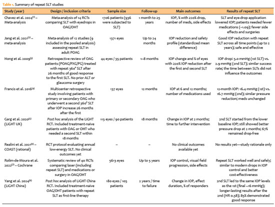
PURPOSE: This study aimed to report the surgical outcomes and success predictors of micropulse transscleral cyclophotocoagulation in eyes with refractory glaucoma.
METHODS: This was a noncomparative, interventional case series. Patients with refractory glaucomas, defined as eyes with prior incisional glaucoma surgery failure and uncontrolled intraocular pressure, who underwent micropulse transscleral cyclophotocoagulation between March 2017 and June 2021 were enrolled. A minimum follow-up period of 6 months was required. Preoperative and postoperative intraocular pressure, number of hypotensive medications, surgical complications, and any subsequent related events were recorded. Success criteria were as follows: 1) intraocular pressure reduction ≥20% and intraocular pressure ≤18 mmHg; 2) intraocular pressure reduction ≥30% and intraocular pressure ≤15 mmHg. The need for topical hypotensive medications was not considered a failure.
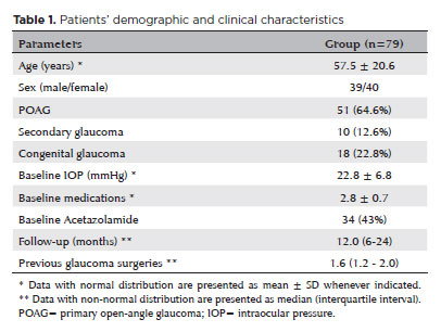
RESULTS: Seventy-nine (79) eyes (79 patients; mean age, 57.5 ± 20.6 years) were included. Overall, the median follow-up duration was 12.0 (interquartile interval, 6–24) months, and the mean intraocular pressure was reduced from 22.8 ± 6.8 mmHg to 15.5 ± 5.6 mmHg at the last follow-up visit (p<0.001). The mean number of medications was reduced from 2.8 ± 0.7 to 2.0 ± 1.0 (p<0.01). At 12 months postoperatively, the success rates for criteria 1 and 2 were 54.9% and 49.7%, respectively. Aside from one case of corneal ulcer, which fully resolved with clinical treatment, and two cases of persistent hypotony (with no visual acuity loss during follow-up), no other vision-threatening complications were observed during the postoperative period. The magnitude of intraocular pressure reduction at 1 month (adjusted to preoperative intraocular pressure; HR=1.01; p=0.002).
CONCLUSION: Our findings suggest that micropulse transscleral cyclophotocoagulation is a relatively effective alternative for managing refractory glaucomas, with minor postoperative complications. In addition, the initial intraocular pressure reduction was a statistically significant predictor of 1-year success in patients undergoing micropulse transscleral cyclophotocoagulation.
Keywords: Intraocular pressure/physiology; Glaucoma, open-angle/surgery; Trabeculectomy; Laser coagulation/methods; Tonometry, ocular/methods; Postoperative complications; Antihypertensive agents/therapeutic use.
 ABO is licensed under a Creative Commons Attribution-NonComercial 4.0 Internacional.
03-tab01.jpg)
13-tab01tb.jpg)
09-tab01tb.jpg)
02-fig01.jpg)
01-fig01tb.jpg)
13-fig01tb.jpg)