Arq. Bras. Oftalmol. 2024;87 (1 )
:1-6
| DOI: 10.5935/0004-2749.2021-0536
Abstract
Objetivo: Avaliar os segmentos anterior e posterior em recém-nascidos a termo durante um período de 1,5 anos.
Métodos: Foram analisados recém-nascidos a termo que tiveram os olhos examinados entre junho de 2019 e dezembro de 2020, e os resultados foram registrados retrospectivamente.
Resultados: O estudo foi composto por 2.972 recém-nascidos com média de uma semana de nascimento de 38,7 ± 1,2 semanas e um peso médio ao nascer de 3235 ± 464 g. Os recém-nascidos foram examinados em média pós-natal de 49,3 ± 18,9 dias. Dos recém-nascidos, 185 (6,2%) apresentaram resultados oculares anormais. Os achados oculares anormais mais prevalentes foram hemorragia da retina em 2,3% (n=68) e alterações brancas na retina periférica em 1,9% (n=55) dos recém-nascidos. Casos de patologias de disco óptico (n=20), nevo de coroide (n=10), coloboma iris-coroide (n=5), hemorragia subconjuntival (n=6), alteração pigmentar da retina não específica (n=4), catarata congênita (n=3), Sinequia posterior (n=3), nevo da íris (n=3), opacidade da córnea (n=1), coloboma de coroide (n=1), coloboma de íris (n=1), buftalmos (n=1), anoftalmia (n=1), microftalmia (n=1), hemangioma de pálpebra (n=1) e hemorragia vítrea (n=1) contabilizaram coletivamente cerca de 2% dos recém-nascidos. As patologias que potencialmente prejudicam a visão, detectadas por exame ocular, representaram 1,2% dos recém-nascidos (n=37).
Conclusão: O achado mais prevalente de exames oculares de recém-nascidos neste estudo foi hemorragia da retina. Exames oftalmológicos em recém-nascidos podem ser úteis na identificação de doenças que podem impactar a visão deles, podendo ser curáveis ou levar à ambliopia no longo prazo.
Keywords: Anormalidades do olho/diagnóstico; Hemorragia retiniana; Triagem neonatal; Seleção visual; Humanos; Recém-nascido.
Arq. Bras. Oftalmol. 2025;88 (6 )
:1-9
| DOI: 10.5935/0004-2749.2024-0411
Abstract
PURPOSE: This study evaluated macular thickness using spectral-domain optical coherence tomography in healthy participants from a population-based eye survey.
METHODS: The Brazilian Amazon Region Eye Survey was a population-based study assessing the prevalence and causes of visual impairment, blindness, and ocular diseases in adults aged ≥45 years from urban and rural areas of Parintins. A subgroup was selected based on inclusion criteria for both eyes: best-corrected visual acuity ≥20/32, normal eye examination results, and no prior ocular surgery. Scans were performed using the iVue optical coherence tomography device. Measurements were taken from the nine subfields defined by the Early Treatment Diabetic Retinopathy Study, examining the full retina as well as the inner and outer retinal layers. Associations of retinal thickness with age and sex were also analyzed. Statistical significance was set at p≤0.05.
RESULTS: In total, 70 healthy participants (25 males), aged 45–65 years (mean=52 ± 5), were included. Mean central foveal thickness was 248.71 ± 18.73 μm. A significant age-related reduction in macular thickness was observed, particularly in the inner superior parafovea (p=0.036), nasal perifovea (p=0.001), superior perifovea (p=0.028), outer layer of inferior parafovea (p=0.049), and the inferior perifovea of the full retina (p=0.029). Males showed significantly greater thickness in the outer layer, especially in the outer parafovea (p=0.004) and perifovea (p<0.0001).
CONCLUSIONS: This study established normative macular thickness values for healthy older adults in the Brazilian Amazon region using spectral-domain optical coherence tomography. Age and sex were found to significantly influence macular thickness and should be considered when interpreting measurements. These data will support future studies of retinal diseases in this population.
Keywords: Retinal diseases/diagnosis; Macula lutea/pathology; Macular degeneration/diagnosis; Diabetic retinopathy/diagnosis; Vision, low; Vision tests; Tomography, optical coherence/methods; Young adult; Cross-sectional studies; Brazil/epidemiology
Arq. Bras. Oftalmol. 2026;89 (1 )
:1-6
| DOI: 10.5935/0004-2749.2025-0049
Abstract
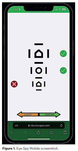
PURPOSE: This cross-sectional study compared best-corrected visual acuity obtained using Cloudscaper symbols, a novel optotype developed according to ETDRS specifications for children's virtual screening, with that obtained using LEA symbols.
METHODS: A total of 560 children aged 3-16 yr underwent visual acuity test with both Cloudscaper symbols and LS. The test application was standardized using the EyeSpy algorithm. Additionally, 147 participants were tested with the standard Snellen E paper chart. Paired t tests were performed to assess the clinical significance of logMAR visual acuity differences.
RESULTS: The mean logMAR visual acuity with LEA symbols was 0.12 (standard deviation [SD]=0.18; range, -0.10 to 0.80), while with Cloudscaper symbols it was 0.18 (SD=0.19; range, -0.10 to 0.80). The mean difference between Cloudscaper symbols and LEA symbols was 0.099 logMAR (approximately 0.5 optotypes; SD=0.08; range, 0.0-0.14; p<0.0001). Cloudscaper symbols slightly underestimated visual acuity compared to LEA symbols. Visual acuity measured by both methods was highly correlated (Spearman's r=0.74, p<0.0001). The mean visual acuity difference between Cloudscaper symbols and the Snellen E chart was 0.0045 (p=0.805; 95% confidence interval [95% CI]), whereas the difference between LEA symbols and Snellen E was 0.0883 (p<0.001; 95% CI).
CONCLUSIONS: Cloudscaper symbols provide a reliable tool for visual screening in children. Although they slightly underestimate visual acuity compared to LEA symbols – a finding also reported when comparing ETDRS letters with LEA symbols – Cloudscaper symbols show strong agreement with Snellen E chart measurements. This suggests that Cloudscaper symbols allow precise visual acuity assessment comparable to the gold standard.
Keywords: Vision screening; Vision tests; Visual acuity; Mobile applications; Eye health; Child health; Diagnostic techniques, Ophthalmological; Child; Preschool child; Adolescent
Arq. Bras. Oftalmol. 2025;88 (5 )
:1-7
| DOI: 10.5935/0004-2749.2024-0074
Abstract
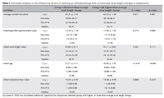
PURPOSE: This study aimed to identify factors influencing axial length changes in adolescents wearing orthokeratology lenses.
METHODS: A retrospective analysis was conducted on 84 adolescents (aged 9-17 yr) who wore orthokeratology lenses at our hospital. Axial length changes were calculated as the difference between the first and last visits. Patients were categorized into two groups based on axial length change: lower-than-average and higher-than-average. Data on sex, age at orthokeratology lens initiation, family history, initial equivalent spherical lens value, initial cylindrical lens value, initial average K value, and initial axial length were collected. Univariate and mixed-effects model analyses were performed to assess their influence on axial length changes.
RESULTS: Age (p<0.05) and initial equivalent spherical value (p<0.05) were significant predictors of axial length changes in both eyes and the left eye. For the right eye, only age was a significant factor (p<0.05). The mixed-effects model revealed that the difference between the left and right eyes, duration of orthokeratology lens use, age, initial equivalent spherical lens value, and initial axial length significantly influenced axial length changes in adolescents (p<0.05).
CONCLUSION: The factors influencing axial length changes in adolescents wearing orthokeratology lenses differ between the left and right eyes. These changes depend on the duration of lens wear, age, initial equivalent spherical lens value, and initial axial length. This study provides a theoretical basis for evaluating the clinical efficacy of orthokeratology lenses in managing myopia progression in adolescents.
Keywords: Orthokeratology; Contact lens; Myopia; Adolescent; Axial length, eye
 ABO is licensed under a Creative Commons Attribution-NonComercial 4.0 Internacional.
08-fig01tb.jpg)
08-fig01.jpg)
13-fig01.jpg)
14-fig01.jpg)
13-fig01.jpg)
03-fig01.jpg)
03-fig01.jpg)
02-fig01.jpg)