Arq. Bras. Oftalmol. 1999;62 (6 )
:701-703
| DOI: 10.1590/S0004-27491999000600009
Abstract
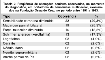
Objetivo: Determinar a freqüência e o tipo de comprometimento ocular em pacientes portadores de hanseníase no momento do diagnóstico. Metodologia: O estudo foi realizado na Fundação Oswaldo Cruz (Rio de Janeiro) e avalia 77 casos classificados como multibacilares. O exame oftalmológico foi realizado de acordo com o sistema de classificação de discapacidades recomendado pela Organização Mundial de Saúde. Resultados: Referiam queixas especificas 36,3%: dor e ardência ocular, lacrimejamento e dificuldade para enxergar. Em 55,8% dos pacientes foram detectadas alterações oculares no exame oftalmológico. A diminuição da sensibilidade da córnea, que predispõe a ulceração e opacificação, foi a alteração mais freqüente (29,3%). Conclusão: Os achados desta pesquisa demonstram a gravidade e a alta freqüência das lesões oculares nos casos avaliados e alertam para a importância do exame oftalmológico como rotina em portadores de hanseníase multibacilar.
Keywords: Infecções oculares bacterianas; Mycobacterium leprae
Arq. Bras. Oftalmol. 2025;88 (6 )
:1-8
| DOI: 10.5935/0004-2749.2025-0077
Abstract
PURPOSE: Standard intravitreal medication dosages are based on an assumed vitreous cavity volume of 4.0-4.5 mL. However, individual variations in vitreous cavity volume may influence both the efficacy and safety of these medications. This study proposes dosage adjustments for intravitreal medications and gases according to axial length and the corresponding vitreous cavity volume.
METHODS: This descriptive study employed reference guidelines that use axial length to estimate the Axial Length-based Volume of the Vitrectomized Space and the Vitreous Volume EXact table for determining dose adjustments across varying eye sizes. Small eyes (axial length 19-22 mm) have an average vitreous cavity volume of 3.5 mL at an axial length of 20.5 mm; standard-sized eyes (22-25 mm) have 4.8 mL at 23.5 mm; large eyes (25-28 mm) have 6.4 mL at 26.5 mm; and extra-large eyes (28-32 mm) have 8.4 mL at 29.5 mm. The medications considered included anti-infectives, anti-VEGFs, complement inhibitors, recombinant proteases, chemotherapy agents, corticosteroids, and medical gases.
RESULTS: Analysis of intravitreal drug concentrations relative to vitreous cavity volume demonstrated notable variability when a standard dose was administered. Small eyes received about 135% of the concentration intended for a standard-sized eye; large eyes received around 75%; and extra-large eyes received under 60%. The recommended dose adjustments are as follows: for small eyes, administer 70-80% of the standard dose; for large eyes, 130-140%; and for extra-large eyes, 170-180%.
CONCLUSIONS: Tailoring intravitreal drug and gas dosages according to axial length and vitreous cavity volume may enhance intraocular drug distribution, potentially improving both safety and therapeutic outcomes.
Keywords: Intravitreal injections; Axial length; Vitreous body; Drug dosage calculations; Pharmacokinetics; Anti-infective agents
Arq. Bras. Oftalmol. 2026;89 (1 )
:1-4
| DOI: 10.5935/0004-2749.2024-0402
Abstract
PURPOSE: This study evaluated the proportion of corneas discarded by the Eye Bank of Londrina, Paraná, due to positive serology over a 5-year period and its impact on transplant availability.
METHODS: A cross-sectional study was conducted, analyzing 1,968 corneas from 1,056 donors collected between January 2014 and December 2018 at the Eye Bank of Londrina. Serological tests for hepatitis B (HBsAg and anti-HBc), hepatitis C (anti-HCV), and HIV (anti-HIV 1 and 2) were performed using chemiluminescent microparticle immunoassays. Data were analyzed descriptively and presented in tables and graphs.
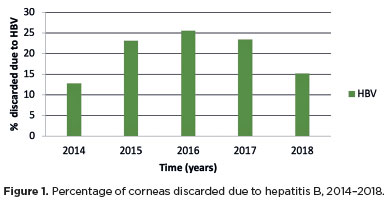
RESULTS: Of the 1,968 corneas processed, 897 (45.57%) were discarded. Among these, 333 (37.12%) tested positive for serological markers. Hepatitis B accounted for 34.67% of positive cases (15% of total donations), hepatitis C for 1.11% (0.50% of total), and HIV for 0.89% (0.4% of total). Hepatitis cases remained stable between 2014 and 2016, with a marked decline in 2017 and 2018. Most discarded corneas were positive for anti-HBc (31.88%) and negative for HBsAg; however, the anti-HBs test was not performed to confirm immunity to the hepatitis B virus.
CONCLUSION: The findings highlight the importance of serological testing to identify and eliminate contaminated corneas, thereby preventing the transmission of infectious diseases to recipients. Positive serology for hepatitis, particularly hepatitis B, was the leading cause of corneal disposal.
Keywords: Cornea; Corneal transplantation; Corneal donation; Eye banks; Hepatitis B virus; Hepatitis C virus; HIV infections; Seropositivity; Serologic tests
Arq. Bras. Oftalmol. 2025;88 (4 )
:1-7
| DOI: 10.5935/0004-2749.2024-0229
Abstract
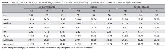
PURPOSE: The volume of the vitreous chamber varies with the size of the eye. The space created in the vitreous cavity by a vitrectomy is called the vitrectomized space. The volume of the vitrectomized space is strongly correlated with the axial length of the eye. This study aims to present guidelines for estimating the using participants stratified by axial length, sex, and history of cataract surgery.
METHODS: This retrospective, observational, cross-sectional study included 144 randomly selected participants who underwent vitrectomies between 2013 and 2023. Before surgery, the axial lengths of participants' eyes were measured using optical biometrics. The axial lengths of the eyes in our sample were between 20-32 mm. In all cases, a complete vitrectomy was performed, followed by complete fluid-air exchange and injection of a balanced saline solution. The volume infused was recorded.
RESULTS: The median (interquartile range; range) volume of the vitrectomized space was 6.1 (3.8; 3.1-11.3) mL in men and 6.1 (3.3; 3.2-11.2) mL in women (p=0.811). The median volume of the vitrectomized space was 5.9 (3.6; 3.1-11.2) mL in patients with phakic lenses and 6.25 (3.6; 3.3-11.3) mL in those with pseudophakic lenses (p=0.533). A positive correlation was found between the axial length and the volume of the vitrectomized space in this sample (r=0.968; p<0.001). In a cubic polynomial regression, the coefficient of determination was 0.948. Similar results were observed in both sexes and in both phakic and pseudophakic patients. The estimated cubic polynomial regression equation for this sample was VVS = 0.000589052857847605 × AL3 - 0.025114926401582700 × AL2 + 0.685961117595624000 × AL - 5.088226672620790000.
CONCLUSION: We developed this axial length estimation of the volume of vitrectomized space as a guideline for the determination of vitrectomized space volume using axial length.
Keywords: Cataract extraction; Retinal perforations/surgery; Epiretinal membrane/surgery; Vitreous body; Axial length, eye; Vitrectomy; Biometry/methods; Diagnostic techniques, ophthalmological; Guidelines as topic.
Arq. Bras. Oftalmol. 2022;85 (4 )
:377-381
| DOI: 10.5935/0004-2749.20220016
Abstract
Objetivo: Determinar a incidência de emergências oculares em um centro oftalmológico de referência no Brasil.
Métodos: O banco de dados de prontuários da Fundação Altino Ventura, Recife, Brasil, foi analisado retrospectivamente e incluiu pacientes atendidos, entre janeiro de 2017 e janeiro de 2018, na sala de emergência oftalmológica. Foram excluídos os prontuários com dados incompletos e com quadros ambulatoriais. Apenas o primeiro atendimento na emergência foi considerado para análise.
Resultados: Em um ano, 134.788 pacientes (idade média de 38,7 ± 22 anos [0-99 anos]) foram admitidos na emergência da Fundação Altino Ventura. Os diagnósticos mais frequentes foram conjuntivite (52.732 casos [37,3%]), blefarite (7.213 casos [5,1%]) e corpo estranho na córnea/conjuntiva (6.925 casos [4,9%]). Corpo estranho na córnea/conjuntiva e trauma ocular foram cerca de 8 vezes e 2 vezes mais incidente em indivíduos do sexo masculino, respectivamente (p<0,001 em ambos). Triquíase e blefarite afetaram ~2 vezes mais pacientes do sexo feminino, respectivamente (p<0,001 em ambos). Corpo estranho na córnea/conjuntiva e trauma ocular afetaram mais pacientes em idade produtiva (>15 anos), enquanto úlcera, blefarite e triquíase da córnea afetaram mais pacientes idosos. Todos os grupamentos de diagnóstico (doenças infecciosas, trauma ocular, corpos estranhos, retinopatias, doenças das pálpebras, doenças da córnea, crise glaucomatosa e doenças neurooftalmológicas) foram mais incidentes na primavera (valor de p<0,001).
Conclusão: As emergências oftalmológicas mais comuns no presente estudo foram as doenças infecciosas e o corpo estranho. Porém, a incidência das emergências oculares são fluências pela faixa etária e gênero do paciente, além da época do ano.
Keywords: Emergência; Oftalmopatia; Transtorno da visão; Conjuntivite; Corpo estranho; Traumatismo oculare; Estações do ano; Brasil
Arq. Bras. Oftalmol. 2024;87 (5 )
:1-10
| DOI: 10.5935/0004-2749.2022-0215
Abstract
Objetivo: Descrever os achados clínicos, tratamentos, e desfechos em uma série de pacientes com metástases vítreas de melanoma cutâneo.
Métodos: Série retrospectiva de casos de único centro com intervenção. Pacientes incluídos tiveram seu diagnóstico de MVMC confirmado por biópsia entre 1997 e 2020. Vitrectomia via pars plana com 23 ou 25 gauge foram realizadas para obter espécimens. Esclerotomias foram tratadas com crioterapia em duplo ou triplo congelamento. Injeção intravítrea perioperatória de melfalano (32 ug/0,075 mL) foi administrada quando necessário. Foram relatados acuidade visual, pressão intraocular, resposta terapêutica sistêmica e ocular.
Resultados: Cinco olhos de 5 pacientes com metástases vítreas de melanoma cutâneo unilateral foram identificados. Idade média de diagnóstico foi 84 anos (variando de 37-88). Seguimento médio após diagnóstico oftalmológico foi 28 (8,5-36) meses; 1 paciente não teve acompanhamento. Acuidade visual inicial variou de 20/30 a movimentos de mão. Achados clínicos iniciais incluíram infiltração de células pigmentadas e não-pigmentadas no vítreo (5/5), segmento anterior (4/5), e retina (3/5). Quatro pacientes tiveram glaucoma secundário. Tratamento sistêmico incluiu imunoterapia com inibidores da via de sinalização (3 - todos com resposta parcial/completa), quimioterapia sistêmica (2), ressecção cirúrgica (3), e irradiação (2). Intervalo médio entre diagnóstico primário e metástases vítreas foi 2 (2-15) anos. Um paciente teve doença sistêmica ativa simultânea as metástases vítreas. Acuidade visual final variou entre 20/40 e SPL. Tratamento oftalmológico incluiu vitrectomia nos 5 pacientes, melfalano intravítreo em 3 e metotrexato intravítreo em 1. Um paciente precisou de enucleação. A histopatologia revelou invasão celular extensa de melanoma.
Conclusões: Metástases vítreas de melanoma cutâneo pode se manifestar como uma infiltração difusa de células pigmentadas e não-pigmentadas no vítreo e erroneamente diagnosticada como uveites. Vitrectomia diagnóstica e quimioterapia intravítrea periódica podem estar indicadas.
Keywords: Melanoma; Neoplasias oculares; Neoplasias cutâneas; Corpo vítreo; Metástase neoplásica; Inibidores de checkpoint imunológico; Imunoterapia; Injeções intravítreas; Melfalan; Metotrexato
Arq. Bras. Oftalmol. 2025;88 (3 )
:1-6
| DOI: 10.5935/0004-2749.2024-0112
Abstract
PURPOSE: To describe the ophthalmological findings of dry eye disease and its relation to the quality of life of COVID-19 survivors.
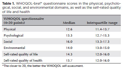
METHODS: COVID-19 survivors who had previously been hospitalized at Hospital das Clínicas de Ribeirão Preto complex underwent an ophthalmological evaluation, which included a dry eye disease questionnaire, break-up time, fluorescein staining, and Schirmer test. We collected the presenting and best-corrected visual acuity, sociodemographic data, personal medical history, and scores from a self-reported quality of life questionnaire (WHOQOL-bref). According to the severity of the acute phase of the disease, the patients were classified into mild-to-moderate, severe, and critical groups.
RESULTS: Ninety-five patients (190 eyes) were evaluated 100 ± 44 days after the onset of COVID-19 symptoms. Of these, 83 patients (87.3%) completed the WHOQOL-bref questionnaire. Ten patients (12.0%) had mild-to-moderate COVID-19, 41 (49.4%) had severe COVID-19, and 32 (38.6%) had critical COVID-19. The median best-corrected visual acuity was logMAR 0 (0-1). Approximately 26.3% patients had a history of dry eye disease or severe dry eye symptoms (frequent or constant ocular dryness and irritation). There was an association between the proportion of patients with dry eye disease and the quality of life (p=0.014) and health (p=0.001). Furthermore, there was a significant trend between the proportion of patients with dry eye disease and how they rated their health and quality of life (p=0.0004 and 0.0027, respectively.
CONCLUSIONS: There is a significant negative correlation between the proportion of patients with dry eye disease and their self-reported quality of life.
Keywords: COVID-19; Coronavirus infections; SARS-CoV-2; Eye diseases; Epidemiology; Ocular surface; Public health
Arq. Bras. Oftalmol. 2024;87 (2 )
:1-5
| DOI: 10.5935/0004-2749.2021-0395
Abstract
Objetivos: Avaliar a segurança e eficácia a longo prazo da vitreólise com Nd:YAG laser para moscas volantes sintomáticas, uma vez que permanece como um procedimento controverso devido a falta de evidência científica robusta sobre a manutenção dos resultados e ocorrência de efeitos adversos.
Métodos: Este estudo é uma extensão observacional de um ensaio clínico prospectivo, randomizado, duplo cego, previamente publicado. Oito de treze pacientes que foram submetidos a vitreólise com YAG laser foram acompanhados para uma reavaliação tardia, dezoito meses após o procedimento, para avaliar a eficácia e segurança do procedimento.
Resultados: Todos os pacientes mantiveram a melhora na sintomatologia notada ao final do procedimento original, com 25% dos casos apresentando melhora completa, e uma proporção semelhante (37,5%) demonstrando melhora significativa ou parcial. A melhora objetiva na opacidade foi similar ao achado no seguimento original de 6 meses. O questionário de qualidade de vida NEI-VFQ 25 não demonstrou diferença estatisticamente significativa nas respostas entre o sexto e o décimo oitavo mês de acompanhamento. Nenhum efeito adverso foi notado no exame clínico ou reportado pelos pacientes.
Conclusão: A eficácia da vitreólise observada ao sexto mês do acompanhamento foi mantida até o décimo oitavo mês, com todos os pacientes notando algum grau de melhora quando comparado ao estado pré procedimento. Nenhum efeito adverso tardio foi notado. Um ensaio clínico randomizado maior é necessário para confirmar a segurança do procedimento.
Keywords: Terapia a laser; Lasers de estado sólido; Vitrectomia; Corpo vítreo; Cirurgia vitreorretiniana; Acuidade visual; Doenças oculares; Qualidade de vida; Inquéritos e questionários
Arq. Bras. Oftalmol. 2024;87 (2 )
:1-6
| DOI: 10.5935/0004-2749.2022-0334
Abstract
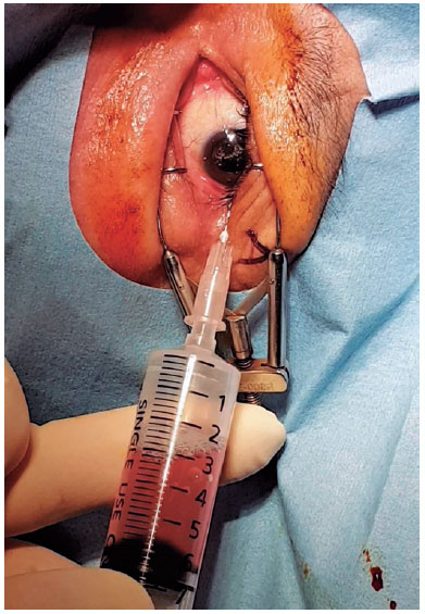
PURPOSE: This study aimed to evaluate the efficacy and clinical outcomes of a one-way fluid-air exchange procedure for the treatment of postvitrectomy diabetic vitreous hemorrhage in patients with proliferative diabetic retinopathy.
METHODS: This retrospective study included 233 patients with proliferative diabetic retinopathy, who underwent vitrectomy. A one-way fluid-air exchange procedure was performed in 24 eyes of 24 (10.30%) patients with persistent vitreous cavity rebleeding after the operation. Preprocedural and postprocedural best-corrected visual acuity values were achieved. Complications occurring during and after the procedure were analyzed.
RESULTS: Significant visual improvement was observed 1 month after the one-way fluid-air exchange procedure (2.62 ± 0.60 LogMAR at baseline vs. 0.85 ± 0.94 LogMAR at postprocedure, p<0.0001). Moreover, 19 (79.17%) eyes needed the procedure once, and 5 (20.83%) eyed had the procedure more than twice. In 3 (12.50%) eyes, reoperation was eventually required because of persistent rebleeding despite several fluid-air exchanges. No complication was observed during the follow-up.
CONCLUSIONS: The one-way fluid-air exchange procedure can be an excellent alternative to re-vitrectomy for patients with proliferative diabetic retinopathy suffering from postvitrectomy diabetic vitreous hemorrhage by removing the hemorrhagic contents directly and achieving fast recovery of visual function without apparent complications.
Keywords: Diabetic retinopathy; Vitrectomy; Vitreous body; Vitreous hemorrhage; Hemostatic techniques


 ABO is licensed under a Creative Commons Attribution-NonComercial 4.0 Internacional.



01-tab01.jpg)
06-tab01tb.jpg)
14-fig01tb.jpg)
01-fig01.jpg)
03-fig01.jpg)