Arq. Bras. Oftalmol. 1999;62 (6 )
:739-746
| DOI: 10.1590/S0004-27491999000600016
Abstract
Objetivo: Avaliar o sucesso na redução da pressão intra-ocular, com a utilização de trabeculectomia simples e quando associada a 5-fluorouracil ou mitomicina-C e avaliar se o tempo de exposição à mitomicina-C modifica este índice de sucesso. Métodos: Analisaram-se retrospectivamente 171 olhos consecutivos, entre janeiro de 1989 e março de 1998, sendo 16 olhos submetidos à trabeculectomia simples, 38 olhos à trabeculectomia e aplicação pós-operatória de 5-fluorouracil e 117 olhos à trabeculectomia e aplicação intra-operatória de mitomicina-C. Resultados: Os resultados mostraram uma redução significativa da PIO (pressão intra-ocular), com o uso de antifibroblásticos (p < 0,05). O tempo de exposição à mitomicina-C não apresentou diferença estatisticamente significativa (p > 0,05). Conclusões: Os autores concluem que a trabeculectomia associada a drogas antifibroblásticas reduz a PIO de maneira significativa e promove estabilidade a curto prazo.
Keywords: Trabeculectomia; 5-Fluorouracil; Mitomicina C
Arq. Bras. Oftalmol. 2023;86 (5 )
:1-6
| DOI: 10.5935/0004-2749.20230064
Abstract
Objetivo: Avaliar a resposta tecidual e clínica a um implante orbitário de polimetilmetacrilato, oco e multiperfurado em sua porção posterior em modelo animal após evisceração.
Métodos: Dezesseis coelhos da raça Nova Zelândia foram submetidos à evisceração do globo ocular direito. Todos receberam implante oco de polimetilmetacrilato de 12 mm de diâmetro, multiperfurado em sua semiesfera posterior. O estudo foi dividido em avaliação clínica e histopatológica. A avaliação clínica foi diária até 14 dias pós-evisceração e, a cada sete dias, até completar 180 dias. Os animais foram divididos em grupos de quatro animais e cada um foi submetido à exenteração com 07, 30, 90 e 180 dias e depois à eutanásia. A análise histopatológica teve por fim caracterizar o padrão inflamatório, a estrutura do colágeno e o grau de neovascularização. Para isso, além da tradicional coloração pela hematoxilina-eosina, utilizou-se o corante Picrosirius Red (PSR) e imuno-histoquímica com o marcador CD 34.
Resultados: Não houve sinais de infecção, afinamento conjuntival ou escleral, exposição ou extrusão do implante em nenhum animal durante o estudo. Já no sétimo dia, o tecido neoformado migrou para dentro do implante formando uma rede fibrovascular através dos canais posteriores. A resposta inflamatória diminuiu ao longo do tempo avaliado e não foram encontradas células gigantes multinucleadas.
Conclusão: O implante analisado permite a sua integração aos tecidos orbitários pelo crescimento fibrovascular em seu interior. Os autores acreditam que este modelo de implante orbital pode fazer parte de testes com humanos.
Keywords: Implantes orbitários; Polimetilmetacrilato; Evisceração ocular; Anoftalmia; Procedimentos cirúrgicos oftalmológicos; Coelhos.
Arq. Bras. Oftalmol. 2021;84 (3 )
:220-224
| DOI: 10.5935/0004-2749.20210030
Abstract
OBJETIVO: Avaliar a morfologia da córnea e da câmara anterior em olhos fácicos com inflamação intraocular não infecciosa.
MÉTODOS: Esse estudo incluiu 59 olhos com uveíte ativa, 62 olhos com uveíte inativa e 95 olhos saudáveis. A densidade de células endoteliais da córnea, a proporção de células hexagonais, o coeficiente de variação, o volume e a espessura da córnea, a ceratometria máxima e o volume e profundidade da câmara anterior foram medidos com um microscópio especular e uma Pentacam HR.
RESULTADOS: A duração média da uveíte foi de 24,6 ± 40,5 (0-180) meses. O número médio de crises de uveíte foi de 2,8 ± 3,0 (1-20). O coeficiente de variação foi significativamente maior no grupo com uveíte ativa do que no grupo com uveíte inativa (p=0,017, Tukey post-hoc). Não houve diferença significativa nos demais parâmetros do segmento anterior entre os grupos com uveíte ativa, com uveíte inativa e controle (p>0,05). A análise de regressão linear múltipla demonstrou que o coeficiente de variação foi maior na uveíte ativa do que na uveíte inativa, após ajustes para a duração e tipo de uveíte e a presença ou não de doença reumática e de tratamento imunossupressor (p=0,003). A duração da uveíte e o número de crises não demonstraram correlação significativa com os parâmetros oculares (p>0,05, correlação de Spearman). A diferença nos parâmetros não demonstrou correlação significativa com o tipo de uveíte (p>0,05).
CONCLUSÕES: O coeficiente de variação foi maior nos olhos com uveíte ativa do que naqueles com uveíte inativa, ao passo que a densidade de células endoteliais e a morfologia da câmara anterior não mostraram diferenças significativas entre os grupos com uveíte ativa, com uveíte inativa e controle.
Keywords: Câmara anterior; Inflamação; Epitélio posterior; Contagem de células; Uveites
Arq. Bras. Oftalmol. 2020;83 (4 )
:299-304
| DOI: 10.5935/0004-2749.20200051
Abstract
Objetivo: Investigar a utilidade de marcadores inflamatórios sistêmicos (ou seja, contagem de glóbulos brancos e plaquetas, volume médio de plaquetas e suas proporções) como marcadores de diagnóstico da patogênese do edema macular diabético.
Métodos: A coorte do estudo incluiu 80 pacientes com edema macular diabético (40 com retinopatia diabética e 40 sem) e 40 controles saudáveis de acordo com a idade e sexo. As contagens de neutrófilos, linfócitos, monócitos, plaquetas e valores do volume plaquetário médio foram determinados a partir de amostras de sangue periféricdo, e as proporções de monócitos/linfócitos, plaquetas/linfócitos, volume plaquetário médio/linfócitos e neutrófilos/linfócitos foram calculadas e comparadas entre os grupos.
Resultados: A proporção média de neutrófilos/linfócitos dos grupos com edema macular diabético e não-diabético foi maior que a do grupo controle, e o valor do grupo com edema macular diabético foi maior que o do grupo com edema macular não diabético (p<0,001 no com edema macular diabético vs. controle, p=0,04 no com edema macular não diabético vs. controle e p=0,03 no com edema macular diabético vs. o com edema macular não-diabético). Um valor de corte de neutrófilos/linfócitos ≥2,26 foi identificado como um indicador da patogênese do edema macular diabético com sensibilidade de 85% e especificidade de 74%. O volume plaquetário médio do grupo com edema macular diabético foi maior que o dos grupos com edema macular não-diabético e controle, enquanto os do grupo de edema macular não-diabético e controle foram semelhantes (edema macilar diabético vs. Edema macular não-diabético, p=0,08; com edema macular diabético vs. controle, p=0,02; e com edema macular não-diabético vs. controle, p=0,78). Todos os outros parâmetros foram semelhantes entre os grupos (todos p>0,05).
Conclusão: A proporção de neutrófilos/linfócitos e o volume plaquetário médio do grupo com edema macular diabético foram superiores aos do grupo com edema macular não-diabético e controle. Um valor de corte da razão neutrófilos/linfócitos ≥2,26 foi identificado como um indicador da patogênese do edema macular diabético com alta sensibilidade e especificidade. Além disso, a proporção de neutrófilos/linfócitos do grupo com edema macular não-diabético foi superior à do grupo controle.
Keywords: Edema macular; Retinopatia diabética; Volume plaquetário médio; Contagem de linfócitos; Neutrófilos; Inflamação
Arq. Bras. Oftalmol. 2021;84 (5 )
:481-489
| DOI: 10.5935/0004-2749.20210049
Abstract
OBJETIVO: Otimizar a dose efetiva de mitomicina C, 5fluorouracil e da combinação de ambos em culturas de células de carcinoma basocelular (CBC).
MÉTODOS: Culturas de células de células de carcinoma basocelular e de fibroblastos foram tratadas com diferentes concentrações de mitomicina C, 5fluorouracil e combinação de ambos. Além disto, foram investigados a viabilidade celular, o ciclo celular, a apoptose e a expressão dos genes TP53, CDKN1A e CDK6. O medicamento mais eficaz, em sua dosagem otimizada, foi administrado em últiplas injeções intralesionais em uma mulher de 65 anos com carcinoma basocelular nódulo-ulcerativo periorbital avançado.
RESULTADOS: A concentração de 0,00312 mg/mL de mitomicina C e a de 0,312 mg/mL de 5fluorouracil foram consideradas as ideias. A viabilidade média das células de carcinoma basocelular tratadas com mitomicina C isoladamente e em combinação foi significativamente menor que nas células de controle (respectivamente, p=0,002 e p=0,04). Todos os grupos de carcinoma basocelular tratados demonstraram interrupção do ciclo celular na fase S. As células de carcinoma basocelular tratadas com mitomicina C mostraram maiores taxas de apoptose (p=0,002) e significativa regulação positiva do gene TP53 (p=0,0001). Além disso, o gene CDKN1A foi positivamente regulado e o gene CDK6 foi negativamente regulado em células de carcinoma basocelular tratadas com 5fluorouracil (respectivamente, p=0,0001 e p=0,01) ou com a combinação de medicamentos (respectivamente, p=0,007 e p=0,001). Injeções posteriores de mitomicina C na paciente em questão levaram à melhora significativa da lesão do carcinoma basocelular.
CONCLUSÃO: Nossos resultados in vitro otimizaram as doses efetivas de mitomicina C e 5fluorouracil na cultura de células de carcinoma basocelular e mostraram que a mitomicina C tem mais eficácia na apoptose de células de carcinoma basocelular do que o 5fluorouracil e a combinação de ambos. Injeções intralesionais de doses otimizadas de mitomicina C podem ser propostas para o tratamento não cirúrgico do células de carcinoma basocelular avançado de pálpebra.
Keywords: Carcinoma basocelular; mitomicina C; 5fluorouracil; TP53; CDKN1A; CDK6
Arq. Bras. Oftalmol. 2025;88 (4 )
:1-6
| DOI: 10.5935/0004-2749.2024-0278
Abstract
PURPOSE: This study aimed to evaluate the prevalence of orbital conditions in a tertiary ophthalmic outpatient hospital in Sao Paulo, Brazil, with a focus on the main diagnoses and their distribution.
METHODS: A retrospective chart review was conducted involving patients registered and admitted to the orbital disease unit at the Department of Ophthalmology, University of São Paulo Medical School, from January 2004 to March 2018. A total of 838 medical charts were analyzed, of which 37 were excluded due to incomplete data. The remaining charts were categorized into eight diagnostic groups: Graves’ orbitopathy , inflammatory disorders, tumors, vascular lesions, acquired structural abnormalities, congenital structural abnormalities, infectious diseases, and others.
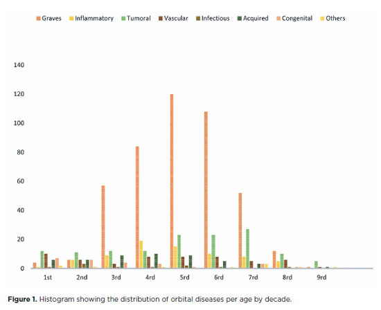
RESULTS: Of the 837,300 ophthalmological appointments, 3,372 (0.4%) were related to orbital diseases. The study included 801 patients, of whom 63.45% were women. The patients’ mean age was 42.86 years. Graves’ orbitopathy was the most common (55%), followed by tumor (17%), inflammatory disorders (9%), vascular lesions (7%), acquired structural abnormalities (5%), congenital structural abnormalities (4%), others (2%), and infectious diseases (1%). The study found significant differences in the incidence and types of orbital diseases, indicating the specialized nature of tertiary care and referral biases.
CONCLUSION: Published data on epidemiological orbital diseases is scarce. Therefore, this study focused on the diverse nature of orbital diseases and their low incidence among ophthalmology appointments. The major trends align with other epidemiological studies, demonstrating a preponderance of Graves’ orbitopathy in middle-aged adults and a bimodal distribution of tumors. These findings are essential in shaping resident training programs and healthcare policies, particularly in tertiary settings. Understanding the epidemiology of orbital diseases can improve diagnostic accuracy, treatment approaches, and patient outcomes as well as support future systemic prospective studies.
Keywords: Orbital diseases; Orbital tumors; Neoplasms; Inflammation; Graves’ ophthalmopathy; Outpatients
Arq. Bras. Oftalmol. 2025;88 (6 )
:1-7
| DOI: 10.5935/0004-2749.2025-0006
Abstract
PURPOSE: This study aimed to evaluate the practices employed by oculoplastic surgeons in the assessment and management of anophthalmic sockets and external ocular prostheses.
METHODS: Oculoplastic surgeons from two countries, who specialized in the management of anophthalmic sockets, participated in a web-based survey. Data collected included demographics, types of surgery, implant use, external ocular prostheses management (including fabrication and cleaning), complications encountered, and follow-up times. The frequencies and distributions of the responses were statistically analyzed.
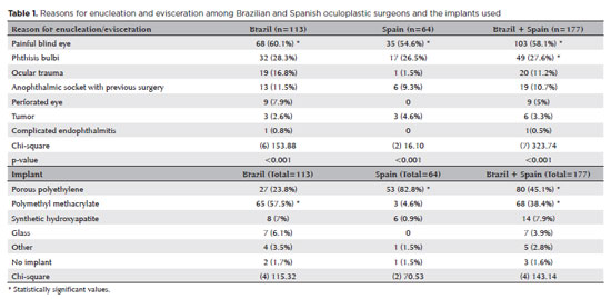
RESULTS: A total of 177 oculoplastic surgeons participated, 113 (63.8%) from Brazil, the remainder from Spain. Evisceration was the preferred surgical procedure of 149 (84.2%) surgeons. The most commonly reported indication for enucleation was a painful blind eye (n=103, 58.1%; both Brazil and Spain, p<0.001). Brazilian surgeons preferred polymethyl methacrylate implants (n=65, 57.5%), while Spanish surgeons favored porous polyethylene implants (n=53, 82.8%; p<0.001). Discharge was the most frequently observed clinical feature during socket evaluation (n=164, 92.6%; p<0.001). Brazilian surgeons recommended daily (n=53, 46.9%) or weekly (n=41, 36.2%) cleaning of external ocular prostheses, while Spanish surgeons more commonly recommended monthly cleaning (n=31, 48.4%; p<0.001). The majority of Brazilian surgeons (n=83, 73.4%) advised patients to remove their external ocular prostheses at night. Only a small number of Spanish surgeons (n=3, 4.6%) suggested this practice (p<0.001). Overall, the follow-up recommendations varied, with 70 (39.5%) surgeons recommending follow-up based on individual case needs, and 59 (33.3%) suggesting annual visits (p<0.001). The primary indications for external ocular prostheses replacement were edge damage (n=75, 42.3%) and loss of volume (n=68, 38.4%). The replacement intervals given typically ranged from 1 to 5 years (n=92, 51.9%; p<0.001).
CONCLUSION: Oculoplastic surgeons in Brazil and Spain demonstrated similar practices in the management of anophthalmic sockets. However, notable differences were observed in the choice of implant materials, cleaning protocols, and recommendations regarding external ocular prostheses removal during sleep.
Keywords: Anophthalmos; Eye, artificial; Polymethyl methacrylate; Polyethylene; Surgeons; Surveys and questionnaires; Brazil; Spain.
Arq. Bras. Oftalmol. 2026;89 (5 )
:1-6
| DOI: 10.5935/0004-2749.2025-0154
Abstract
PURPOSE: This study aimed to evaluate the serum levels of thrombospondin-1, growth differentiation factor-15, resolvin D1, and resolvin E1 in patients with keratoconus.
METHODS: Thirty-five patients with keratoconus and 35 control subjects of similar age and sex were evaluated. The keratoconus group was divided into four stages according to the modified Krumeich classification. Commercial kits were used to determine thrombospondin-1, growth differentiation factor-15, resolvin D1, and resolvin E1 levels in serum samples using enzyme-linked immunosorbent assay (ELISA). Serum thrombospondin-1, growth differentiation factor-15, resolvin D1, and resolvin E1 levels were compared between the patient and control groups.
RESULTS: The keratoconus group consisted of 14 females and 21 males, and the control group consisted of 14 females and 21 males, with mean ages of 29.4±8.8 and 30.0±7.8 yr, respectively. There were significant differences in serum thrombospondin-1, growth differentiation factor-15, resolvin D1, and resolvin E1 levels between the serum of the keratoconus and control groups (p=0.006, p<0.001, p<0.001, and p=0.001, respectively). No significant correlations were found between keratoconus stage and serum thrombospondin-1, growth differentiation factor-15, resolvin D1 and resolvin E1 levels (r=−0.025, p=0.892; r=−0.019, p=0.913; r=−0.036, p=0.838; and r=−0.108, p=0.538, respectively).
CONCLUSION: Serum levels of thrombospondin-1, growth differentiation factor-15, resolvin D1, and resolvin E1 were significantly lower in patients with keratoconus. These results suggest that these parameters may be associated with the pathogenesis of keratoconus.
Keywords: Keratoconus/pathology; Thrombospondin 1/blood; Growth differentiation factor 15/blood; Docosahexaenoic acids/blood; Eicosapentaenoic acid/blood; Inflammation mediators
Arq. Bras. Oftalmol. 2026;89 (4 )
:1-7
| DOI: 10.5935/0004-2749.2025-0216
Abstract
PURPOSE: To compare the efficacy and safety of trabeculectomy with mitomycin C, performed with versus without adjunctive Ologen collagen matrix in a multicenter real-world setting.
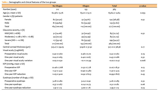
METHODS: This multicenter retrospective comparative study included 277 patients (365 eyes) who underwent trabeculectomy with mitomycin C, with or without Ologen, between 2017 and 2022 across five Brazilian centers. Extracted data comprised demographic characteristics, glaucoma subtype, intraocular pressure, number of glaucoma medications, postoperative interventions, complications, and additional surgical procedures. Surgical success was defined using two criteria: (1) intraocular pressure ≤21mmHg with ≥20% reduction from baseline and (2) final intraocular pressure between 6 and 21mmHg. Outcomes were categorized as complete (without medications) or qualified (with medications).
RESULTS: Both groups achieved substantial reductions in intraocular pressure and medication burden at 12 months, with high rates of complete and qualified success under both definitions. The Ologen group required fewer suture lysis procedures (49.0% vs. 60.8%; p=0.025), suggesting a modulatory effect on early postoperative fibrosis. Rates of other postoperative interventions, complications, and additional glaucoma surgeries were comparable between groups. Kaplan–Meier analysis demonstrated no significant difference in cumulative success over time.
CONCLUSION: Trabeculectomy with mitomycin C is effective and safe regardless of adjunctive Ologen use. Although Ologen did not confer superior long-term efficacy, its association with reduced suture lysis suggests a potential role in modulating early wound healing.
Keywords: Trabeculectomy, Intraocular pressure, Mitomycin C; Wound healing; Sutures
Arq. Bras. Oftalmol. 2026;89 (5 )
:1-8
| DOI: 10.5935/0004-2749.2026-0041
Abstract
PURPOSE: To evaluate the efficacy of autologous fat grafting for treating enophthalmos in anophthalmic sockets.
METHODS: In this prospective interventional case series, consecutive patients with anophthalmic sockets and enophthalmos were enrolled between August 2013 and July 2025. Clinical ophthalmologic examination, Hertel exophthalmometry, and orbital computed tomography scans were performed. Enophthalmos was assessed clinically and radiologically before and after orbital lipofilling with autologous fat. Fat was harvested from the inner thigh and injected into the inferolateral orbital compartment. The volume of injected fat was determined according to the degree of enophthalmos measured by Hertel exophthalmometry. Follow-up ranged from 2 to 60 months (median, 7 months).
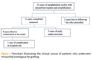
RESULTS: Thirteen patients (13 sockets) underwent orbital autologous fat grafting using Coleman's technique. The injected fat volume ranged from 3.7 to 6 mL per procedure (median, 4.2 mL). Clinically, a reduction in enophthalmos of 1–5 mm was observed in 12 cases (median, 2 mm), which was associated with a decrease in prosthesis thickness. One patient developed a fat cyst due to anterior fat migration, and three patients required repeat grafting because of undercorrection. Computed tomography scans, available for nine patients, demonstrated a 6.8%–10.8% increase in intraorbital volume, particularly in the inferolateral quadrant. Conclusions: Clinical and radiological assessments demonstrated that lipofilling is an effective technique for restoring orbital volume in anophthalmic sockets with enophthalmos.
Keywords: Anophthalmos; Enophthalmos; Orbital volumetry; Fat grafting; Orbital reconstruction
Arq. Bras. Oftalmol. 2025;88 (5 )
:1-8
| DOI: 10.5935/0004-2749.2024-0312
Abstract
PURPOSE: To evaluate the changes in the rates and indications of eye removal procedures during the recent COVID-19 pandemic.
METHODS: The medical records of all patients who underwent eye removal from 2007 to 2022 were retrospectively reviewed. The patient demographic data and indications for surgery were collected. Data from two groups of patients (prepandemic surgery and postpandemic surgery) were compared. Statistical significance was set at p<0.05.
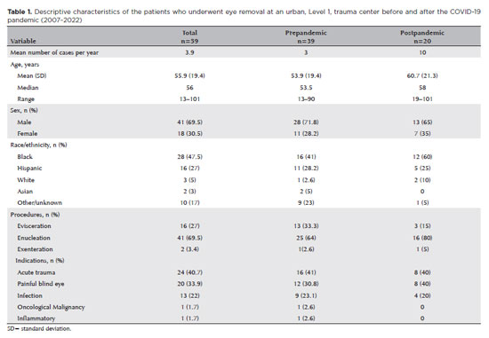
RESULTS: Fifty-nine patients underwent enucleation (69%), evisceration (27%), or exenteration (3%). The mean (SD) age of the patients was 55.9 (19.4) years, and most (69%) of the patients were males. Most (47%) of the study population were Black. The common indications for eye removal were trauma (41%), painful blind eye (34%), and infection/inflammation (24%). The types of trauma were assault (55%), accidental (39%), and self-inflicted (6%). The mean (SD) monthly rates of eye removal increased from 0.25 (0.50) in the prepandemic period to 0.77 (0.91) during the pandemic (p<0.001). These increases were noted in both males (p=0.003) and females (p=0.001) and were the highest among Black patients [0.42 (0.76); p<0.001]. Among the indications of eye removal, painful blind eyes [0.35 (0.75); p<0.001] and ocular trauma [0.31 (0.47); p=0.051] exhibited the greatest increases following the pandemic.
CONCLUSION: The rate of eye removal procedures increased during the recent pandemic. Although delayed care of chronic eye conditions may have contributed to the increased rates of painful blind eyes, the increased trauma-related eye removals may be attributed to the simultaneous spike in violent assaults in New York City.
Keywords: Eye injuries; Eye enucleation; COVID-19; Pandemics; Ethinicity; Inflammation, Trauma centers
Arq. Bras. Oftalmol. 2026;89 (3 )
:1-6
| DOI: 10.5935/0004-2749.2025-0332
Abstract
PURPOSE: To quantitatively compare eyebrow and eyelid positions in anophthalmic sockets reconstructed with conical or spherical orbital implants combined with customized external ocular prostheses.
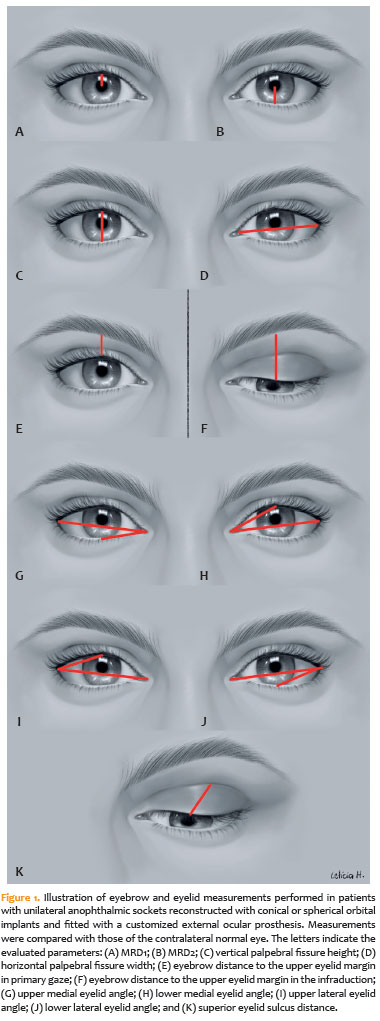
METHODS: This cross-sectional observational study included 38 patients with unilateral anophthalmic sockets, of whom 21 received conical implants, and 17 received spherical implants. Eyelid and eyebrow parameters—including margin reflex distance 1 and 2, vertical and horizontal palpebral fissure dimensions, eyebrow-to-upper-eyelid margin distance in primary gaze and infraduction, medial and lateral eyelid angles in primary gaze, and superior eyelid sulcus depth —were quantitatively assessed using standardized digital photographs analyzed with Image J software. The contralateral healthy eye served as the control. Statistical analyses were performed to compare measurements between groups.
RESULTS: In the primary gaze position, conical and spherical implants showed comparable margin-reflex distance1, margin-reflex distance2, vertical palpebral fissure height, eyelid margin position, and medial and lateral eyelid angles. During infraduction, the upper eyelid margin was significantly lower in sockets reconstructed with conical implants. Compared with contralateral normal eyes, anophthalmic sockets exhibited a reduced horizontal palpebral fissure and a deeper superior eyelid sulcus, irrespective of implant shape.
CONCLUSION: Anophthalmic sockets reconstructed with conical or spherical implants demonstrate similar eyebrow and eyelid positioning in primary gaze. However, conical implants are associated with a lower eyelid margin during infraduction. Independent of implant format, anophthalmic sockets show a narrower horizontal palpebral fissure and increased superior sulcus depth compared with normal eyes.
Keywords: Anophthalmos; Prosthesis implantation; Anophthalmic socket; Conical implants; Spherical implants; Orbital implants; Eyelid measurements


 ABO is licensed under a Creative Commons Attribution-NonComercial 4.0 Internacional.


06-fig01.jpg)
04-tab01.jpg)
06-tab01tb.jpg)
06-fig01.jpg)

08-fig01.jpg)
12-fig01.jpg)
08-tab01.jpg)