

Arq. Bras. Oftalmol. 2026; 89 (5): 10.5935/0004-2749.2026-0041
Total: 927
Maria Antonieta da A. Ginguerra-Nascimento1; Laura Goldfarb Cyrino1; Mario Padula4; Carolina Passareli Diniz1; Vitor Expedito Alves Ribeiro2; Andrea Alves Morato1; Yolanda de Souza Mares1; Carlos Augusto Ferraresi Sampaio3; Maria Eugenia B. Gorski1; Mariana Nobrega Meireles Baptista1; Jose Byron Vicente Dias Fernandes1; Eloisa Maria Mello Santiago Gebrim4; Suzana Matayoshi1
DOI: 10.5935/0004-2749.2026-0041
ABSTRACT
PURPOSE: To evaluate the efficacy of autologous fat grafting for treating enophthalmos in anophthalmic sockets.
METHODS: In this prospective interventional case series, consecutive patients with anophthalmic sockets and enophthalmos were enrolled between August 2013 and July 2025. Clinical ophthalmologic examination, Hertel exophthalmometry, and orbital computed tomography scans were performed. Enophthalmos was assessed clinically and radiologically before and after orbital lipofilling with autologous fat. Fat was harvested from the inner thigh and injected into the inferolateral orbital compartment. The volume of injected fat was determined according to the degree of enophthalmos measured by Hertel exophthalmometry. Follow-up ranged from 2 to 60 months (median, 7 months).
RESULTS: Thirteen patients (13 sockets) underwent orbital autologous fat grafting using Coleman's technique. The injected fat volume ranged from 3.7 to 6 mL per procedure (median, 4.2 mL). Clinically, a reduction in enophthalmos of 1–5 mm was observed in 12 cases (median, 2 mm), which was associated with a decrease in prosthesis thickness. One patient developed a fat cyst due to anterior fat migration, and three patients required repeat grafting because of undercorrection. Computed tomography scans, available for nine patients, demonstrated a 6.8%–10.8% increase in intraorbital volume, particularly in the inferolateral quadrant. Conclusions: Clinical and radiological assessments demonstrated that lipofilling is an effective technique for restoring orbital volume in anophthalmic sockets with enophthalmos.
Keywords: Anophthalmos; Enophthalmos; Orbital volumetry; Fat grafting; Orbital reconstruction
INTRODUCTION
Loss or surgical removal of the eyeball, either by evisceration or enucleation, results in the formation of an anophthalmic socket, which is typically associated with an approximately one-third reduction in orbital volume(1).
To address orbital volume loss, minimally invasive techniques using injectable materials have emerged as viable treatment options, reducing the risks associated with additional surgery and scarring in complex sockets(2). Alloplastic fillers, including hyaluronic acid and calcium hydroxyapatite, have demonstrated short-term efficacy(3,4).
Autologous fat grafting has been proposed as an effective alternative for orbital volumization(5-9). Previous studies have reported potential benefits, including improved cosmetic outcomes and a lower risk of complications than alloplastic fillers. However, limitations related to long-term graft stability and variability in outcomes have also been reported. Despite these findings, few studies have correlated both clinical and radiological outcomes in patients with anophthalmic sockets and orbital implants who undergo this procedure. This lack of comprehensive evidence highlights the need for further investigation to optimize the technique and evaluate its long-term efficacy in orbital rehabilitation.
This study aimed to evaluate the clinical and radiological outcomes of orbital fat grafting in patients with anophthalmic sockets and enophthalmos.
METHODS
A prospective study was conducted to clinically and radiologically evaluate 13 patients with anophthalmic sockets and enophthalmos who underwent intraorbital autologous fat grafting.
The study was conducted in accordance with the Declaration of Helsinki and was approved by the Hospital Ethics Committee.
The inclusion criteria were age >18 years, the presence of an anophthalmic socket with enophthalmos, and a history of previous surgery involving either an alloplastic orbital implant or a dermoadipose graft. The exclusion criteria were a history of orbital fracture or ongoing oncologic treatment. All participants underwent comprehensive ophthalmologic examinations. Clinical data collected included sex, age, etiology of anophthalmia, affected side, type of primary surgery, implant type, and preoperative and postoperative relative enophthalmos measurements.
Enophthalmos was measured using a Hertel exophthalmometer before surgery and during follow-up. Standardized photographs were obtained with patients seated in the primary gaze position using a 48-MP camera positioned 1 m away under consistent lighting and ambient conditions.
Preoperative and postoperative orbital computed tomography (CT) scans were acquired using a 64-slice CT scanner with 1-mm collimation, a 0.625-mm reconstruction interval, 120 kVp, 80 mA, and a 0.5-s acquisition time. The images were postprocessed, and orbital volume measurements were performed using segmentation and thresholding techniques after excluding the ocular prosthesis.
Autologous fat grafting was performed according to Coleman's technique, which emphasizes atraumatic fat harvesting to preserve adipocyte viability, purification by centrifugation, and transplantation of small aliquots to promote adequate graft vascularization(10-13).
All procedures were performed in the operating room under local anesthesia. Retrobulbar anesthesia was administered using 3 mL of a 1:1 mixture of 2% lidocaine and 0.25% bupivacaine with epinephrine. The donor site on the inner thigh was infiltrated with a modified Klein tumescent solution consisting of 500 mL of 0.9% saline, 20 mL of 2% lidocaine, 20 mL of 0.25% bupivacaine, and 0.5 mL of 1:1000 epinephrine. Sixty milliliters of the solution was distributed among three 20-mL syringes.
Fat was manually aspirated using 10-mL syringes attached to 2- and 3-mm-diameter, 15-cm-long cannulas. The target harvest volume was determined according to the preoperative difference in Hertel exophthalmometry measurements between the orbits, with an additional 50% to compensate for volume loss during centrifugation. The donor-site incisions were closed with 5-0 mononylon sutures.
After centrifugation, the blood and oil layers were discarded, and the purified fat was transferred to 1-mL syringes for injection.
Fat grafting was performed through transcutaneous cannulation at the inferolateral orbital rim, an area associated with a lower risk of intravascular injection(6,14). A 14- or 16-gauge Jelco™ catheter was used to access the postseptal space. Fat was injected using 1.2-mm-diameter, 7-cm blunt-tip cannulas connected to 1-mL syringes. The graft was injected in small aliquots (0.03-0.1 mL) into the postseptal and extraconal spaces along the orbital floor using a fanning technique until symmetry with the contralateral orbit was achieved or the patient reported discomfort.
Perioperative antibiotic prophylaxis consisted of intravenous cefalotin administered during surgery, followed by oral cephalexin (2g/day) for 7 days postoperatively. Patients were instructed to avoid manipulating the treated area and to refrain from applying cold compresses.
Postoperative follow-up included clinical examination, standardized photography, and Hertel exophthalmometry at 1 and 6 months. Orbital CT was repeated 6 months after the fat grafting procedure. Preoperative and postoperative enophthalmos measurements were compared using the Wilcoxon signed-rank test. A p-value <0.05 was considered statistically significant.
RESULTS
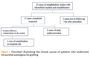
A total of 13 anophthalmic sockets in 13 patients underwent intraorbital autologous fat grafting. Of these, 11 patients completed clinical follow-up, whereas two were lost to follow-up and excluded from further analysis (Figure 1). Radiological assessment was available for 9 cases, with a minimum follow-up of 2 months after the procedure (median, 7 months). The majority of patients were female (10/13), and the age ranged from 22 to 75 years (median, 51 years). Ten sockets had previously undergone evisceration, and three had undergone enucleation. At the time of grafting, 54% of the sockets contained alloplastic orbital implants, whereas 46% contained dermoadipose grafts. The left orbit was affected in 8 cases and the right orbit in 5. The etiologies of anophthalmia included ocular trauma (38.5%), glaucoma (23.1%), infection (15.4%), intraocular tumor (7.7%), and other causes, including inflammatory conditions (ocular burn or retinal detachment; 15.4%).
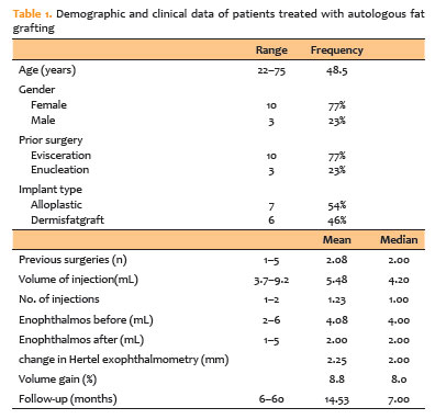
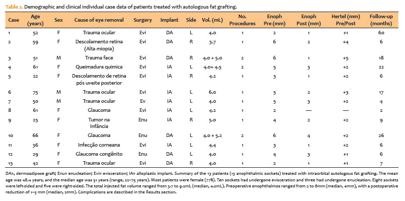
The injected fat volume per procedure ranged from 3.7 to 6mL (median, 4.2mL). Relative enophthalmos, defined as the difference in Hertel exophthalmometry measurements between the anophthalmic and contralateral orbits, ranged from 2 to 6mm preoperatively (median, 4mm). Postoperatively, the reduction in enophthalmos ranged from 1 to 5mm (median, 2mm) in the 12 evaluable patients (Table 1). Individual patient data are presented in table 2. Complications were limited: three patients required a second fat grafting procedure because of undercorrection (cases 3, 4, and 10), and one patient developed a transient fat cyst with local inflammation that resolved after oral antibiotic therapy (case 6).
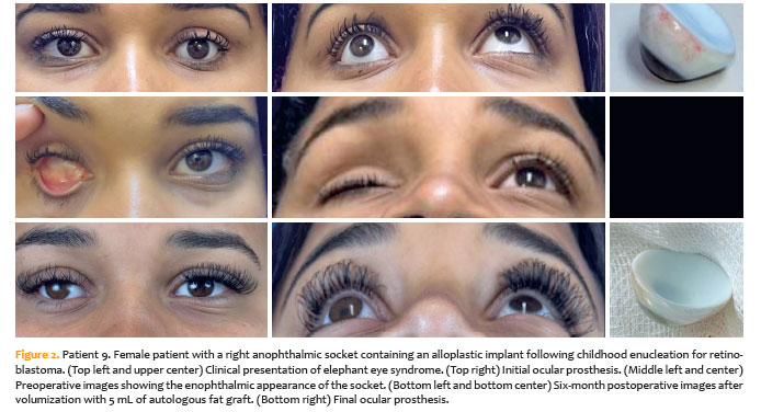
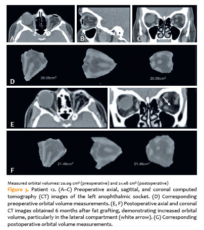
Representative clinical and radiological images are shown in Figures 2 and 3. Figure 2 illustrates the clinical course of patient 9, who presented with elephant eye syndrome (EES) after childhood enucleation with an alloplastic implant. Six months after fat grafting, marked aesthetic improvement and a better prosthetic fit were observed. Similarly, patient 12 (Figure 3) demonstrated an increase in orbital volume of approximately 1.37cm3 at the 6-month follow-up.
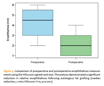
Preoperative and postoperative enophthalmos measurements were compared using the Wilcoxon signed-rank test because of the non-normal distribution of the data. The analysis demonstrated a significant reduction in relative enophthalmos after autologous fat grafting (median reduction, 2mm; Wilcoxon V=0, p<0.001), indicating consistent clinical improvement across the cohort (Figure 4).
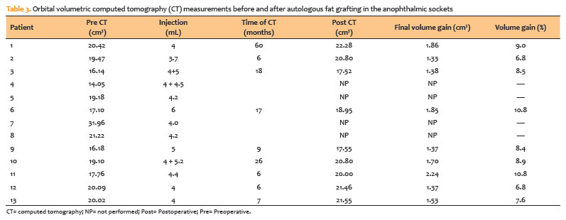
Quantitative CT volumetry confirmed a consistent postoperative increase in orbital volume in all evaluated sockets. Across the cohort, postoperative orbital volumes were consistently greater than baseline values, with mean relative volumetric gains ranging from approximately 6.8% to 10.8%. Table 3 summarizes the preoperative and postoperative volumetric measurements obtained by CT, demonstrating measurable fat retention over follow-up periods of 6-60 months.
No consistent trend toward lower volumetric gains was observed in sockets that had undergone multiple previous surgeries. The mean volumetric gain was 10%±1.3% in sockets with one previous surgery, 8.2%±0.8% in those with two previous procedures, and approximately 9%-10% in sockets with three or more previous surgeries. Spearman's correlation analysis showed no significant association between the number of previous surgeries and postoperative volumetric gain (ρ=-0.19, p>0.05). Likewise, no significant differences were observed according to the primary reconstruction procedure (evisceration vs. enucleation) or implant type (dermoadipose graft vs. alloplastic implant).
No significant clinical complications, including thromboembolic events, were observed during or after the procedure. Most patients reported mild intraoperative discomfort, described as pressure at the recipient site during fat injection, which resolved within 24-48 h with oral analgesics.
Undercorrection of enophthalmos was observed in three patients (cases 3, 4, and 10) between the first and third postoperative months. These patients underwent a second fat grafting procedure 6 months later to correct the residual volume deficit.
One patient (case 6) developed a fat cyst associated with local inflammation and suspected secondary infection; however, culture results were negative. The complication occurred approximately 1 month after surgery and resolved completely after a 14-day course of oral cephalexin.
Donor-site complications were minor and infrequent. Mild contour irregularities of the medial thigh were observed in a few patients and improved with conservative management and local massage.
DISCUSSION
The main causes of anophthalmic sockets include ocular trauma, intraocular infections, refractory glaucoma, and intraocular tumors(15,16). In this series, trauma was the most frequent cause (38.5%), followed by glaucoma (23.1%), infection (15.4%), inflammatory conditions (15.4%), and intraocular tumors (7.7%). In addition to its disfiguring effects, this condition impairs social reintegration, self-esteem, and quality of life, particularly among working-age adults(1,4).
The primary goal of rehabilitation is to restore orbital symmetry, ensure prosthesis stability, and minimize periocular deformities(17,18). Ideally, this is achieved at the time of globe removal by placing a primary orbital implant, either alloplastic or autologous (e.g., a dermis-fat graft), to restore orbital volume(5,15,17). However, many patients develop progressive orbital soft-tissue atrophy over time, resulting in postenucleation or postevisceration socket syndrome. This syndrome is characterized by enophthalmos, upper eyelid ptosis, deep superior sulcus deformity, inferior fornix foreshortening, prosthesis instability, and lower eyelid laxity(6,19). Progressive thickening of the prosthesis to compensate for the volumetric deficit often leads to EES, which further exacerbates lower eyelid malposition and orbital asymmetry(15,20,21). This condition not only increases cosmetic deformity but also contributes to functional impairment. In the present series, classic features of EES were observed in cases 6 (Figure 2) and 9, highlighting the syndrome as a clinical marker of progressive socket atrophy and a strong indication for secondary volumetric reconstruction.
Injectable fillers, such as hyaluronic acid and calcium hydroxyapatite, provide temporary orbital volume restoration with favorable short-term outcomes(17,21,22). However, these materials are associated with high resorption rates, potential migration, and an increased risk of chronic inflammation after repeated procedures, limiting their long-term utility(5).
Autologous fat grafting has emerged as a promising alternative because it provides autologous tissue with excellent biocompatibility, low immunogenicity, cost-effectiveness, and regenerative properties attributed to adipose-derived stem cells(6). The technique was first described by Neuber in 1893 for facial reconstruction and was subsequently refined by Coleman in the 1990s, who emphasized atraumatic fat harvesting, purification by centrifugation, and micro-aliquot injection with blunt cannulas to optimize graft survival through neovascularization(19).
In orbital surgery, the inferolateral postseptal extraconal approach is commonly used to minimize vascular risk and achieve appropriate three-dimensional graft distribution(23). Outcome assessment includes standardized clinical photography, Hertel exophthalmometry, and thin-slice CT with volumetric segmentation, enabling quantitative evaluation of orbital volume gain(17,21,22). Radiological studies have reported volume increases of up to 25% after fat grafting, primarily within the inferolateral compartment, and these increases correlate with clinical improvement in enophthalmos (mean reduction, 1-5 mm) and prosthesis stability(24-26). In the present study, relative volumetric gains ranged from 6.8% to 10.8%.
Final volumetric gain in anophthalmic sockets can be monitored using orbital CT. In this series, CT-based volumetric measurements demonstrated a persistent increase in orbital volume during follow-up periods ranging from 2 to 60 months (Table 3).
Previous studies have also validated orbital CT volumetry for assessing structural changes in pathological conditions, such as thyroid eye disease, and age-related orbital remodeling(27,28).
Collectively, these findings indicate that fat grafting is clinically effective for orbital volumization and that its results can be objectively confirmed and monitored over time using imaging. However, the current literature is limited by heterogeneous surgical techniques, small sample sizes, the lack of standardized radiological protocols, and the scarcity of long-term randomized controlled trials(6,29,30). In the present study, clinical measurements obtained using a standard scleral shell showed a stronger correlation with the overall degree of enophthalmos, thereby minimizing the confounding effect of variations in prosthesis thickness.
The significant reduction in relative enophthalmos observed in this series (median decrease, 2mm; p<0.001) supports the clinical efficacy of autologous fat grafting for orbital volume restoration. This finding, illustrated in figure 4, demonstrates consistent postoperative improvement across the cohort. These results are consistent with previous reports by Hardy et al. and Cervelli et al.(6,7), who also demonstrated measurable improvement in enophthalmos after orbital fat grafting. The present study adds to this evidence by integrating clinical and radiological assessments and further supports autologous fat grafting as a minimally invasive and repeatable technique for socket volumization.
This study helps address these gaps by combining clinical and radiological outcomes and using standardized photography, Hertel exophthalmometry, and quantitative CT volumetry to objectively evaluate orbital volume restoration after autologous fat grafting in anophthalmic sockets with diverse etiologies and surgical histories.
Volumetric gain after orbital lipofilling appeared to be independent of the socket's surgical history. Neither the number of previous procedures, the type of primary surgery (evisceration or enucleation), nor the implant material (alloplastic or dermis-fat graft) significantly influenced the postoperative increase in orbital volume. These findings suggest that even extensively manipulated sockets retain sufficient vascular and stromal support for partial graft survival(10,11). Similar observations were reported by Hardy and Fox, who demonstrated reliable fat retention regardless of the underlying etiology or reconstructive history(10,11). Thus, meticulous fat harvesting, purification, and micro-aliquot injection appear to play a more important role in graft integration than the socket's previous surgical complexity(9,14).
Despite the small sample size, autologous fat grafting was clinically and radiologically effective for restoring volume in anophthalmic sockets with enophthalmos. Favorable outcomes were observed even in patients with a history of trauma, infection, burns, or multiple previous procedures and were associated with few procedure-related complications.
Clinically, enophthalmos decreased in all patients after orbital fat grafting, as demonstrated by Hertel exophthalmometry. Radiologically, the benefits of fat grafting were also evident, with partial maintenance of the grafted fat volume observed over follow-up periods ranging from 6 to 60 months.
AUTHORS' CONTRIBUTIONS
Significant contribution to conception and design: Maria Antonieta da A. Ginguerra-Nascimento, Mario Padula, Suzana Matayoshi.
Data Acquisition: Maria Antonieta da A. Ginguerra-Nascimento, Mario Padula, Jose Byron Vicente Dias Fernandes, Maria Eugenia B. Gorski, Mariana Nobrega Meireles Baptista, Andrea Alves Morato, Yolanda de Souza Mares.
Data Analysis and interpretation: Mario Padula, Laura Goldfarb Cyrino, Carolina Passareli Diniz, Vitor Expedito Alves Ribeiro, Carlos Augusto Ferraresi Sampaio, Eloisa Maria Mello Santiago Gebrim.
Manuscript Drafting: Maria Antonieta da A. Ginguerra-Nascimento, Laura Goldfarb Cyrino.
Significant intellectual content revision of the manuscript: Maria Antonieta da A. Ginguerra-Nascimento, Suzana Matayoshi.
Final approval of the submitted manuscript: Maria Antonieta da A. Ginguerra-Nascimento, Laura Goldfarb Cyrino, Mario Padula, Carolina Passareli Diniz, Vitor Expedito Alves Ribeiro, Andrea Alves Morato, Yolanda de Souza Mares, Carlos Augusto Ferraresi Sampaio, Maria Eugenia B. Gorski, Mariana Nobrega Meireles Baptista, Jose Byron Vicente Dias Fernandes,, Eloisa Maria Mello Santiago Gebrim, Suzana Matayoshi.
Statistical analysis: Vitor Expedito Alves Ribeiro, Suzana Matayoshi.
Obtaining funding: Not applicable.
Supervision of Administrative, technical, or material support: Maria Antonieta da A. Ginguerra-Nascimento, Jose Byron Vicente Dias Fernandes.
Research group leadership: Maria Antonieta da A. Ginguerra-Nascimento, Eloisa Maria Mello Santiago Gebrim, Suzana Matayoshi.
REFERENCES
1. Sagoo MS, Rose GE. Mechanisms and treatment of extruding intraconal implants: socket aging and tissue restitution (the "Cactus Syndrome"). Arch Ophthalmol. 2007;125(12):1616-20.
2. Vagefi MR, McMullan TF, Burroughs JR, Georgescu D, McCann JD, Anderson RL. Orbital augmentation with injectable calcium hydroxylapatite for correction of postenucleation/evisceration socket syndrome. Ophthalmic Plast Reconstr Surg. 2011;27(2):90-4.
3. Vagefi MR, McMullan TF, Burroughs JR, White GL Jr, McCann JD, Anderson RL. Injectable calcium hydroxylapatite for orbital volume augmentation. Arch Facial Plast Surg. 2007;9(6):439-42.
4. Zamani M, Thyagarajan S, Olver JM. Adjunctive use of hyaluronic acid gel (Restylane Sub-Q) in anophthalmic volume deficient sockets and phthisical eyes. Ophthalmic Plast Reconstr Surg. 2010;26(4):250-3.
5. Hunter PD, Baker SS. The treatment of enophthalmos by orbital injection of fat autograft. Arch Otolaryngol Head Neck Surg. 1994;120(8):835-9.
6. Hardy TG, Joshi N, Kelly MH. Orbital volume augmentation with autologous micro-fat grafts. Ophthalmic Plast Reconstr Surg. 2007;23(6):445-9.
7. Cervelli D, Gasparini G, Moro A, Grussu F, Boniello R, Pelo S. Retrobulbar lipofilling to correct the enophthalmos. J Craniofac Surg. 2011;22(5):1918-22.
8. Fox DM. Orbital fat injection: technique and 5-year follow-up. Aesthetic Plast Surg. 2019;43(1):123-32.
9. Murta F, Guevara GF, Hyer JN, Ezra DG. Quantitative assessment of periocular autologous fat transfer survival using 3D stereophotogrammetric imaging. Ophthalmic Plast Reconstr Surg. 2024;40(1):55-60.
10. Coleman SR. The technique of periorbital lipoinfiltration. Oper Tech Plast Reconstr Surg. 1994;1(3):120-6.
11. Coleman SR. Long-term survival of fat transplants: controlled demonstrations. Aesth Plast Surg. 1995;19(5):421-5.
12. Coleman SR. Structural fat grafts: the ideal filler? Clin Plast Surg. 2001;28(1):111-9.
13. Coleman SR. Avoidance of arterial occlusion from injection of soft tissue fillers. Aesthet Surg J. 2002;22(6):555-7.
14. Bergen MP. A literature review of the vascular system in the human orbit. Acta Morphol Neerl Scand. 1981;19(4):273-305.
15. Quaranta-Leoni FM, Fiorino MG, Quaranta-Leoni F, Di Marino M. Anophthalmic socket syndrome: prevalence, impact and management strategies. Clin Ophthalmol. 2021;15:3267-81.
16. Vagefi MR. Minimally invasive approaches to orbital volume augmentation. Int Ophthalmol Clin. 2013;53(3):67-86.
17. Aletaha M, Salour H, Yadegary S, Fekri Y, Tavakoli M. Orbital volume augmentation with calcium hydroxyapatite filler in anophthalmic enophthalmos. J Ophthalmic Vis Res. 2017;12(4):397-401.
18. Calluaud G, Amelot A, Kulker D, Laure B, Pare A. Management of post enucleation socket syndrome patients - A retrospective study. J Craniomaxillofac Surg. 2022;50(2):163-9.
19. Coleman SR. Structural fat grafting: more than a permanent filler. Plast Reconstr Surg. 2006;118(3 Suppl):108S-20S.
20. McGrath D. Patient expectations key to socket surgery success. EuroTimes. Feb 2018 [cited 2026 Jul 22]. Avalable from: https://www.escrs.org/eurotimes/socket-surgery-issues
21. Musch DC, Frueh BR, Landis JR. The reliability of Hertel exophthalmometry. Observer variation between physician and lay readers. Ophthalmology. 1985;92(9):1177-80.
22. Lam AK, Lam CF, Leung WK, Hung PK. Intra-observer and inter-observer variation of Hertel exophthalmometry. Ophthalmic Physiol Opt. 2009;29(4):472-6.
23. Musch DC, Gillespie BW, Niziol LM, Lichter PR, Varma R; CIGTS Study Group. Intraocular pressure control and long-term visual field loss in the Collaborative Initial Glaucoma Treatment Study. Ophthalmology. 2011;118(9):1766-73.
24. Fontdevila J, Serra-Renom JM, Raigosa M, Berenguer J, Guisantes E, Prades E, et al. Assessing the long-term viability of facial fat grafts: an objective measure using computed tomography. Aesthet Surg J. 2008;28(4):380-6.
25. Ye L, Zhang L, Zhu Y, Zhang Y, Wu W, Zhang Y. Enophthalmos: Exploration of Quantitative Treatment With Retro-Orbital Fat Globules Injection. J Craniofac Surg. 2020;31(1):54-7.
26. Strong AL, Cederna PS, Rubin JP, Coleman SR, Levi B. The Current State of Fat Grafting: A Review of Harvesting, Processing, and Injection Techniques. Plast Reconstr Surg. 2015;136(4):897-912.
27. Gonçalves AC, Silva LN, Gebrim EM, Matayoshi S, Monteiro ML. Predicting dysthyroid optic neuropathy using computed tomography volumetric analyses of orbital structures. Clinics (Sao Paulo). 2012;67(8):891-6.
28. Zhang KR, Chon BH, Hwang CJ, Jellema LM, Perry JD. Comparison of orbital volume in young versus senescent human skulls. Ophthalmic Plast Reconstr Surg. 2020;36(2):139-43.
29. Lv Q, Li X, Qi Y, Gu Y, Liu Z, Ma GE. Volume retention after facial fat grafting and relevant factors: a systematic review and meta-analysis. Aesthetic Plast Surg. 2021;45(2):506-20.
30. Yang F, Ji Z, Peng L, Fu T, Liu K, Dou W, et al. Efficacy, safety and complications of autologous fat grafting to the eyelids and periorbital area: A systematic review and meta-analysis. PLoS One. 2021;16(4):e0248505.
Submitted for publication:
March 12, 2026.
Accepted for publication:
July 18, 2026.
Data Availability Statement: The datasets generated and/or analyzed during the current study are already available.
Edited by
Editor-in-Chief-in-Chief: Newton Kara-Júnior
Associate Editor: Tammy H. Osaki
Funding: This study received no specific financial support.
Disclosure of potential conflicts of interest: The authors declare no potential conflicts of interest.

How to cite this article:
 ABO is licensed under a Creative Commons Attribution-NonComercial 4.0 Internacional.

