Arq. Bras. Oftalmol. 2018;81 (5 )
:401-407
| DOI: 10.5935/0004-2749.20180078
Abstract
Objetivo: Estudar a acuidade visual, erros de refração, fixação excêntrica e desempenho de leitura em pacientes com retinocoroidite macular por Toxoplasmose.
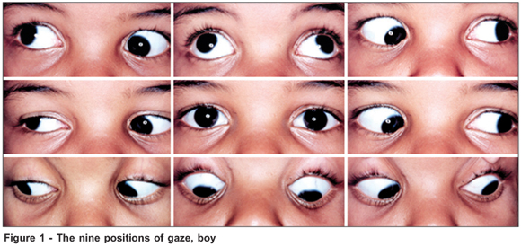
Métodos: Vinte e três pacientes com retinocoroidite macular por Toxoplasmose bilateral e quatro com retinocoroidite macular por Toxoplasmose no seu único olho foram avaliados. Os participantes relataram sua dominância ocular, confirmada pelo teste de Portus e Miles. A acuidade visual melhor corrigida, refração em equivalente esférico, magnificação necessária e velocidade de leitura foram medidas. A microperimetria (MAIA, Centervue - Padova, Italy) registrou a estabilidade preferida do locus e da fixação da retina por meio da área da elipse de contorno bivariada. Quatorze olhos de 14 pacientes com boa visão serviram como controles.
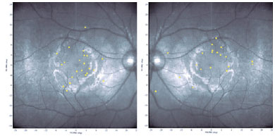
Resultados: A média ± DP da acuidade visual melhor corrigida foi melhor no olho dominante do que no não dominante: 0,9 ± 0,2 (logMAR 0,5 a 1,4) vs. 1,2 ± 0,3 (logMAR 0,6 a 1,7) (p<0,0001, teste t pareado). Miopia em equivalente esférico de -4,00 ou maior estava presente em 42% dos olhos. Microperimetria foi realizada em 42 olhos. Fixação excêntrica foi observada em todos os olhos examinados. Em 14 olhos (33%), o locus retiniano preferencial estava localizado, na retina, na região súpero-temporal à lesão macular, em 10 (24%) súpero-nasal, em 6 (14%) ínfero-temporal, e em 12 olhos (29%) ínfero-nasal. Não houve diferença significativa na distribuição da posição do locus retiniano preferencial entre olhos dominantes e não-dominantes (p=0,85, teste de Pearson). Não houve correlação entre velocidade de leitura e distância entre o locus retiniano preferencial e a posição foveal original estimada (r=-0,09; p=0,73), a área bivariada de contorno elipsóide (r=-0,19; p=0,44) ou acuidade visual melhor corrigida (r=0,024; p=0,92).
Conclusões: A miopia é mais prevalente em pacientes com retinocoroidite macular por Toxoplasmose. A velocidade de leitura não é dependente da posição do locus retiniano preferencial, da estabilidade ou da acuidade visual. A documentação do padrão de fixação excêntrica, entretanto, oferece novos dados no impacto da deficiência visual nesses pacientes e pode ser útil em estratégias de reabilitação.
Keywords: Coroidite; Coriorretinite; Miopia; Toxoplasmose ocular; Leitura
Arq. Bras. Oftalmol. 2025;88 (3 )
:1-6
| DOI: 10.5935/0004-2749.2024-0112
Abstract
PURPOSE: To describe the ophthalmological findings of dry eye disease and its relation to the quality of life of COVID-19 survivors.
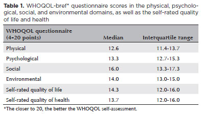
METHODS: COVID-19 survivors who had previously been hospitalized at Hospital das Clínicas de Ribeirão Preto complex underwent an ophthalmological evaluation, which included a dry eye disease questionnaire, break-up time, fluorescein staining, and Schirmer test. We collected the presenting and best-corrected visual acuity, sociodemographic data, personal medical history, and scores from a self-reported quality of life questionnaire (WHOQOL-bref). According to the severity of the acute phase of the disease, the patients were classified into mild-to-moderate, severe, and critical groups.
RESULTS: Ninety-five patients (190 eyes) were evaluated 100 ± 44 days after the onset of COVID-19 symptoms. Of these, 83 patients (87.3%) completed the WHOQOL-bref questionnaire. Ten patients (12.0%) had mild-to-moderate COVID-19, 41 (49.4%) had severe COVID-19, and 32 (38.6%) had critical COVID-19. The median best-corrected visual acuity was logMAR 0 (0-1). Approximately 26.3% patients had a history of dry eye disease or severe dry eye symptoms (frequent or constant ocular dryness and irritation). There was an association between the proportion of patients with dry eye disease and the quality of life (p=0.014) and health (p=0.001). Furthermore, there was a significant trend between the proportion of patients with dry eye disease and how they rated their health and quality of life (p=0.0004 and 0.0027, respectively.
CONCLUSIONS: There is a significant negative correlation between the proportion of patients with dry eye disease and their self-reported quality of life.
Keywords: COVID-19; Coronavirus infections; SARS-CoV-2; Eye diseases; Epidemiology; Ocular surface; Public health
 ABO is licensed under a Creative Commons Attribution-NonComercial 4.0 Internacional.
12-fig01.jpg)
14-fig01.jpg)
07-fig01.jpg)