Arq. Bras. Oftalmol. 2026;89 (1 )
:1-8
| DOI: 10.5935/0004-2749.2025-0025
Abstract
PURPOSE: Diabetic retinopathy screening in low- and middle-income countries is limited by restricted access to specialized care. Portable retinal cameras offer a practical alternative; however, image quality – affected by mydriasis – directly influences the performance of artificial intelligence models. This study evaluated the effect of mydriasis on image gradability and AI-based diabetic retinopathy detection in real-world, resource-limited settings.
METHODS: The proportions of gradable images were compared between mydriatic and non-mydriatic groups. Generalized estimating equations were used to identify factors associated with image gradability, including age, sex, race, diabetes duration, and systemic hypertension. A ResNet-200d model was trained on the mobile Brazilian Ophthalmological dataset and externally validated on both mydriatic and non-mydriatic images. Model performance was evaluated using accuracy, F1 score, area under the curve, and confusion matrix metrics. Sensitivity differences were assessed using the McNemar test, and area under the curves were compared using DeLong's test. The Youden index was used to determine optimal classification thresholds. Agreement between macula- and disc-centered images was analyzed using Cohen's κ.
RESULTS: The mydriatic group demonstrated a higher proportion of gradable images compared with the non-mydriatic group (82.1% vs. 55.6%; p<0.001). In non-mydriatic images, lower gradability was associated with systemic hypertension, older age, male sex, and longer diabetes duration. The AI model achieved better performance in mydriatic images (accuracy, 85.15%; area under the curve, 0.94) than in non-mydriatic images (accuracy, 79.68%; area under the curve, 0.93). The McNemar test showed a significant difference in sensitivity (p=0.0001), whereas DeLong's test revealed no significant difference in area under the curve (p=0.4666). The Youden index indicated that optimal classification thresholds differed based on mydriasis status. Agreement between image fields was moderate to substantial and improved with mydriasis.
CONCLUSION: Mydriasis significantly improves image gradability and enhances AI performance in diabetic retinopathy screening. Nonetheless, in low- and middle-income countries where pharmacologic dilation may be impractical, optimizing model calibration and thresholding for non-mydriatic images is essential to ensure effective AI implementation in real-world clinical environments.
Keywords: Artificial intelligence; Bias; Diabetic retinopathy; Portable camera; Retina
Arq. Bras. Oftalmol. 2026;89 (3 )
:1-14
| DOI: 10.5935/0004-2749.2025-0248
Abstract
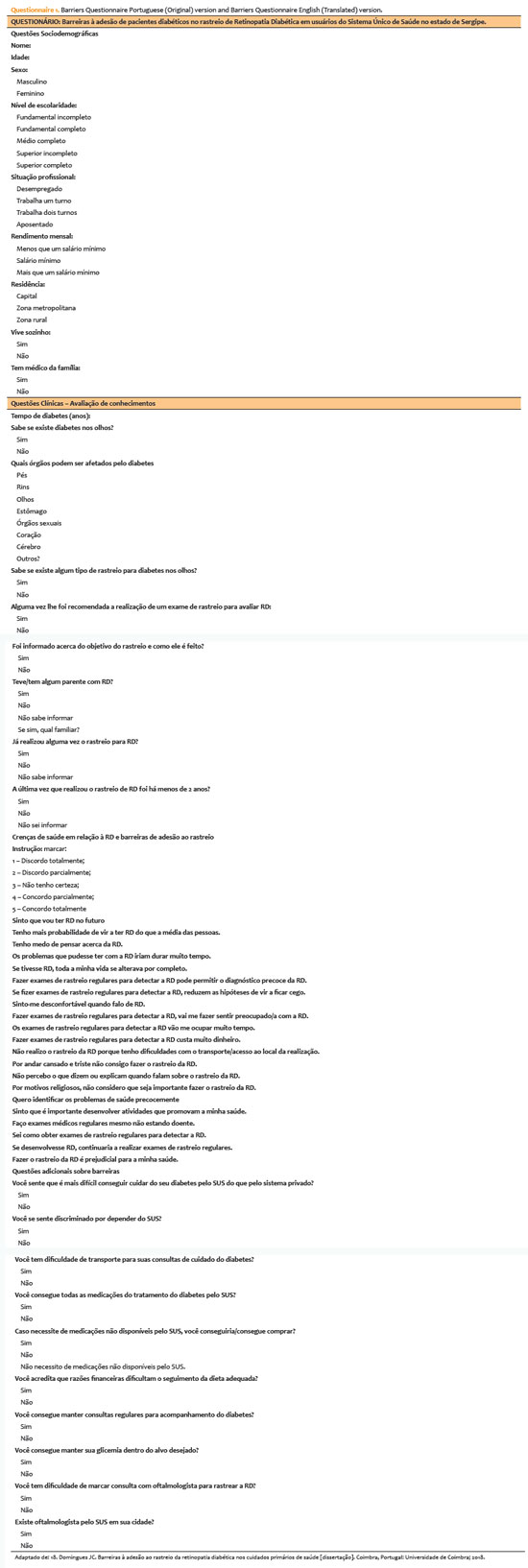
PURPOSE: This study aimed to identify barriers to diabetic retinopathy screening among a socioeconomically vulnerable urban population in northeast Brazil.
METHODS: A cross-sectional study was conducted during a diabetic retinopathy screening campaign at primary healthcare units. Ninety-five patients with diabetes underwent retinal examinations and completed a structured interview. Clinical, demographic, and socioeconomic data were collected.
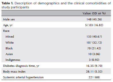
RESULTS: The study population consisted predominantly of older adults (mean age: 60.7 ± 10.5 years), with a high prevalence of type 2 diabetes (99.0%) and low educational attainment. Most participants were economically inactive (81.1%) and reported low income (83.2%). Diabetic retinopathy and maculopathy were highly prevalent, affecting 50.0% and 22.9% of participants, respectively. Longer duration of diabetes was significantly associated with greater awareness of diabetic retinopathy (p=0.035), higher HbA1c levels (p<0.001), and increased prevalence of diabetic retinopathy (p=0.013) and maculopathy (p=0.002). Notably, 33.3% of participants reported difficulties attending medical appointments for diabetes management. In addition, 78.1% experienced challenges scheduling ophthalmologic evaluations, and 76.3% reported that no ophthalmologist was available in their city through the public healthcare system. Financial constraints also limited adherence to recommended dietary practices (90.4%) and impaired glycemic control, with more than half of participants reporting difficulty maintaining target glucose levels.
CONCLUSION: Major barriers to diabetic retinopathy screening included limited awareness of the importance of screening, financial hardship, and transportation challenges. Targeted educational initiatives and structural interventions such as expanded screening programs incorporating telemedicine and subsidized transportation—may improve screening adherence among vulnerable populations.
Keywords: Diabetic retinopathy; Mass screening; Health services accessibility; Health knowledge, attitudes, practices; Socioeconomic factors
Arq. Bras. Oftalmol. 2024;87 (4 )
:1-6
| DOI: 10.5935/0004-2749.2023-0200
Abstract
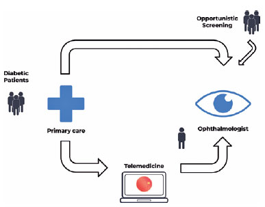
PURPOSE: Timely screening and treatment are essential for preventing diabetic retinopathy blindness. Improving screening workflows can reduce waiting times for specialist evaluation and thus enhance patient outcomes. This study assessed different screening approaches in a Brazilian public healthcare setting.
METHODS: This retrospective study evaluated a telemedicine-based diabetic retinopathy screening implemented during the COVID-19 pandemic and compared it with in-person strategies. The evaluation was conducted from the perspective of a specialized referral center in an urban area of Central-West Brazil. In the telemedicine approach, a trained technician would capture retinal images by using a handheld camera. These images were sent to specialists for remote evaluation. Patient variables, including age, gender, duration of diabetes diagnosis, diabetes treatment, comorbidities, and waiting time, were analyzed and compared.
RESULTS: In total, 437 patients with diabetes mellitus were included in the study (mean age: 62.5 ± 11.0 years, female: 61.7%, mean diabetes duration: 15.3 ± 9.7 years, insulin users: 67.8%). In the in-person assessment group, the average waiting time between primary care referral and specialist evaluation was 292.3 ± 213.9 days, and the referral rate was 73.29%. In the telemedicine group, the average waiting time was 158.8 ± 192.4 days, and the referral rate was 29.38%. The telemedicine approach significantly reduced the waiting time (p<0.001) and significantly lowered the referral rate (p<0.001).
CONCLUSION: The telemedicine approach significantly reduced the waiting time for specialist evaluation in a real-world setting. Employing portable retinal cameras may address the burden of diabetic retinopathy, especially in resource-limited settings.
Keywords: Telemedicine/methods; Diabetic retinopathy; Diagnostic screening programs; Vision screening; Practice patterns, physicians
 ABO is licensed under a Creative Commons Attribution-NonComercial 4.0 Internacional.