Arq. Bras. Oftalmol. 200265
| DOI: 10.1590/S0004-27492002000300002
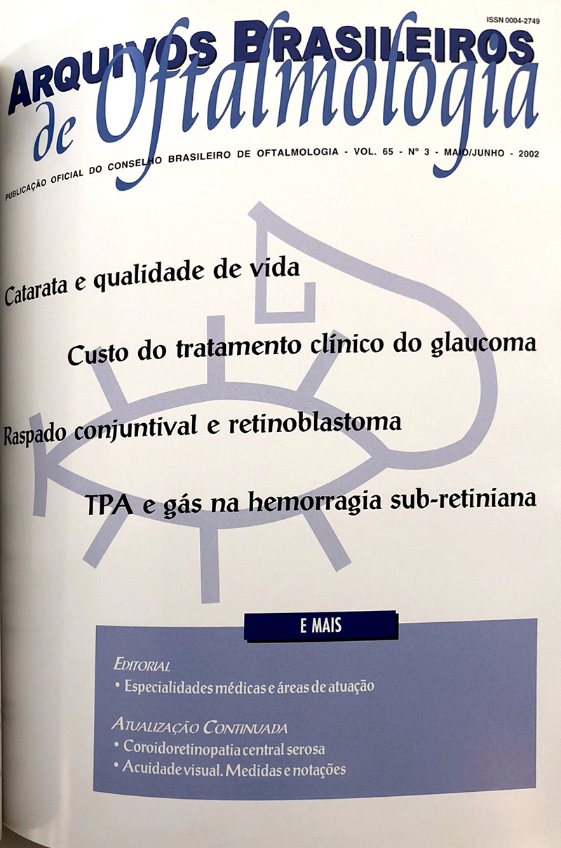
Purpose: Cataract represents a public health problem because patients presenting the disease are unable to adequately perform routine activities, with a negative influence on their quality of life. The purpose of this research is to evaluate the impact of reduced visual acuity on the quality of life of these patients. The authors adjusted a specific questionnaire to the Brazilian social reality and tested its response adequacy and reliability.
Methods: 30 cataract patients with surgical indication were interviewed. The questionnaire was applied and the same interview was done twice within a one-hour interval.
Results: More than 80% of questions were answered the first time they were asked. Consistence analysis showed that only one patient answered four questions in a different manner during the interviews.
Conclusion: The authors concluded that the adjusted questionnaire is applicable to a population with a low level of formal education, being a reliable instrument in the evaluation of quality of life in cataract patients.
Keywords: Cataract; Cataract; Quality of life; Questionnaires; Public health; Activities of daily living
Arq. Bras. Oftalmol. 200265
| DOI: 10.1590/S0004-27492002000300003
Purpose: To verify the social characteristics and the impact of glaucoma treatment on the familial income of patients followed at a university hospital.
Methods: One hundred and forty six glaucomatous patients were interviewed at the Hospital das Clínicas da Universidade de Campinas to evaluate their social economic profile. The questionnaire investigated the occupation, the individual and familial income, as well as the type and frequency of the antiglaucomatous drugs used by each patient. Knowing the monthly cost of antiglaucomatous drugs available in Brazil, we were able to calculate the monthly cost of glaucoma treatment and the percentage of committed familial income.
Results: The mean monthly cost of glaucoma treatment was 36.09 ± 31.99 reais, which corresponded to 15.5% of the familial income. Thirty-six (24%) patients had 25 percent or more of the familial income spent on their treatment. Sixty-six (45.2%) patients had difficulty in buying their medications. Factors associated with this difficulty included low familial income (p=0.0001), and high percentage of the income used to buy the drugs (p=0.0002).
Conclusion: The cost of glaucoma treatment is high compared to the income of patients treated at a public institution. This population has a low familial income, of which a high percentage is required to acquire antiglaucoma medications. We suggest that these patients may be at risk for low compliance due to economical limitations.
Keywords: Glaucoma; Health care costs
Arq. Bras. Oftalmol. 200265
| DOI: 10.1590/S0004-27492002000300004
Purpose: Human conjunctival cytology has been extensively studied. Lack of data about the characteristics of conjunctival smears in patients treated for retinoblastoma who underwent enucleation and radiotherapy motivated this study.
Methods: A previously studied group of 114 orbits of 58 patients was examined. The authors present the results of conjunctival cytology in 4 different situations, with and without enucleation, with and without radiotherapy.
Results: In the leukocytic infiltrate in orbits with ocular bulbi predominated the lymphocyte, while in the others predominated the polymorphonuclear neutrophil. In general, the majority of epithelial cell alterations seen in the conjunctival smears of irradiated orbits were: cell with large nuclei, prominent nucleoli, and cytoplasmatic hyalinization.
Conclusion: The cytological findings of the conjunctival smears do not indicate a characteristic and distinct pattern for each group.
Keywords: Eye; Conjunctiva; Anophthalmia; Retinoblastoma; Eye enucleation
Arq. Bras. Oftalmol. 200265
| DOI: 10.1590/S0004-27492002000300005
Purpose: The assessment of the epidemiological profile of patients victims of lime ocular burns, by the Ophthalmology Department of the Evangelic University Hospital of Curitiba, with emphasis on "Lime Bomb" burn cases.
Methods: A study on 88 ocular lime burn patients (lime bomb or not), who came to the HUEC Emergency Service from September 1999 to September 2000.
Results: Of 88 patients that were evaluated, 73 (82.95%) had suffered ocular lime burn in their working place and 15 (17.24%) had suffered it due to the `Lime Bomb" itself (local custom). Most of the patients that were examined were males, aged 20-40 years, with predominance of unilateral ocular burn and degree I in case of accident at the workplace, and bilateral and degree IV in case of "Lime Bomb".
Conclusion: By comparing the studied groups, it is noticed that those with "Lime Bomb" burn show greater ocular health damage which was demonstrated in this study by means of bilaterality, injury severity and occurring in young patients.
Keywords: Eye burns; Eye; Chemical burns; Alkalies; Occupational accidents; Hospital emergency service; University hospitals
Arq. Bras. Oftalmol. 200265
| DOI: 10.1590/S0004-27492002000300006
Purpose: To evaluate the frequency of anterior chamber contamination during phacoemulsification surgery with intraocular lens implant. Method: A prospective study was done with 30 patients submitted to phacoemulsification surgery with intraocular lens implant. Two samples of the aqueous humor were removed from the anterior chamber, one at the beginning and another one at the end of the surgery and were inoculated in culture plates for anaerobic and aerobic bacteria and fungi.
Results: Intraocular aspirates yielded negative cultures.
Conclusion: The absence of microorganisms in the samples evaluated allowed us to conclude that contamination during the phacoemulsification surgery with lens implant is unusual.
Keywords: Cataract extraction; Phacoemulsification; Contamination; Aqueous humor; Cell culture
Arq. Bras. Oftalmol. 200265
| DOI: 10.1590/S0004-27492002000300007
Purpose: To study the aerobic bacterial conjunctival flora in patients with clinical diagnosis of acute viral conjunctivitis. Method: Thirty patients between 18 and 30 years with acute adenoviral conjunctivitis and 30 normal subjects underwent conjunctival culture examination. Material from the conjunc-tiva of patients with conjunctivitis was collected within the first 3 days of onset of the symptoms. Blood-agar and chocolate-agar plates were used for culture. Patients under any topical or systemic medication, contact lens wearers, patients with ocular or systemic disease were excluded.
Results: Significant positivity in culture results of patients with adenoviral conjunctivitis was observed (33.3%, among which 50% were Haemophylus influenzae and 50% were Streptococcus pneumoniae), when compared to conjunctival culture of normal subjects (6.6% of coagulase-negative staphylococci). There was no clinical difference related to the isolation of bacteria in the conjunctival sample of all patients.
Conclusion: Patients with clinically diagnosed viral conjunctivitis have a higher rate of positive cultures when compared to controls. Nevertheless, this finding does not correlate with clinical differences. Microorganisms isolated in the conjunctiva of patients with adenoviral conjunctivitis were different from those isolated in the conjunctiva of normal subjects.
Keywords: Adenoviridae infections; Adenoviridae infections; Conjunctivitis, viral; Bacteria; aerobic
Arq. Bras. Oftalmol. 200265
| DOI: 10.1590/S0004-27492002000300008
Purpose: To identify the microorganisms of the ocular conjunctiva in healthy dogs in Araçatuba (SP) city, in summer and winter.
Methods: Forty dogs, male and female, age ranging from 2 to 5 years were used. After ocular wash, samples were collected from the inferior conjunctival sac in order to isolate and identify anaerobic, aerobic bacteria and fungi.
Results: Staphylococcus aureus and Sthaphylococcus beta-haemolyticus and Penicilium sp. were the most frequently observed microorganisms.
Conclusion: There was ocular conjunctiva microbiota variation between the studied seasons. Staphylococcus beta-haemolyticus was observed only during the winter.
Keywords: Conjunctiva; Staphylococcus; purification; Seasons; Dogs
Arq. Bras. Oftalmol. 200265
| DOI: 10.1590/S0004-27492002000300009
Purpose: To describe the clinical features of patients with dermolipoma and orbital fat prolapse.
Methods: A retrospective 12-years survey was done concerning patients with dermolipoma and orbital fat prolapse, at the Faculdade de Medicina de Botucatu - UNESP.
Results: During that time we found 20 patients with dermolipoma and 10 with orbital fat prolapse. Dermolipoma occurred mainly in children, more in female, and was frequently located at the external canthus, 7 patients had other ocular associated diseases. The orbital fat prolapse occurred also in Caucasians, more in males, the elderly, and in all but one occurred temporally.
Conclusion: Dermolipoma and orbital fat prolapse have similar clinical appearances, but they are clearly distinct. Dermolipoma affects children, female, and orbital fat prolapse, elderly and males.
Keywords: Lipoma; Adipose tissue; Choristoma; Prolapse; Orbital disease; Differential diagnosis; Retrospective studies
Arq. Bras. Oftalmol. 200265
| DOI: 10.1590/S0004-27492002000300010
Purpose: To determine glaucoma epidemiology in a region of southern Brazil having as a group of study the population of Piraquara city -- PR (patients over 40 years old), and analyzing the importance of the screening examination.
Methods: A total of 1953 patients were screened in the Glaucoma Project (first phase) between June 1999 to December 2000. The screening process included a specific interview, examination of anterior chamber, applanation tonometry and fundoscopy. The patients screened as glaucoma suspects were submitted to a complete ophthalmologic examination at the Hospital das Clínicas -- UFPR.
Results: Of the 1953 screened patients, 282 were classified as glaucoma suspects, but only 184 returned for the complete ophthalmologic examination. There were 73 confirmed cases of chronic open angle glaucoma; 35 (1.79%) high-tension glaucoma cases and 38 (1.95%) patients with normal tension glaucoma, and other 6 cases of secondary glaucoma or angle closure glaucoma. In both groups we determined gender, age and race distributions; the prevalence of diabetic and systemic hypertense population; and the IOP and CCT (central corneal thickness) averages, observing a statistically significant difference between the two groups.
Conclusion: The prevalence of high-tension glaucoma and normal tension glaucoma was similar in this population sample, and this alerted us to the fact that fundoscopy is a very important examination in detecting glaucomatous patients.
Keywords: Glaucoma; Open angle glaucoma; Glaucoma; Open angle glaucoma; Fundus oculi; Triage
Arq. Bras. Oftalmol. 200265
| DOI: 10.1590/S0004-27492002000300011
Purpose: To establish the most frequently isolated agents in keratitis, conjunctivitis and endophthalmitis by the Laboratory of Microbiology/ Santa Casa of São Paulo, and to determine their antimicrobial sensitivity.
Methods: A retrospective study including all ophthalmologic charts of the Laboratory of Microbiology/ Santa Casa of São Paulo was conducted. All ocular material collected during the last five years was analyzed. We included in this study only patients whose diagnoses were conjunctivitis, keratitis and endophthalmitis. Susceptibility tests of drugs with ophthalmological application were analyzed.
Results: In a total of 568 cases, we found 282 of bacterial keratitis (49.6%), 214 of conjunctivitis (37.7%) and 72 of endophthalmitis (12.7%); three hundred thirty-three negative culture cases were found (58.9%). S. aureus was the most frequently isolated agent (n=73; 31.0%). Conjunctivitis showed Candida sp in 6.6% of all cases, and 38.3% of the cultures were negative. When the diagnosis was keratitis, P. aeruginosa was positive in 3.9% and 71.6% of the cultures were negative. Endophthalmitis showed only 32% of positivity.
Conclusion: Inconclusive cultures were the majority. Facing an ocular infection, there is a considerable chance to find S. aureus.
Keywords: Eye infections; Microbial sensitivity tests; Conjuntivitis bacterial; Keratitis; Endophthalmitis; Drug resistance
Arq. Bras. Oftalmol. 200265
| DOI: 10.1590/S0004-27492002000300012
Purpose: To describe and analyze the eyelash abnormalities diagnosed at the Hospital of the School of Medicine of Ribeirão Preto.
Methods: A total of 59 patients who presented during 1999 with eyelash problems at the oculoplastic clinic were studied. Patients with previous treatments were excluded. Data collection included systemic or local disease, symptom duration, eyelid and eye surface abnormalities as well lash morphologic and location characteristics.
Results: Trichiasis was the main diagnosed abnormality (94.9% of the cases). Trachoma was the main disease associated with trichiasis. Trachomatous trichiasis differed from nontrachomatous trichiasis according to the number of affected eyelid segments, laterality, affected eyelid (upper or lower) and magnitude of corneal damage. Lash abnormalities regarding color, size and thickness were seen in both trachomatous and nontrachomatous trichiasis. Conclusions: Trachomatous trichiasis continues to be the main lash problem in the Hospital of the School of Medicine of Ribeirão Preto. Color and thickness lash abnormalities can not be used as criteria for the distinction between trichiasis and acquired distichiasis.
Keywords: Eyelash; Trachoma; Eyelid diseases; Eyelid
Arq. Bras. Oftalmol. 200265
| DOI: 10.1590/S0004-27492002000300013
Purpose: To determine the quality of life in patients after penetrating keratoplasty using the VF-14 test and correlating it with final visual acuity.
Methods: A total of 58 penetrating keratoplasty operated patients (1 year after surgery) were evaluated by the VF-14 test. Ophthalmological examination data were collected from their charts. Student's t test and linear regression were applied.
Results: The VF-14 ratio was 69.3. Best scores were obtained in activities such as cooking (90.9), card games (88.0), fine handworks (83.9). On the contrary, worst scores were found for car driving (at night, 22.5; and during the day, 40.9). Results were statistically significant (p< 0.01) and linear regression showed weak correlation between scores and final visual acuity (r=0.41).
Conclusion: We observed a weak correlation between the quality of life and final visual acuity and suggest changes in this questionnaire including activities that are more adequate for our patients.
Keywords: Corneal transplantation; Keratoplasty; Quality of life; Patient satisfaction; Health status indicators
Arq. Bras. Oftalmol. 200265
| DOI: 10.1590/S0004-27492002000300014
Purpose: The author describes a case of bilateral subretinal and sub-RPE hemorrhage due to age related macular degeneration treated with injections of tPA and perfluoropropane gas. Case report: Two eyes of a patient with subretinal and sub-RPE macular hemorrhage received injections of 25 mug of tPA and 0,3 mL of C3F8. The patient was assessed regarding visual acuity, retinography and fluoresceinography before and after the procedure.
Results: One week after treatment, both eyes presented with complete resolution of subfoveal hemorrhage, however traces of subretinal hemorrhage were found in both eyes, mainly in the left eye. Sub-RPE hemorrhage remained the same during the treatment of the left eye. Visual acuity improved in both eyes, specifically in the right eye.
Conclusion: Intravitreal injection of tPA and perfluoropropane gas seems to be an alternative procedure in the treatment of subretinal hemorrhage due to age related macular degeneration, however, sub-RPE hemorrhage remains a therapeutic challenge. Further studies are necessary.
Keywords: Tissue plasminogen activator; Retinal hemorrhage; Macular degeneration; Aging
Arq. Bras. Oftalmol. 200265
| DOI: 10.1590/S0004-27492002000300015
The authors report a case of nasal furuncle that progressed to septic bilateral and asymmetric thrombosis of cavernous, transverse, sigmoid sinus and internal jugular vein, associated with bacterial meningitis, in a previously healthy patient. In spite of the extensive thrombosis, the patient presented a good evolution, after an aggressive clinical treatment with antibiotics, corticosteroids and anticoagulants. However, there remained paresis of the VI nerve on the left and partial lesion of the homolateral optic nerve.
Keywords: Cavernous sinus thrombosis; Furunculosis; Nose diseases; Antibiotics; Adrenal cortex hormones; Paresis; Bacterial meningitis; Case report
Arq. Bras. Oftalmol. 200265
| DOI: 10.1590/S0004-27492002000300016
Purpose: To describe the role of the optical coherence tomography, fluorescein and indocyanine green angiography findings in the diagnosis of idiopathic polypoidal choroidal vasculopathy (IPCV).
Methods: A series of four eyes of three patients thought to have IPCV were examined and evaluated by optical coherence tomography, fluorescein and indocyanine green angiographies.
Results: Fluorescein angiography was highly important for the diagnosis only in case 1. Indocyanine green angiography showed a branching network of the vessels with marginal vascular dilatations in all cases. Optical coherence tomography showed hemorrhagic detachment of the retinal pigment epithelium contiguous with the anterior delimitation of the polypoidal elements in two cases.
Conclusion: The association of clinical manifestations with fluorescein and indocyanine green angiographies and optical coherence tomography can be useful in the differential diagnosis of exudative maculopathies, particularly IPCV with serohemorrhagic manifestations.
Keywords: Choroid; Choroid hemorrhage; Choroid diseases; Fluorescein angiography; Indocyanine green; Fundus oculi; X-ray computed tomography
Arq. Bras. Oftalmol. 200265
| DOI: 10.1590/S0004-27492002000300017
Purpose: To describe a case of persistent retinopathy after bone marrow transplantation in the absence of radiation therapy.
Methods: Case Report.
Results: A 42 year-old man developed bilateral visual loss 15 months after receiving a bone marrow transplant for acute leukemia. The patient was treated with a high dose of cyclosporin A and oral corticosteroids. No radiation therapy was given. Late-onset, multiple, bilateral cotton-wool spots developed 15 months after the bone marrow transplantation and still persist. After three years other cotton-wool spots arose in the absence of any immunosuppressive therapy. Conclusions: Bone marrow transplantation microvasculopathy of the retina may be related to certain combinations of chemotherapy drugs or immunosuppression itself and may persist in the absence of these immunosuppressive drugs.
Keywords: Bone marrow transplantation; Adrenal cortex hormones; Cyclosporins; Retinal diseases; Drug combinations; Case report; Follow-up
Arq. Bras. Oftalmol. 200265
| DOI: 10.1590/S0004-27492002000300018
The authors describe 3 cases of optic disc pit, showing by optic coherence tomography images the different phases of secondary retinal detachment, from the internal retinal layer split, intraretinal cyst formation, until the neurosensorial retinal serous detachment.
Keywords: Optic nerve; Optic disc; Abnormalities; Tomography
Arq. Bras. Oftalmol. 200265
| DOI: 10.1590/S0004-27492002000300019
Evaluations of the visual function are very intricate since they depend on afferent, efferent and cognitive mechanisms, besides external factors of the examined subject, such as the type of the stimulus and of its presentation. Testing the visual acuity is discussed in its formal aspects related to definitions, quantifications (criteria for the measurements of an angle, size of optotypes), notations (decimal or fractionary), scales (representing angular, linear or logarithmic relationships) and units in which the values are expressed (reciprocal of minutes of arc, pure number, spacial frequency, decibels, octaves). As a consequence, numerical references about visual acuity and respective operations (e.g., calculations of average values, determinations of variations, relationships between them) may lead to very different interpretations of a study, sometimes even opposite, according to the criteria which are used.
Keywords: Visual acuity; Vision tests; Vision tests; Reproducibility of results; Visual fields; Vision disorders; Visual perception; Differential threshold
Arq. Bras. Oftalmol. 200265
| DOI: 10.1590/S0004-27492002000300020
The author describes the clinical features and new diagnostic methods of central serous chorioretinopathy. He discusses experimental and clinical studies related to the pathogenesis of the disease and presents therapy choices and prognosis of central serous chorioretinopathy, based on reference survey.
Keywords: Choroid diseases; Choroid diseases; Retinal detachment; Fluorescein angiography; Differential diagnosis; Pigment epithelium of eye; Prognosis


 ABO is licensed under a Creative Commons Attribution-NonComercial 4.0 Internacional.

