Arq. Bras. Oftalmol. 2022;85 (4 )
:344-350
| DOI: 10.5935/0004-2749.20220052
Abstract
Objetivo: Investigar a redução na densidade celular endotelial corneana associada à trabeculotomia transluminal assistida por gonioscopia (GATT) em curto prazo.
Métodos: Análise retrospectiva de prontuários médicos de pacientes com glaucoma de ângulo aberto que foram submetidos à trabeculotomia transluminal assistida por gonioscopia isolada ou combinada com facoemulsificação. Pacientes que foram submetidos à facoemulsificação isolada foram incluídos como controles. Dados da densidade celular endotelial corneana (avaliada através de microscópio especular) pré-operatória e ao primeiro mês pós-operatório foram coletados e comparados.
Resultados: Sessenta e dois olhos que foram submetidos à trabeculotomia transluminal assistida por gonioscopia (trabeculotomia transluminal assistida por gonioscopia=39 olhos; faco com trabeculotomia transluminal assistida por gonioscopia=23 olhos) passaram pelos critérios de inclusão. A idade média dos pacientes estudados era 61,3 ± 18,4 anos no grupo trabeculotomia transluminal assistida por gonioscopia isolada e 60,4 ± 11,9 anos no grupo faco com trabeculotomia transluminal assistida por gonioscopia. Homens eram 66,6% do grupo trabeculotomia transluminal assistida por gonioscopia isolada e 56,5% do grupo faco com trabeculotomia transluminal assistida por gonioscopia. O defeito perimétrico médio (Mean Deviation) era -13,9 ± 9,2 dB e -10,3 ± 7,7 dB nos grupos trabeculotomia transluminal assistida por gonioscopia isolada e faco com trabeculotomia transluminal assistida por gonioscopia respectivamente. O grupo que fora submetido à trabeculotomia transluminal assistida por gonioscopia isolada apresentou redução média da densidade celular endotelial corneana de 28,8 células/mm2 (1,31%; p=0,467). No grupo faco com trabeculotomia transluminal assistida por gonioscopia, a redução média da densidade celular endotelial corneana foi de 89,4 células/mm2 (4,36%; p=0,028). Olhos controle (23 olhos) apresentaram redução média da densidade celular endotelial corneana de 114,1 ± 159,8 células/mm2 (4,41%; p=0,505). A redução na densidade celular endotelial corneana no grupo faco com trabeculotomia transluminal assistida por gonioscopia não foi significativamente diferente do grupo controle (p=0,81).
Conclusões: A trabeculotomia transluminal assistida por gonioscopia parece ser segura para a camada endotelial corneana em um curto prazo quando realizada de forma isolada ou combinada com cirurgia de catarata.
Keywords: Glaucoma de ângulo aberto; Perda de células endoteliais da córnea; Extração de catarata; Malha trabecular; Gonioscopia/métodos; Trabeculectomia/métodos
Arq. Bras. Oftalmol. 2022;85 (1 )
:25-29
| DOI: 10.5935/0004-2749.20210091
Abstract
Objetivo: Avaliar e comparar a variação do diâmetro pupilar antes e após a cirurgia de catarata por facoemulsificação convencional versus cirurgia de catarata assistida por laser de femtossegundo, usando o LDV Z8 (Ziemer Ophtalmic). Também avaliamos a relação entre o diâmetro pupilar com o tempo da cirurgia e o tempo de ultrassom.
Métodos: Estudo comparativo prospectivo, realizado no Centro de Excelência em Oftalmologia, Brasil. Foram incluídos 79 olhos de 67 pacientes com opacidade nuclear. Os mesmos foram divididos em Grupo Controle, que foi submetido a cirurgia de catarata com facoemulsificação manual, e Grupo Estudo, com catarata assistida por laser de femtossegundo. Todas as cirurgias foram realizadas pelo mesmo cirurgião experiente. Todos os pacientes receberam antiinflamatório não esteróide tópico no dia anterior à cirurgia e o mesmo colírio midriático no pré-operatório. Para quantificar o tamanho da pupila, as medidas foram realizadas usando um compasso cirúrgico: anterior ao procedimento de facoemulsificação e ao final da cirurgia. No grupo de estudo, medidas após o laser foram adicionadas. O tempo cirúrgico e o tempo de facoemulsificação também foram analisados.
Resultados: Não foi encontrada diferença significativa entre o tamanho da pupila pré-femto x pré-faco (8,69 ± 0,44 mm x 8,63 ± 0,72 mm; p=0,643), bem como o tamanho da pupila no final da cirurgia (7,96 ± 0,98 mm x 7,78 ± 0,95 mm; p=0,480) e o tempo médio de cirurgia (p=0,780). No entanto, no grupo de catarata assistida por laser de femtossegundo, houve um aumento transitório do diâmetro pupilar após o laser, indicando uma tendência para maior variação no grupo femto.
Conclusões: Embora o diâmetro pupilar fosse semelhante ao final da cirurgia, o grupo com catarata assistida por laser de femtossegundo apresentou maior variação intraoperatória da pupila. Portanto, para uma cirurgia de catarata assistida por laser de femtossegundo mais eficiente e segura, o cirurgião deve estar ciente do tamanho do diâmetro pupilar antes do procedimento.
Keywords: Catarata; Miose; Facoemulsificação; Laser; Pupila
Arq. Bras. Oftalmol. 2021;84 (5 )
:454-461
| DOI: 10.5935/0004-2749.20210071
Abstract
Objetivo: Comparar a estrutura da córnea e as alterações morfológicas endoteliais após cirurgia de catarata por facoemulsificação sem intercorrências entre pacientes com diabetes mellitus tipo 2 e não diabéticos; e determinar quais fatores pré e intra-operatórios relacionados com a maior redução da densidade celular endotelial.
Métodos: Quarenta e cinco diabéticos (45 olhos) e 43 (43 olhos) controlos com catarata relacionada à idade foram incluídos neste estudo observacional prospectivo. Os parâmetros da córnea (espessura e volume) e do segmento anterior foram medidos pela tomografia Scheimpflug; a densidade e morfologia celular endotelial (coeficiente de variação do tamanho das células, células hexagonais) foram registrados usando microscopia especular não contato. Os pacientes foram avaliados no pré-operatório, 1 e 6 meses após a cirurgia. Foi realizada uma análise de regressão linear uni e multivariada para avaliar a relação entre os parâmetros demográficos, clínicos, oculares e intra-operatórios com a redução da densidade celular endotelial aos 6 meses.
Resultados: Nos dois grupos houve uma perda significativa de células endoteliais ao 1º mês pós-operatório (p<0,001), que permaneceu estável até ao 6º mês; sem diferenças estatisticas entre os grupos diabetes mellitus e não diabetes mellitus em qualquer avaliação. A espessura média da córnea no pós-operatório central aos 1 e 6 meses não mudou significativamente em relação ao valor médio pré-operatório nos dois grupos (p>0.05). A análise de regressão multivariada linear mostrou que a idade avançada (p=0.042) e os graus mais elevados de catarata (p=0.001) foram significativamente associados à maior redução densidade celular endotelial aos 6 meses de seguimento.
Conclusão: Este estudo mostrou que a idade avançada e as cataratas mais densas podem predispor a uma maior redução densidade celular endotelial após a cirurgia de catarata. Outros fatores, como diabetes mellitus e parâmetros pré-operatórios do segmento anterior, não influenciaram significativamente as alterações pós-operatórias da densidade celular endotelial.
Keywords: Catarata; Facoemulsificação; Diabetes mellitus tipo 2; Retinopatia diabética; Epitélio posterior; Paquimetria corneana; Perda de células endoteliais da córnea
Arq. Bras. Oftalmol. 2023;86 (3 )
:1-8
| DOI: 10.5935/0004-2749.20230034
Abstract
Purpose: To assess the outcomes of the trabecular bypass as replacement therapy for medications in pharmacologically controlled vs. pharmacologically uncontrolled open-angle glaucoma patients.
Methods: This was a retrospective study of eyes treated with first- (iStent) or second-generation (iStent inject) trabecular bypass. Group 1 consisted of eyes with pharmacologically controlled intraocular pressure <18 mmHg and Group 2 consisted of eyes with pharmacologically controlled intraocular pressure ≥18 mmHg. The main outcomes measured were qualified (with or without medications) and unqualified or complete (without medications) success rates at different target intraocular pressures, mean reduction (%) in medication use, and proportion of medication-free eyes.
Results: The mean age was 70.4 years in Group 1 (n=105) and 68.1 years in Group 2 (n=65). Qualified success rates for intraocular pressure <18 mmHg, intraocular pressure <15 mmHg, and intraocular pressure <12 mmHg were similar between the groups (Group 1: 96.2%, 88.6%, and 32.4%, respectively; Group 2: 93.8%, 78.5%, and 21.5%, respectively; all p>0.05). Complete success rates were significantly higher in Group 1 than in Group 2: for intraocular pressure <18 mmHg (76.2% vs. 47.7%), intraocular pressure <15 mmHg (73.3% vs. 40.0%), and intraocular pressure <12 mmHg (14.3% vs. 4.6%). The mean reduction in medication use was higher in Group 1 than in Group 2. At the end of follow-up, 79.0% of eyes in Group 1 and 47.7% of eyes in Group 2 became medication-free.
Conclusions: Both groups showed high qualified success rates, but eyes with baseline pharmacologically controlled intraocular pressure <18 mmHg showed higher complete success rates and greater chances of achieving no need for medications.
Keywords: Procedimentos cirúrgicos oftalmológicos; Extração de catarata; Glaucoma, ângulo aberto; Glaucoma/terapia; Glaucoma/cirurgia
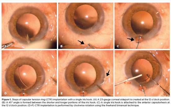
Arq. Bras. Oftalmol. 2026;89 (1 )
:1-5
| DOI: 10.5935/0004-2749.2025-0045
Abstract
PURPOSE: To evaluate the effect of using a single iris retractor, affixed to the anterior capsulorhexis at the 12 o'clock position, on the ease of capsular tension ring implantation.
METHODS: This prospective comparative study comprised 37 patients with zonular weakness attributed to pseudoexfoliation syndrome who underwent capsular tension ring implantation during cataract surgery. In Group 1, a single iris retractor was inserted into the anterior capsulorhexis at the 12 o'clock position. Group 2 did not receive this intervention. Zonular weakness was graded on a scale of 1–5, and the subjective difficulty of capsular tension ring implantation was categorized as easy, medium, or difficult.
RESULTS: Group 1 and 2 comprised 20 and 17 patients, respectively. There were no significant differences between the groups in age, sex distribution, and presence of glaucoma (p=0.53, p=0.28, and p=1.00, respectively). The mean zonular weakness score was significantly higher in Group 1 (3.35 ± 0.45) than in Group 2 (2.71 ± 0.59; p=0.02). Capsular tension ring implantation was significantly easier in the iris retractor group (p<0.001).
CONCLUSIONS: Placement of a single iris retractor attached to the anterior capsulorhexis at the 12 o'clock position may facilitate easier capsular tension ring implantation, even in patients with greater zonular weakness. This technique could reduce the risk of capsular tension ring displacement into the iridocorneal angle or ciliary sulcus.
Keywords: Capsular tension ring; Cataract; Iris hook; Pseudoexfoliation syndrome; Zonular weakness; Cataract extraction; Phacoemulsification; Capsulorhexis.
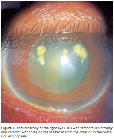
Arq. Bras. Oftalmol. 2021;84 (2 )
:158-162
| DOI: 10.5935/0004-2749.20210024
Abstract
OBJETIVO: Avaliar o momento apropriado para implante de anel de tensão capsular em casos de fraqueza zonular devida à síndrome pseudoesfoliativa.
MÉTODOS: Este foi um estudo prospectivo e comparativo realizado no Departamento de Oftalmologia da Universidade İnönü. Foram incluídos 43 pacientes, sendo 16 no grupo 1 e 27 no grupo 2. O grupo 1 era composto de pacientes que se submeteram ao implante precoce do anel de tensão capsular, enquanto no grupo 2 os pacientes tiveram implante tardio. Foram incluídos pacientes com síndrome pseudoesfoliativa submetidos à cirurgia de facoemulsificação e ao implante de lente intraocular na câmara posterior e anel de tensão capsular. Em cada olho, foram avaliadas as complicações intraoperatórias e as dificuldades tanto com a implantação do anel de tensão capsular quanto com a remoção do córtex.
RESULTADOS: Não houve diferença significativa entre os grupos quanto à dificuldade de implante do anel de tensão capsular (p=0,124). Ao se comparar as remoções do córtex, observou-se diferença significativa entre os grupos (p=0,003). Complicações intraoperatórias foram observadas em 3 pacientes do grupo 1 e 11 pacientes do grupo 2; porém, não houve diferença significativa entre os grupos (p=0,18). No grupo 2, observaram-se flutuações da cápsula posterior em 8 pacientes (29,5%), com ruptura da cápsula posterior em dois deles.
CONCLUSÕES: A remoção do córtex é mais difícil no implante precoce do anel de tensão capsular e flutuações da cápsula posterior podem causar problemas no implante tardio do anel de tensão capsular. O cirurgião deve ponderar a relação risco/benefício do implante precoce e tardio ao avaliar o momento ideal para implante de anel de tensão capsular.
Keywords: Catarata; Facoemulsificação; Anel de tensão capsular
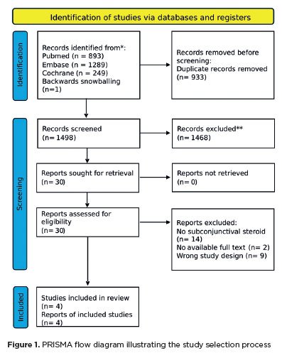
Arq. Bras. Oftalmol. 2025;88 (6 )
:1-8
| DOI: 10.5935/0004-2749.2024-0394
Abstract
The advantages and disadvantages of using perioperative subconjunctival steroid injections in dropless cataract surgery continue to be debated. A systematic review of PubMed, EMBASE, and the Cochrane Central database identified five studies—two randomized controlled trials and three non-randomized studies—encompassing 70,751 eyes. Among these, 12,319 eyes (17.4%) received subconjunctival steroid injections, while 58,432 eyes (82.6%) were managed with topical steroids. The Cochrane Collaboration’s RoB 2 tool was applied for bias assessments in randomized controlled trials, and heterogeneity was assessed using the I² statistics. No statistically significant differences were found between the two groups regarding macular edema (p=0.249), visual acuity (p=0.73), or laser flare count (p=0.45). Both subconjunctival injections and topical steroids demonstrated comparable efficacy and safety in controlling postoperative inflammation after cataract surgery. Additional research is warranted to validate these conclusions.
Keywords: Cataract extraction; Phacoemulsification; Lens implantation, intraocular; Postoperative care; Intravitreal injections; Anti-inflammatory agents, non-steroidal/administration & dosage; Glucocorticoids; Triamcinolone acetonide; Research design; Randomiz
Arq. Bras. Oftalmol. 2024;87 (3 )
:1-8
| DOI: 10.5935/0004-2749.2022-0076
Abstract
MÉTODOS: Córneas humanas de treinamento disponibilizadas foram randomizadas em quatro grupos: Pachy-100 (profundidade de incisão = espessura corneana central - margem de segurança de 100 µm), Pachy-50 (margem de segurança de 50 µm), Pachy-0 (sem margem de segurança) e Pachy+50 (profundidade de incisão = espessura corneana central + 50 µm). Todas as lamelas foram dissecadas através um método padronizado e já publicado (Pachy-DSEK). As espessuras das lamelas (centro, zona de 3,0mm e zona de 6,0mm) foram medidas com tomografia de coerência óptica. A razão de espessura centro-periferia foi calculada aos 3,0 e 6,0 mm de diâmetro.
RESULTADOS: Perfuração endotelial ocorreu apenas no grupo Pachy+50 (n=3, 30%). A espessura central da lamela nos grupos Pachy-100, Pachy-50, Pachy-0 e Pachy+50 foi de 185 ± 42 µm, 122 ± 29 µm, 114 ± 29 µm, e 58 ± 31 µm, respectivamente (p<0,001). As razões C/P aos 3,0 e 6,0 mm foram de 0,97 ± 0,06 e 0,92 ± 0,14, respectivamente. Os parâmetros de características do doador não se correlacionaram com os resultados de espessura de lamela. A profundidade planejada de incisão se correlacionou com a maioria dos parâmetros de espessura de lamela (p<0,001). A espessura de lamela se correlacionou negativamente com a profundidade planejada da incisão (p<0.001, r=-0,580). O melhor resultado foi observado no grupo Pachy-0, em que 75% das lamelas mediram abaixo de 130 µm e não houve perfuração endotelial.
CONCLUSÃO: Através de um método padronizado de dissecção, a maioria das lamelas endoteliais apresentou uma configuração planar. O planejamento de profundidade de incisão igual à espessura corneana central resultou em alta porcentagem de lamelas ultrafinas sem ocorrência de perfuração.
Keywords: Transplante de córnea; Ceratoplastia lamelar; Endotélio corneano; Dissecção; Tomografia de coerência óptica
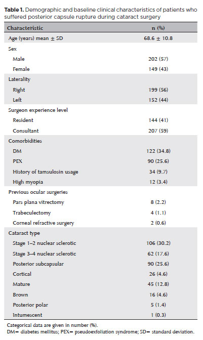
Arq. Bras. Oftalmol. 2025;88 (5 )
:1-8
| DOI: 10.5935/0004-2749.2024-0328
Abstract
PURPOSE: Posterior capsule rupture is defined as an intraoperative posterior capsule tear resulting in vitreous loss. This study aimed to analyze the clinical characteristics, preoperative risk factors, intraoperative management strategies, and postoperative complications associated with posterior capsule rupture during phacoemulsification surgery.
METHODS: This was a retrospective observational cohort study of the medical records for 25,224 phacoemulsification surgeries performed at our tertiary eye care center between 2017 and 2022. We collected and collated the demographic characteristics and clinical findings of the patients in our cohort. Intraoperative management strategies and postoperative outcomes over a 1-year followup period were also recorded.
RESULTS: Posterior capsule rupture occurred in 351 eyes (351 patients), giving an overall posterior capsule rupture rate of 1.3%. The mean patient age was 68.6 ± 10.8 years. Pseudoexfoliation syndrome, mature cataracts, brown cataracts, and surgery performed by a resident were identified as risk factors for posterior capsule rupture (p<0.05 for each; the risk ratios were 2.70, 2.15, 2.44, 1.34, respectively). The most common intraoperative complications were dislocated lens fragments in the vitreous (8%) and iris damage (7.1%). The mean best-corrected visual acuity improved from 1.31 ± 0.84 (logMAR) postoperatively to 0.51 ± 0.56 at the end of the 1-year follow-up period (p<0.001). Corneal edema (55.6%) and elevated intraocular pressure (33.3%) were the most common early postoperative complications. Persistently elevated intraocular pressure (11.1%) and cystoid macular edema (5.1%) were the most common late postoperative complications.
CONCLUSION: Posterior capsule rupture is a common complication of phacoemulsification surgery that requires prolonged postoperative follow-up and a multidisciplinary approach. Despite the increased incidence of complications when rupture occurs, appropriate intraoperative and postoperative management can lead to satisfactory visual outcomes.
Keywords: Cataract extraction; Phacoemulsification; Posterior capsule rupture; Corneal edema; Risk factors; Postoperative complications; Intraoperative complications
Arq. Bras. Oftalmol. 2025;88 (5 )
:1-7
| DOI: 10.5935/0004-2749.2024-0368
Abstract
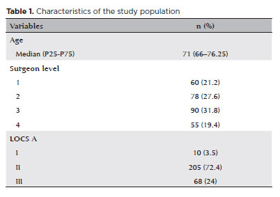
PURPOSE: To compare endothelial corneal cell changes following cataract surgery performed by phacoemulsification with intraocular lens implantation, conducted by surgeons with varying levels of experience.
METHODS: Two hundred and eighty-three eyes diagnosed with cataract were included. Lens opacity was classified into three categories (I, II, and III). Surgeons were categorized into four experience levels (1, 2, 3, and 4), based on years of practice and lifetime surgeries performed. Corneal endothelial characteristics were assessed using non-contact specular microscopy, with measurements taken before surgery and 30-60 days post-surgery.
RESULTS: Pre- and postoperative endothelial analysis showed no significant differences between surgeon levels regarding visual acuity achieved, corneal thickness, and endothelial hexagonality. However, the central endothelial cell density index showed a significantly greater reduction among level 1 surgeons (p=0.026). Grade II cataracts exhibited significant variations in the central endothelial cell density (p=0.011) and average cell size, with level 1 surgeons showing the largest increases (p=0.024).
CONCLUSIONS: The analysis revealed significant differences in visual acuity and endothelial indices between surgeon experience levels, with less experienced surgeons showing greater variations and poorer performance. Clinical protocols should consider these data to establish safer training protocols.
Keywords: Cataract extraction; Phacoemulsification; Endothelium; corneal; Lens implantation, intraocular; Visual acuity; Internship and residency; Surgeons
Arq. Bras. Oftalmol. 2025;88 (3 )
:1-5
| DOI: 10.5935/0004-2749.2024-0084
Abstract
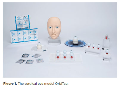
PURPOSE: The OrbiTau surgical simulator is a synthetic eye model developed to enhance cataract surgical training. Herein, we aimed to describe the perspectives of Harvard’s Ophthalmology faculty and residents regarding the effectiveness of OrbiTau.
METHODS: A cross-sectional study was conducted in which 11 surgeons from the Massachusetts Eye and Ear Infirmary, with prior experience utilizing simulated phacoemulsification platforms, conducted cataract surgery with the OrbiTau. Subsequently, they completed a satisfaction questionnaire using the Likert scale.
RESULTS: Regarding the various OrbiTau components, 90.90% of the participants reported that the OrbiTau lens capsule was comparable to that of the human lens during capsulotomy. Furthermore, 72.72% of the participants found that the OrbiTau lens consistency was analogous to that of the human lens nucleus. Approximately 63.63% of the participants reported that the model’s posterior lens capsule resembled the native posterior capsule, and 72.72% of the participants noted that the model’s red reflex was similar to that of the dilated human pupil. Most participants believed that the OrbiTau was easier to use and more realistic than other commercially available simulators.
CONCLUSION: Our single-institution survey of the Orbitau demonstrated that this model realistically replicates ocular structures and may be a viable option for cataract surgery training.
Keywords: Cataract extraction/education; Simulation training/methods; Ophthalmology/education; Phacoemulsification/education; Ophthalmologists/education; Surgeons/education; High fidelity simulation training
Arq. Bras. Oftalmol. 2024;87 (3 )
:1-5
| DOI: 10.5935/0004-2749.2023-0038
Abstract
PURPOSE: To assess the effect of the coronavirus disease 2019 (COVID-19) pandemic on cataract surgery by residents who had mandatory surgical simulator training during residency.
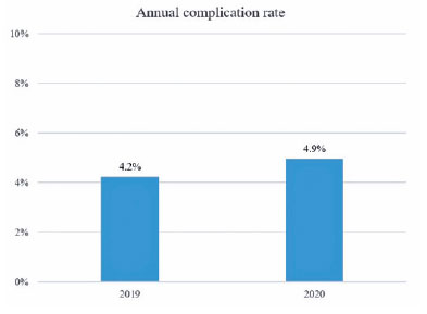
METHODS: In this retrospective, observational analytical study, the total number of cataract surgeries and surgical complications by all senior residents of 2019 (2019 class; prepandemic) and 2020 (2020 class; affected by the reduced number of elective surgeries due to the COVID-19 pandemic) were collected and compared. All residents had routine mandatory cataract surgery training on a virtual surgical simulator during residency. The total score obtained by these residents on cataract challenges of the surgical simulator was also evaluated.
RESULTS: The 2020 and 2019 classes performed 1275 and 2561 cataract surgeries, respectively. This revealed a reduction of 50.2% in the total number of procedures performed by the 2020 class because of the pandemic. The incidence of surgical complications was not statistically different between the two groups (4.2% in the 2019 class and 4.9% in the 2020 class; p=0.314). Both groups also did not differ in their mean scores on the simulator’s cataract challenges (p<0.696).
CONCLUSION: Despite the reduction of 50.2% in the total number of cataract surgeries performed by senior residents of 2020 during the COVID-19 pandemic, the incidence of surgical complications did not increase. This suggests that surgical simulator training during residency mitigated the negative effects of the reduced surgical volume during the pandemic.
Keywords: COVID-19; Pandemics; Cataract extraction/education; Internship and residency/methods; Simulation training/methods; Phacoemulsification/education; Surgery, computer-assisted; Computer simulation; Clinical competence; Ophthalmology/education
 ABO is licensed under a Creative Commons Attribution-NonComercial 4.0 Internacional.
09-tab01.jpg)
03-tab01tb.jpg)
06-tab01tb.jpg)
14-tab01.jpg)
11-tab01tb.jpg)
06-tab01tb.jpg)
12-fig01.jpg)