Showing of 1 until 14 from 104 result(s)
Search for: Esotropia; Depth perception; Strabismus; Visual acuity; Treatment outcome
Abstract
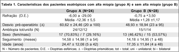
Objetivo: Análise dos resultados cirúrgicos da correção dos estrabismos horizontais em portadores de alta miopia, em pacientes do Departamento de Oftalmologia da Santa Casa de Misericórdia de São Paulo. Métodos: Foram estudados os prontuários de 24 pacientes esotrópicos e 17 exotrópicos, portadores de miopia maior que 6,00 DE operados para correção do estrabismo. Consideramos como bons resultados cirúrgicos desvios residuais entre esotropia e exotropia de 10delta. Resultados: Observou-se grande incidência de maus resultados entre os pacientes esotrópicos altos míopes. Conclusão: Concluímos que existe uma tendência a piores resultados cirúrgicos nos pacientes esotrópicos com miopia maior que -6,00 DE, em comparação com esotrópicos com erro refrativo entre -0,75 DE e +3,50 DE.
Keywords: Estrabismo; Miopia; Exotropia; Esotropia; Cirurgia
Abstract
PURPOSE: To analyze the quality of life and treatment adherence of patients with glaucoma at different disease stages, considering factors such as sex, visual acuity, disease severity, and treatment characteristics.
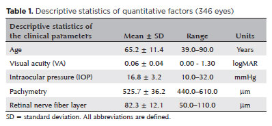
METHODS: This cross-sectional study included 174 patients (346 glaucomatous eyes) recruited from clinical records and routine follow-ups at a specialized ophthalmology center. Their mean age was 39–90 years, and 60.9% of them were women. Their quality of life and adherence were assessed using the NEI-VFQ25 and MMAS-8 questionnaires, respectively. Complementary tests included 24:2 visual field test, retinography, and optical coherence tomography. Patients diagnosed with glaucoma for at least 6 months were included, whereas pregnant patients and those with ocular diseases were excluded.
RESULTS: Among the participants, 59.2% adhered to the treatment whereas 40.8% showed low adherence. The mean quality of life score was 81.87. Patients with low adherence had slightly higher quality of life scores (mean 83.1) than those with good adherence (mean 81.0), but the difference was not statistically significant. Disease severity was associated with increased optic nerve cupping, reduced thickness of the nerve fiber and ganglion cell layers, and great visual field loss. No significant correlation was observed between adherence and quality of life, indicating the independence of these factors and the influence of psychological or social elements.
CONCLUSION: The absence of a correlation between quality of life and treatment adherence highlights the need for tailored interventions for psychological and social aspects. These findings indicate the importance of a comprehensive approach to managing glaucoma, preserving visual function, strengthening doctor–patient relationships, and considering psychosocial factors to enhance quality of life and treatment adherence.
Keywords: Glaucoma; Quality of life; Patient health questionnaire; Patient acuity; Antiglaucoma agents; Visual acuity; Treatment adherence and compliance; Surveys and questionnaires
03-tab01.jpg)
Abstract
Objetivo: Investigar o impacto de diferentes tamanhos de incisões em córnea clara com meridiano íngreme para facoemulsificação com aberrações de mais alta ordem da córnea anterior.
Métodos: Foram retrospectivamente revisados os prontuários médicos de pacientes que se submeteram a cirurgias de catarata com microincisões coaxiais de 2,2 mm ou com incisões coaxiais pequenas de 2,75 mm. Foram apenas incluídos pacientes com astigmatismo preexistente da córnea anterior <2,00 dioptrias (D) e ≥0,50 D, e submetidos a incisões em córnea clara com meridiano íngreme. Os desfechos primários foram aberrações da córnea anterior da 3ª à 6ª ordem com uma pupila de 8 mm. O astigmatismo da córnea anterior e o tempo efetivo de facoemulsificação foram avaliados como desfechos secundários. Os desfechos pré-operatório e pós-operatório aos 3 meses também foram avaliados.
Resultados: O astigmatismo da córnea anterior diminuiu significativamente após ambos os procedimentos, mas não se encontrou nenhuma diferença significativa entre os dois procedimentos quanto ao astigmatismo da córnea anterior, induzido pela cirurgia (p=0,146). Embora as aberrações totais de mais alta ordem não se tenham alterado significativamente após ambos procedimentos, a comparação entre os grupos revelou uma diferença significativa nas aberrações totais de mais alta ordem, induzidas pela cirurgia (uma diminuição de 0,337 ± 1,156 μm na cirurgia de catarata por microincisão coaxial de 2,2 mm e um aumento de 0,106 ± 0,521 μm na cirurgia de catarata por incisão coaxial pequena de 2,75 mm; p=0,046). A aberração esférica diminuiu significativamente após cirurgia de catarata por microincisão coaxial de 2,2 mm (p=0,001), mas não se alterou significativamente após cirurgia de catarata por incisão coaxial pequena de 2,75 mm (p=0,564). A aberração de coma não mudou significativamente após qualquer dos procedimentos. O trifólio não se alterou significativamente após cirurgia de catarata por microincisão coaxial de 2,2 mm (p=0,361), mas aumentou significativamente após cirurgia de catarata por incisão coaxial pequena de 2,75 mm (p<0,001). Nenhuma diferença significativa se evidenciou quanto ao tempo efetivo de faco-emulsificação entre os dois procedimentos. Houve uma correlação positiva significativa entre o astigmatismo da córnea anterior, induzido pela cirurgia e a aberração de coma na cirurgia de catarata por incisão coaxial pequena de 2,75 mm (r=0,387, p=0,006). Não foi encontrada correlação significativa entre as alterações nas aberrações totais de mais alta ordem, induzidas pela cirurgia e o tempo efetivo de faco-emulsificação.
Conclusões: Nem a cirurgia de catarata por microincisão coaxial de 2,2 mm, nem aquela por incisão coaxial pequena de 2,75 mm degradaram significativamente as aberrações totais de mais alta ordem da córnea anterior. Porém, as alterações nas aberrações totais de mais alta ordem, induzidas pela cirurgia mostraram uma diferença significativa entre os dois procedimentos, com uma ligeira redução na cirurgia de catarata por microincisão coaxial de 2,2 mm e um pequeno aumento na cirurgia de catarata por incisão coaxial pequena de 2,75 mm. O tempo de facoemulsificação e a potência utilizada durante a cirurgia não tiveram impacto nas aberrações corneanas.
Keywords: Facoemuslificação; Astigmatismo; Cornea/cirurgia; Ferida cirúrgica; Resultado de tratamento
04-tab01tb.jpg)
Abstract
Objetivo: Verificar se pacientes com dislexia do desenvolvimento (DD) apresentam déficits coerentes com uma disfunção magnocelular visual.
Métodos: Participantes com diagnóstico confirmado de dislexia do desenvolvimento (n=62; faixa etária=8 a 25 anos; Média da idade=13.8 anos, desvio padrão=3.9; 77% homens) foram comparados a um grupo controle com desenvolvimento típico, pareado por idade, sexo, dominância ocular, acuidade visual e compreensão de texto. A perimetria Frequency-Doubling Technology avaliou o limiar de sensibilidade ao contraste do campo visual periférico. O rastreador ocular Visagraph-III registrou os movimentos dos olhos durante leitura de texto.
Resultados: O grupo com dislexia do desenvolvimento apresentou piores limiares de sensibilidade no Frequency-Doubling Technology, com tamanho de efeito forte, do que o grupo controle. O grupo com dislexia do desenvolvimento apresentou mais olhos classificados com déficits na sensibilidade à ilusão de frequência duplicada do que o grupo controle. O grupo com dislexia do desenvolvimento apresentou pior habilidade motora ocular e no desempenho de leitura, revelado pela diferença entre os grupos em relação às fixações oculares, regressões, alcance de reconhecimento, taxa de leitura e eficiência relativa. Foi encontrada correlação significativa entre a sensibilidade ao contraste e as habilidades motoras oculares. Os participantes com boa eficiência relativa apresentaram uma sensibilidade ao contraste significativamente melhor do que os participantes com baixa eficiência relativa.
Conclusões: O grupo com dislexia do desenvolvimento apresentou desempenho inferior nas variáveis visuais relacionadas à função visual magnocelular (i.e., perimetria de frequência duplicada e habilidades motoras oculares), quando comparado ao grupo controle pareado. Os profissionais precisam estar cientes da importância de investigar a visão dos pacientes com dislexia do desenvolvimento além da acuidade visual e incluir nos seus procedimentos diagnósticos instrumentos para avaliar o processamento temporal, com limiar de sensibilidade ao contraste.
Keywords: Dislexia; Leitura; Percepção visual; Transtornos da visão; Músculos oculomotores; Movimentos oculares
Abstract
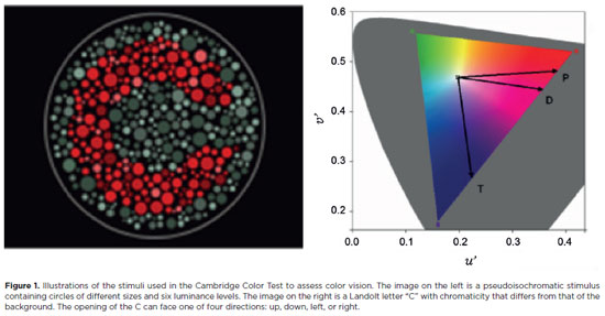
PURPOSE: Amblyopia is a cortical neurological disorder caused by abnormal visual experiences during the critical period for visual development. Recent works have shown that, in addition to the well-known visual alterations, such as changes in visual acuity, several perceptual aspects of vision are affected. This study aims to analyze and compare the effects of different types of amblyopia on visual color processing and determine whether these effects are correlated with visual acuity.
METHODS: Our study sample comprised 42 amblyopic individuals, aged 7-40 years, (strabismus, n=16; anisometropia, n=18; and mixed-cause, n=8) and 33 age-matched controls. Color vision was tested by measuring the chromaticity threshold of each patient on the protan, deutan, and tritan axes using version 02 of the Cambridge Color Test. Spatial stimulation cues were eliminated using spatial noise and luminance.
RESULTS: The color discrimination thresholds on the protan, deutan, and tritan axes were similar between control participants and amblyopic patients (p>0.05). There was no correlation between VA values and color thresholds (p>0.05).
CONCLUSION: Patients with amblyopia have normal color vision in contexts that include luminance and spatial noise. Our results may be indicative of independent neural pathways for spatial and chromatic visual processing.
Keywords: Amblyopia; Anisometropia; Color vision; Strabismus; Vision disorders; Visual acuity
11-fig01.jpg)
Abstract
Objetivo: Reportar a curva de aprendizado dos 2 anos iniciais da trabeculotomia transluminal assistida por gonioscopia, usando a técnica de sutura termicamente atenuada e revisar os fatores que podem afetar o resultado.
Métodos: Este estudo retrospectivo incluiu 100 olhos de 89 participantes com glaucoma resistente ao tratamento clínico máximo, definido como tendo pressão intraocular superior a 21mmHg, além de três ou quatro drogas hipotensoras diferentes. Pressão intraocular inicial, 1 semana, primeiro, segundo, terceiro, sexto, 12 e 24 meses de acompanhamento; necessidade de medicação antiglaucoma; necessidade de mais cirurgias anti-glaucomatosas foram registradas. Olhos que necessitaram de intervenção cirúrgica adicional para o controle da pressão intraocular foram considerados como insucesso.
Resultados: Cinquenta e um olhos foram submetidos à trabeculotomia transluminal assistida por gonioscopia isolado e 49 olhos à trabeculotomia transluminal assistida por gonioscopia associado à extração de catarata no mesmo tempo cirúrgico. Houve diferença estatisticamente significativa entre a pressão intraocular média global no acompanhamento e a pressão intraocular média pré-operatória (p<0,001) em todas as visitas do acompanhamento. Ao avaliar a extensão do tratamento, os pacientes com extensão de 360 graus não apresentaram pressão intraocular média menor estatisticamente significativa em comparação com outras extensões. O hifema foi a única complicação presente em 50 olhos (50%), contudo todos tiveram resolução espontânea em quatro semanas. Um total de 26 olhos (26%) teve que ser submetido a trabeculectomia convencional adicional devido à pressão intraocular descontrolada, principalmente aqueles previamente submetidos à cirurgia vitreorretiniana.
Conclusões: A trabeculotomia transluminal assistida por gonioscopia, além de ser um procedimento aparentemente seguro, apresenta taxas de sucesso satisfatórias, mesmo durante a curva de aprendizado inicial do cirurgião. A técnica foi efetiva em reduzir a pressão intraocular e uso de medicamentos.
Keywords: Trabeculotomia/métdos; Glaucoma de ângulo aberto/cirurgia; Gonioscopia/métodos; Resultado de tratamento
Abstract
PURPOSE: This study aimed to evaluate the outcomes of strabismus surgical correction in patients with Down syndrome.
METHODS: We conducted a retrospective chart review of patients with Down syndrome who underwent strabismus surgery between January 1997 and May 2024 at an Ophthalmology Outpatient Clinic in São Paulo, Brazil. The data collected included age, sex, medical and ocular history, surgical details, and follow-up outcomes. The patients were categorized by strabismus type into esotropia, fourth nerve palsy, and mixed groups. Surgical success was defined as final alignment within 10Δ of orthotropia and, where applicable, whether there was resolution of abnormal head posture of ocular origin. Patients with postoperative follow-up <6 months were excluded.
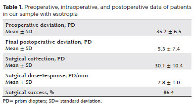
RESULTS: A total of 37 patients (21 females) were included. Of these, 22 (59.5%) were in the esotropia group, 10 (27.0%) in the fourth nerve palsy group, and 5 (13.5%) in the mixed group. The surgical success rate in the esotropia group was 86.4%, with a mean preoperative deviation of 35.2 (± 6.5)Δ, and mean surgical correction of 30.1 (± 10.4)Δ. The success rate in the fourth nerve palsy group was 40.0%, with a mean preoperative deviation of 10.4 (± 4.3)Δ. Overall, success was achieved with a single surgical procedure in 73.0% of the sample. No significant associations were found between surgical success and the clinical and demographic variables, including sex, age at surgery, oblique muscle overaction, pattern strabismus, visual acuity, amblyopia, preoperative deviation, or postoperative follow-up duration (p>0.05).
CONCLUSIONS: When standard surgical tables are applied, strabismus surgery in patients with Down syndrome appears to be safe and effective. We found high success rates, particularly among patients with esotropia. We observed no tendencies toward over- or under-correction. These findings support the use of conventional surgical protocols with this patient population.
Keywords: Down Syndrome/complications; Strabismus/surgery; Esotropia/surgery; Oculomotor nerve diseases/physiopathology; Vision disorders; Humans; Brazil.
Abstract
PURPOSE: Glaucoma is a chronic and progressive disease that requires long-term treatment and continuous monitoring. The Kahook Dual Blade, a device used to perform goniotomy in adults, is designed to improve intraocular pressure control in patients with glaucoma. This study aimed to evaluate the long-term efficacy and safety of kahook dual blade goniotomy in glaucoma patients undergoing cataract surgery over a 36-month follow-up.
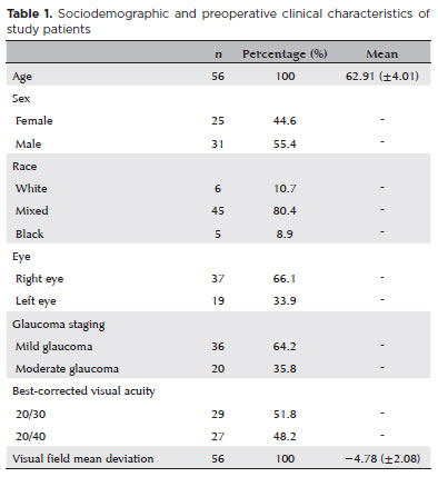
METHODS: This was a retrospective case series including 56 eyes from 56 patients with mild-to-moderate primary open-angle glaucoma who underwent phacoemulsification combined with kahook dual blade goniotomy. Mean intraocular pressure values, number of preoperative and postoperative hypotensive eye drops, procedure survival, and complications were evaluated over 36 months. Surgical success was defined as either a reduction in intraocular pressure of ≥20% with intraocular pressure between 6 and 18 mmHg without additional medication or a reduction of ≥1 eye drop with intraocular pressure between 6 and
18 mmHg.
RESULTS: The mean preoperative intraocular pressure decreased from 15.96 ± 2,83) mmHg to 13.14 ± 2,11) mmHg at 36 months, representing a 14.9% reduction (p<0.001). The mean number of eye drops decreased from 1.91 ± 0,75) to 1.34 ± 0,92), a 29.8% reduction (p<0.001). The overall success rate was 69.6% at 36 months.
CONCLUSION: Kahook dual blade goniotomy combined with cataract surgery significantly reduced intraocular pressure and the number of hypotensive eye drops required in patients with mild-to-moderate primary open-angle glaucoma, with a favorable success rate maintained at 36 months.
Keywords: Glaucoma, open-angle/surgery; Gonioscopy/methods; Intraocular pressure/physiology; Lens implantation, intraocular; Phacoemulsification/methods; Trabeculectomy/instrumentation; Treatment outcome
Abstract
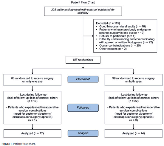
PURPOSE: This prospective, randomized, unmasked, clinical trial aimed to report the visual outcomes of cataract surgery on both eyes versus cataract surgery on one eye in Brazilian patients.
METHODS: This study included patients with bilateral cataracts and binocular visual acuity worse than or equal to 0.3 logarithm of the minimum angle of resolution. The patients were randomly assigned to undergo surgery on one (Control Group) or both eyes (one eye at a time; Intervention Group). Postoperatively, self-reported visual function using Catquest-9SF (primary outcome measure), binocular visual acuity, stereopsis, and ocular dominance (secondary outcome measures) were compared.
RESULTS: A total of 151 patients (77 and 148 eyes in the Control and Intervention Groups, respectively) completed the follow-up. Patients who underwent surgery on both eyes exhibited significantly better self-reported visual function (p=0.036) and stereopsis (p=0.026) than those who underwent surgery on one eye. Binocular visual acuity and ocular dominance did not affect the group comparisons.
CONCLUSIONS: Surgery on both eyes resulted in significantly better self-reported visual function and stereopsis than surgery on one eye.
Keywords: Cataract; Cataract extraction; Quality of life; Treatment outcome; Visual acuity; Binocular vision; Stereopsis
Abstract
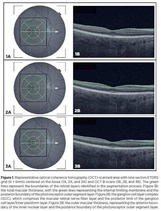
PURPOSE: This study aimed to evaluate the total macular thickness as well as the thickness of the inner and outer retinal layers in patients with Parkinson's disease. It also aimed to verify the correlation of these parameters with motor symptoms and cognitive function.
METHODS: A total of 46 eyes of 23 patients with Parkinson's disease and 40 eyes of 20 healthy controls were included in the study. The patients' cognitive, functional, and nonmotor symptoms were evaluated using the Katz Index of Independence and Pfeffer's Activities of Daily Living, Mini-Mental State Examination, Frontal Assessment Battery, Schwab and England Staging Scales, and Movement Disorders Society Nonmotor Symptoms Scale. The macular thickness measurements obtained via total, inner, and outer optical coherence tomography were recorded. Furthermore, the correlation of the parameters of optical coherence tomography with cognitive, functional, and nonmotor symptoms was assessed.
RESULTS: The scores of the Katz Index of Independence and Pfeffer's Activities of Daily Living as well as the Movement Disorders Society Nonmotor Symptoms Scale were significantly lower in patients with Parkinson's disease than in healthy controls. Moreover, the former had greater total macular thickness. The temporal and inferior outer sectors were significantly greater for the ganglion cell complex thickness in patients. A significant correlation was observed between the total macular thickness and the Movement Disorder Society-Unified Parkinson's Disease Rating Scale, Parte III (MDS-UPDRS-III) values. Contrarily, there was a negative correlation between the outer macular thickness and the MDS-UPDRS-III values. Meanwhile, the total macular thickness and ganglion cell complex thickness were significantly correlated with the scores of the Mini-Mental State Examination, Schwab and England Staging Scale, Frontal Assessment Battery, and Katz Index of Independence and Pfeffer's Activities of Daily Living. In addition, the Schwab and England scale was correlated with the outer macular thickness.
CONCLUSION: The total and inner macular thicknesses at the temporal and inferior outer sectors were greater in patients with Parkinson's disease than in the control group. These findings indicate that macular thickness may be greater in those with Parkinson's disease, particularly when associated with mild motor symptoms. In addition, the parameters of the total, inner, and outer optical coherence tomography were significantly associated with motor and nonmotor symptoms as well as cognitive function impairment.
Keywords: Parkinson's disease; Tomography, optical coherence; Neurodegenerative diseases; Cognitive dysfunction; Cognition; Motor perception; Visual acuity; Retina
11-tab01.jpg)
Abstract
OBJETIVO: Associar os resultados refrativos a longo prazo da cirurgia de catarata e a função visual autorreferida pelo questionário Catquest-9SF.
MÉTODOS: Paciente recrutados no ambulatório de catarata da VER MAIS Oftalmologia, foram submetidos a exame oftalmológico completo. Após diagnóstico de catarata com indicação de tratamento cirúrgico com facoemulsificação e implante de lente intraocular, o questionário foi aplicado antes da intervenção, 30 dias após cirurgia e 1 ano após, novamente.
RESULTADOS: Foram recrutados 133 pacientes. No decorrer do seguimento, 32 pacientes foram perdidos e ao final foram analisados os dados de 101 pacientes, dos quais 48 foram homens e 53 foram mulheres. A variância bruta explicada por dados foi de 69,9% e a inexplicada em primeiro contraste por 2,39 eigenvalores, sendo maior que 2, o que nos mostra que são resultados diferentes dos esperados de dados aleatórios. O índice de separação de pessoas foi de 2.95 (>2) e o valor de confiança de pessoas foi de 0,9 (>0,8). Estes índices são os valores mínimos aceitáveis na diferenciação de níveis de habilidade. Acuidade visual foi a principal variável correlacionada com o score do Catquest.
CONCLUSÕES: O Catquest-9SF traduzido para o português se demonstrou unidimensional e uma ferramenta psicometricamente válida para avaliar disfunção visual em pacientes com catarata, além de ter tido sucesso para quantificar objetivamente melhoras após a intervenção cirúrgica. Essa ferramenta pode ser utilizada como preditiva e concordante da melhora da acuidade visual.
Keywords: Extração de catarata; Acuidade visual; Inquéritos e questionários; Qualidade de vida; Medidas de resultados relatados pelo paciente
13-tab01tb.jpg)
Abstract
OBJETIVO: Avaliar as alterações precoces após a primeira injeção de anticorpos antifator de crescimento endotelial vascular (anti-VEGF) em casos de edema macular secundário à retinopatia diabética e oclusão da veia da retina e a relação entre essas alterações e o resultado a longo prazo.
MÉTODOS: Foram incluídos no estudo pacientes que receberam uma injeção de antifator de crescimento endotelial vascular para edema macular, virgem de tratamento e devido à oclusão da veia retiniana ou a retinopatia diabética. A espessura macular central foi medida no início do tratamento e no 1º dia, 2ª semana e 1º mês após a injeção, bem como na última visita, através de tomografia de coerência óptica de domínio espectral. Definiu-se uma “boa resposta” como uma redução ≥10% na espessura macular central no 1º dia após a injeção. Os pacientes foram reavaliados na última visita com relação à resposta ao tratamento no 1º dia após a injeção, com base em um resultado anatômico favorável, definido como uma espessura macular central <350 µm.
RESULTADO: Foram registrados 26 (44,8%) pacientes com edema macular e oclusão da veia da retina e 32 (55,2%) com edema macular e retinopatia diabética. O tempo médio de acompanhamento foi de 24,0 meses (desvio-padrão de 8,5 meses). Foi observada uma diminuição estatisticamente significativa da espessura macular central após o tratamento antifator de crescimento endotelial vascular tanto em pacientes com edema macular e oclusão da veia retiniana quanto naqueles com edema macular e retinopatia diabética (p<0,001 para ambos). Todos os pacientes com edema macular e oclusão da veia retiniana responderam bem no 1º dia pós-injeção. Todos os que responderam mal no 1º dia pós-injeção pertenciam ao grupo com edema macular e retinopatia diabética (n=16,50%). A presença de manchas hiperrefletivas foi maior nos pacientes que responderam mal do que naqueles que tiveram boa resposta no grupo com edema macular e retinopatia diabética (p=0,03). Um dos 42 (2,4%) pacientes com boa resposta total teve espessura macular central >350 µm, enquanto 5 (31,2%) do total de 16 pacientes com resposta ruim apresentaram espessura macular central >350 µm na última visita (p=0,003).
CONCLUSÃO: O resultado anatômico de longo prazo do edema macular secundário à oclusão da veia retiniana e à retinopatia diabética pode ser previsto pela resposta ao tratamento no 1º dia após a injeção de antifator de crescimento endotelial vascular.
Keywords: Edema macular; Retinopatia diabética; Diabetes mellitus; Oclusão da veia retiniana; Fator A de crescimento do endotélio vascular; Inibidores da angiogênese; Resultado do tratamento
Abstract
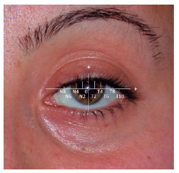
PURPOSE: Evaluation of lid contour and marginal peak point changes to compare outcomes of external levator advancement and Müller’s muscle conjunctival resection surgery in unilateral ptosis.
METHODS: We reviewed the charts of unilateral ptosis patients who underwent external levator advancement or Müller’s muscle conjunctival resection. Eyelid contour analysis was conducted on preoperative and 6-month postoperative digital images. This was performed with the multiple margin reflex distances technique, measuring the vertical distance from a line intersecting the center of the pupil to the eyelid margin at 10 positions at 2 mm intervals. The marginal peak point changes were analyzed digitally using the coordinates of the peak point according to the pupil center. Each position’s mean distance was compared preoperatively, postoperatively, and with the fellow eyelid.
RESULTS: Sixteen patients underwent external levator advancement and 16 patients had Müller’s muscle conjunctival resection. The mean margin reflex distance was improved by both techniques (1.46 vs. 2.43 mm and 1.12 vs. 2.25 mm, p=0.008 and p=0.0001 respectively) and approached that of the fellow eyelid (2.43 vs. 2.88 and 2.25 vs. 2.58 mm, p=0.23 and p=0.19, respectively). However, statistically significant lid margin elevation was limited to between the N6 and T6 points in the external levator advancement group. Whereas, significant elevation was achieved along the whole lid margin in the Müller’s muscle conjunctival resection group. The marginal peak point was shifted slightly laterally in the external levator advancement group (p=0.11).
CONCLUSIONS: Both techniques provide effective lid elevation, however, the external levator advancement’s effect lessens toward the canthi while Müller’s muscle conjunctival resection provides more uniform elevation across the lid margin. The margin reflex distance alone is not sufficient to reflect contour changes.
Keywords: Blepharoptosis; Eyelids; Conjunctiva; Oculomotor muscles; Image processing, computer-assisted; Treatment outcome
Abstract
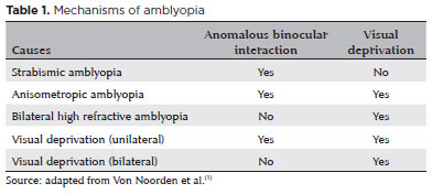
This study aimed to propose a guideline for amblyopia treatment and follow-up. Studies show that amblyopia leads to a series of perceptual deficits, including loss of visual acuity, stereoacuity, and contrast sensitivity. Perceptual changes are also found in the sound eye, such as those involving the types of motion perception. The gold standard of treatment remains the prescription of eyeglasses, when indicated, and patching of the dominant eye. The treatment is mostly effective in patients aged <7 years and must be discontinued gradually, tapering off patching for at least 5 weeks. Atropine may be performed for penalization in hyperopic children whose amblyopic eye has better visual acuity under cycloplegia than the fellow eye. The discovery of significant neural plasticity in the amblyopic brain after the critical period opens possibilities for new treatment modalities even after childhood.
Keywords: Amblyopia; Atropine; Contrast sensitivity; Motion perception; Eyeglasses; Visual acuity; Prescriptions
 ABO is licensed under a Creative Commons Attribution-NonComercial 4.0 Internacional.
About
Issues
Editorial Board
Submission
Arquivos Brasileiros de Oftalmologia
Official publication of Brazilian Council of Ophthalmology - Conselho Brasileiro de Oftalmologia (CBO)
Rua Casa do Ator, 1.117 - 2nd floor - Zip Code: 04546-004
São Paulo - SP, Brazil
TEL: +55 11 3266-4000
E-mail: [email protected]