Arq. Bras. Oftalmol. 2026;89 (2 )
:1-8
| DOI: 10.5935/0004-2749.2025-0105
Abstract
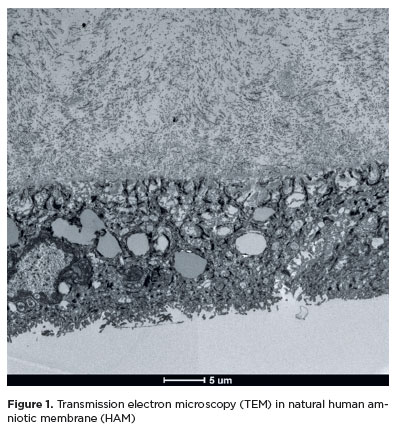
PURPOSE: To evaluate structural differences in amniotic membrane fragments subjected to different preservation techniques for potential ophthalmologic applications.
METHODS: Three placentas were collected from healthy donors, and four amniotic membrane fragments were prepared from each placenta. The fragments were divided into four groups with three samples each: cryopreserved, lyophilized, vacuum-dried using a vacuum concentrator, and fresh (control). After processing, the fragments were fixed, sectioned, and analyzed using scanning transmission electron microscopy to assess tissue morphology.
RESULTS: All samples met the established evaluation criteria. No morphological differences were observed among the groups. The structural characteristics of lyophilized and vacuum-dried membranes were comparable with those of cryopreserved and fresh membranes. However, vacuum drying demonstrated the greatest practicality for ophthalmologic use, as it allows membrane availability at any time and storage at room temperature.
CONCLUSION: Vacuum drying using a vacuum concentrator, lyophilization, and cryopreservation preserve the morphological characteristics of the human amniotic membrane similar to those of fresh tissue. A standardized protocol using a vacuum concentrator may be established owing to its advantages in storage convenience and accessibility.
Keywords: Amnion/transplantation; Cryopreservation/methods; Freeze drying; Lyophilization; Ophthalmologic surgical procedures; Regenerative medicine
Arq. Bras. Oftalmol. 2023;86 (3 )
:1-7
| DOI: 10.5935/0004-2749.20230020
Abstract
Objetivos: Blefaroptose e estrabismo podem ser coexistentes em adultos e ambos afetam a aparência estética e o domínio psicossocial. Ambos também geralmente requerem cirurgia, realizada tradicionalmente em uma abordagem sequencial. O objetivo do presente estudo foi avaliar a eficácia da execução simultânea da ressecção musculoconjuntival de Müller, com ou sem cirurgia de tarsectomia, e da cirurgia de estrabismo em pacientes adultos com ptose e estrabismo coexistentes.
Métodos: Foram retrospectivamente avaliados pacientes com ptose e estrabismo coexistentes submetidos simultaneamente à ressecção musculoconjuntival de Müller, com ou sem tarsectomia, e à cirurgia de estrabismo horizontal. A análise incluiu a mensuração do ângulo de desvio das dioptrias de prisma, a distância do reflexo à margem, a assimetria da altura palpebral e quaisquer complicações após a cirurgia. A ressecção musculoconjuntival de Müller, com ou sem sucesso na tarsectomia, foi considerada bem-sucedida com uma distância reflexo-margem medindo entre 3,5 e 5 mm, e uma diferença entre as duas pálpebras superiores menor que 1 mm. O sucesso da cirurgia de estrabismo foi definido como um alinhamento com ± 10 dioptrias prismáticas de ortotropia.
Resultados: Os pacientes foram 3 mulheres e 5 homens, com média de idade de 37,12 anos (faixa de 22 a 62 anos). A parte de estrabismo da cirurgia foi realizada primeiro em todos os pacientes. Os resultados da simetria palpebral superior foram avaliados como perfeitos (<0,5 mm) em 4 pacientes, bons (≥0,5 mm, <1 mm) em 4 pacientes e regulares (≥1 mm) em nenhum. A ressecção musculoconjuntival de Müller, com ou sem sucesso na tarsectomia, teve sucesso em 6 dos 8 pacientes (75%) e a intervenção para o estrabismo foi bem-sucedida em todos os pacientes. Não foi necessária cirurgia de revisão da pálpebra ou do estrabismo após a cirurgia simultânea em nenhum paciente.
Conclusões: A ressecção musculoconjuntival de Müller, com ou sem tarsectomia, pode ser combinada com a cirurgia de estrabismo em uma abordagem alternativa para pacientes com ptose e estrabismo coexistentes.
Keywords: Blefaroptose/cirurgia; Ambliopia; Estrabismo/cirurgia; Músculos oculomotores/cirurgia; Pálpebras; Procedimentos cirúrgicos oftalmológicos/métodos
Arq. Bras. Oftalmol. 2021;84 (4 )
:374-379
| DOI: 10.5935/0004-2749.20210065
Abstract
Objetivo: Sincinesias são resultado de inervações anômalas e ocasionam movimentos aberrantes dos músculos envolvidos. Apresentamos uma série com casos raros de sincinesias oculares congênitas dos músculos extraoculares e do levantador da pálpebra superior e especulamos a possibilidade de classificá-las dentro do espectro das desordens congênitas da desnervação cranianana.
Métodos: Prontuários de pacientes com diagnóstico de sincinesia ocular congênita foram estudados retrospectivamente. Analisamos sexo, lateralidade e as características completas do exame de motilidade de cada paciente.
Resultados: Nove pacientes com sincinesias oculares congênitas foram incluídos. Houve discreta predominância no sexo feminino. Em termos de lateralidade, o olho direito foi o único envolvido em 4 casos, o olho esquerdo também em 4 casos e 1 caso apresentou acometimento bilateral. 55,5% dos pacientes eram ortotrópicos na posição primária. Os III, VI e IV nervos participaram da sincinesia em 100%, 44,4% e 11,1% dos casos, respectivamente.
Conclusões: Sincinesias oculares congênitas podem se apresentar de modo bastante eclético e incomum. A inervação aberrante presente em cada um desses casos os coloca na lista de candidatos a integrar o grupo das desordens congênitas da desenervação craniana.
Keywords: Sincinesia; Nervo troclear; Nervos cranianos/ anormalidades; Músculos oculomotores; Transtornos da motilidade ocular/congênito
Arq. Bras. Oftalmol. 2026;89 (1 )
:1-6
| DOI: 10.5935/0004-2749.2025-0071
Abstract
PURPOSE: This study aimed to evaluate the outcomes of strabismus surgical correction in patients with Down syndrome.
METHODS: We conducted a retrospective chart review of patients with Down syndrome who underwent strabismus surgery between January 1997 and May 2024 at an Ophthalmology Outpatient Clinic in São Paulo, Brazil. The data collected included age, sex, medical and ocular history, surgical details, and follow-up outcomes. The patients were categorized by strabismus type into esotropia, fourth nerve palsy, and mixed groups. Surgical success was defined as final alignment within 10Δ of orthotropia and, where applicable, whether there was resolution of abnormal head posture of ocular origin. Patients with postoperative follow-up <6 months were excluded.
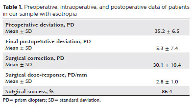
RESULTS: A total of 37 patients (21 females) were included. Of these, 22 (59.5%) were in the esotropia group, 10 (27.0%) in the fourth nerve palsy group, and 5 (13.5%) in the mixed group. The surgical success rate in the esotropia group was 86.4%, with a mean preoperative deviation of 35.2 (± 6.5)Δ, and mean surgical correction of 30.1 (± 10.4)Δ. The success rate in the fourth nerve palsy group was 40.0%, with a mean preoperative deviation of 10.4 (± 4.3)Δ. Overall, success was achieved with a single surgical procedure in 73.0% of the sample. No significant associations were found between surgical success and the clinical and demographic variables, including sex, age at surgery, oblique muscle overaction, pattern strabismus, visual acuity, amblyopia, preoperative deviation, or postoperative follow-up duration (p>0.05).
CONCLUSIONS: When standard surgical tables are applied, strabismus surgery in patients with Down syndrome appears to be safe and effective. We found high success rates, particularly among patients with esotropia. We observed no tendencies toward over- or under-correction. These findings support the use of conventional surgical protocols with this patient population.
Keywords: Down Syndrome/complications; Strabismus/surgery; Esotropia/surgery; Oculomotor nerve diseases/physiopathology; Vision disorders; Humans; Brazil.
Arq. Bras. Oftalmol. 2026;89 (3 )
:1-9
| DOI: 10.5935/0004-2749.2025-0312
Abstract
PURPOSE: To quantitatively assess changes in central corneal sensitivity after phacoemulsification and to characterize recovery patterns up to 90 days using standardized esthesiometry.
METHODS: This prospective observational study included 44 patients (88 eyes) undergoing uncomplicated phacoemulsification with intraocular lens implantation. Central corneal sensitivity was measured using a Cochet-Bonnet® esthesiometer preoperatively and at 30 and 90 days postoperatively. Repeated-measures data were analyzed using Friedman and Wilcoxon signed-rank tests (p<0.05). Inter-eye differences were assessed with a paired Wilcoxon test. Individual changes from baseline (Δ30, Δ90) were calculated, and 90-day recovery was categorized according to thresholds aligned with the 5-mm device resolution. Spearman correlation was used to explore associations between age and Δ90.
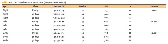
RESULTS: Corneal sensitivity decreased after surgery. In right eyes, mean sensitivity declined from 41.14 ± 7.77 mm at baseline to 36.82 ± 9.03 mm at 30 days and partially recovered to 38.64 ± 7.73 mm at 90 days. In left eyes, sensitivity decreased from 44.11 ± 6.29 mm to 37.39 ± 9.05 mm at 30 days and recovered to 41.82 ± 7.63 mm at 90 days. Left eyes showed higher sensitivity than right eyes at baseline (p=0.023) and at 90 days (p=0.018). At 90 days, complete or near-complete recovery (within ± 5 mm of baseline) occurred in 73.2% of right eyes and 78.0% of left eyes, while improvement above baseline (≥ +5 mm) occurred in 7.3% and 4.9%, respectively. Age showed weak, nonsignificant correlations with Δ90 (p=−0.14 to −0.19; p>0.2).
CONCLUSION: Phacoemulsification with a 2.75-mm clear corneal incision leads to a temporary reduction in central corneal sensitivity, with partial recovery by 90 days. Recovery patterns vary among individuals, highlighting the value of postoperative sensitivity monitoring to identify atypical trajectories and guide ocular surface care during visual rehabilitation.
Keywords: Phacoemulsification; Cornea/innervation; Ophthalmic nerve/physiology; Optometry/instrumentation; Diagnostic techniques, ophthalmological; Neural regeneration; Visual rehabilitation.
 ABO is licensed under a Creative Commons Attribution-NonComercial 4.0 Internacional.
06-tab01tb.jpg)
15-tab01tb.jpg)
12-fig01.jpg)
08-fig01.jpg)
08-fig01.jpg)
14-fig01.jpg)
14-fig01.jpg)
03-fig01.jpg)
04-fig01.jpg)