Showing of 1 until 15 from 202 result(s)
Search for: Mobile; Artificial intelligence; Corneal topography; Astigmatism
06-fig01.jpg)
Abstract
Objetivo: Identificar parâmetros tomográficos de normalidade em córneas de crianças e adolescentes sem a presença de atopias sistêmicas e alergias oculares.
Métodos: Este estudo descritivo transversal avaliou pacientes com idade entre 8 e 16 anos que foram submetidos a exame biomicroscópico completo por lâmpada de fenda e avaliação tomográfica da córnea por tomógrafo dual Scheimpflug, excluindo pacientes com doença ocular (incluindo conjuntivite alérgica) ou prick test positivo para atopias sistêmicas.
Resultados: Cento e setenta pacientes foram avaliados e após cumpridos os critérios de exclusão, 34 (68 olhos) foram analisados. A média etária da amostra foi 10,76 ± 2,31 anos; 19 (55,9%) eram meninos e 15 (44,1%) meninas. A média da ceratometria em dioptrias (D) no meridiano mais plano (Kflat), mais curvo (Ksteep) e máxima (Kmax) foram 42,37 ± 1,63D, 43,53 ± 1,65D e 43,90 ± 1,73D, respectivamente. Os valores médios da asfericidade corneana (ε2) e do ponto mais fino da córnea foram 0,28 ± 0,11 e 550,20 ± 37,90 micras (μm). A assimetria corneana inferior-superior (I-S) e coma foi em média 0,74 ± 0,59D e 0,28 ± 0,12D, respectivamente.
Conclusão: O conhecimento dos valores médios e sua variação de parâmetros tomográficos da córnea em crianças e adolescentes sem atopias sistêmicas ou alergias oculares pode ser útil para o diagnóstico precoce do ceratocone e o seu tratamento em estágio inicial.
Keywords: Córnea; Topografia corneana; Astigmatismo; Conjuntivite alérgica; Tomografia; Ceratocone/diagnóstico; Humanos; Criança; Adolescente.
02-fig01.jpg)
Abstract
OBJETIVO: Determinar o efeito da blefaroplastia superior na topografia corneana e no cálculo do poder das lentes intraoculares usando Galilei e IOLMaster.
MÉTODOS: Trinta pacientes submetidos a blefaroplastia superior de maio de 2014 a março de 2017 no Hospital Oftalmológico de Sorocaba, São Paulo, Brasil foram incluídos neste estudo de série de casos observacional. Todos os pacientes foram submetidos a sessões de imagem com Galilei e IOLMaster antes da cirurgia (exame de base) e no 1º e 6º mês pós-operatório. Os resultados primários utilizando os dois aparelhos incluíram ceratometria, astigmatismo corenano e astigmatismo corneano induzido pela blefaroplastia. O comprimento axial e o cálculo do poder da lente intraocular foram realizados unicamente com o IOLMaster (fórmula de Holladay). Teste-t pareado e análise vetorial foram usados na análise estatística.
RESULTADOS: Sessenta olhos de 30 pacientes foram incluídos prospectivamente. A análise vectorial mostrou que após 6 meses da cirurgia, a blefaroplastia superior induziu na média 0,39 D de astigmatismo corneano medido com o Galilei e 0,31 D com IOLMaster. As medidas com o IOLMaster mostraram que a ceratometria média (44,56 vs 44,64 D, p=0,01), ceratometria máxima (45,17 vs 45,31, p=0,01) e o astigmatismo corneano (1,22 vs 1,34, p=0,03) foram maiores após 6 meses da blefaroplastia. As medidas com IOLMaster mostraram que o poder da lente intraocular foi significativamente menor 6 meses após a blefaroplastia (22,07 vs 21,93, p=0,004). Todos os outros parâmetros não mostraram mudanças entre o pré-operatório e o 6º mês da cirurgia (p>0,05 para todas as comparações).
CONCLUSÕES: A blefaroplastia superior influenciou o cálculo da lente intraocular utilizando o IOLMaster. Contudo, a influência não foi clinicamente significativa. Não foram encontradas mudanças topográficas com o Galilei.
Keywords: Blefaroplastia; Lentes intraoculares; Ceratometria; Topografia da córnea; Biometria
02-fig01tb.jpg)
Abstract
Objetivo: O comprimento da membrana de Descemet e o tamanho do enxerto doador na ceratoplastia lamelar anterior profunda não coincidem em córneas muito íngremes, o que pode levar às dobras da membrana de Descemet. O objetivo deste estudo é estabelecer um modelo teórico para cálculo do tamanho do enxerto para ceratoplastia lamelar anterior profunda e avaliar a sua eficácia na prevenção de dobras da membrana de Descemet.
Métodos: Calculamos o diâmetro do arco do leito receptor usando a fórmula do cosseno e desenvolvemos uma tabela para auxiliar os cirurgiões na seleção do tamanho da punção no doador. Para testar a utilidade dessa fórmula, avaliamos o desenvolvimento das dobras da membrana de Descemet em pacientes com ceratocone com córneas muito íngremes (K>60D). No grupo 1, foram realizadas cirurgias de ceratoplastia lamelar anterior profunda, utilizando tamanhos de enxerto que foram determinados com base em nosso modelo (n=31). No grupo 2, os tamanhos dos enxertos foram determinados com base no julgamento empírico do cirurgião sem qualquer cálculo formal (n=30).
Resultados: Nossos cálculos teóricos demonstraram que o diâmetro dos tamanhos da punção do doador necessários para evitar as dobras na membrana de Descemet aumenta quando a córnea é mais íngreme ou o tamanho da trefina é maior. Testamos a eficácia deste modelo no resultado clínico da ceratoplastia lamelar anterior profunda. A média de idade (28,9 ± 10,1 anos vs. 32,8 ± 8,3 anos, p=0,11) e K1 pré-operatório (59,2 ± 9,3 D vs. 58,1 ± 9,4 D, p=0,67), K2 (66,2 ± 6,0 D vs. 65,7 ± 7,4) D, p=0,81) e Km (62,1 ± 7,7 D vs. 61,8 ± 8,1 D, p=0,88) foram semelhantes entre os dois grupos. Três pacientes desenvolveram dobras na membrana de Descemet no grupo 2, e nenhum dos pacientes desenvolveu dobras na membrana de Descemet no grupo 1. Estes resultados apoiam nossos cálculos teóricos.
Conclusão: O ajuste do tamanho do enxerto doador com base no diâmetro do arco calculado do leito receptor reduziu o desenvolvimento das dobras na membrana de Descemet após ceratoplastia lamelar anterior profunda em córneas íngremes.
Keywords: Membrana de Descemet; Ceratocone; Ceratoplastia penetrante; Topografia da córnea; Córnea/patologia
Abstract
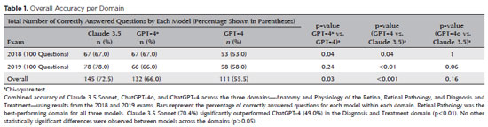
PURPOSE: Natural language models and chatbots, particularly OpenAI’s Generative Pre-Trained Transformer architecture, have transformed human interaction with digital interfaces. The latest versions, including ChatGPT-4o, offer enhanced functionalities compared to their predecessors. This study evaluates the accuracy of ChatGPT-4, ChatGPT-4o, and Claude 3.5 Sonnet in answering questions from the Brazilian Retina and Vitreous Society certification exam.
METHODS: We compiled 200 multiple-choice questions from the Brazilian Retina and Vitreous Society 2018 and 2019 exams. Questions were categorized into three domains: Anatomy and Physiology of the Retina, Retinal Pathology, and Diagnosis and Treatment. Using a standardized prompt developed according to prompt design guidelines, we tested ChatGPT-4, ChatGPT-4o, and Claude 3.5 Sonnet, recording their first responses as final. Three retina specialists performed a qualitative analysis of the answers. Accuracy was determined by comparing responses to the official correct answers. Statistical analysis was conducted using chi-square tests and Cohen’s Kappa.
RESULTS: Claude 3.5 Sonnet achieved the highest overall accuracy (72.5%), followed by ChatGPT-4o (66.0%) and ChatGPT-4 (55.5%). Claude 3.5 Sonnet and ChatGPT-4o significantly outperformed ChatGPT-4 (p<0.01 and p=0.03, respectively), while no significant difference was observed between Claude 3.5 Sonnet and ChatGPT-4o (p=0.16). Model responses agreed 74.5% of the time, with a Cohen’s κ of 0.47. Retinal Pathology was the best-performing domain for all models, whereas Anatomy and Physiology of the Retina and Diagnosis and Treatment were the weakest domains for Claude 3.5 Sonnet and ChatGPT-4, respectively.
CONCLUSIONS: This study is the first to assess Claude 3.5 Sonnet, ChatGPT-4, and ChatGPT-4o in retina specialist certification exams. Claude 3.5 Sonnet and ChatGPT-4o significantly outperformed ChatGPT-4, highlighting their potential as effective tools for studying retina specialist board exams. These findings suggest that the enhanced functionalities of Claude 3.5 Sonnet and ChatGPT-4o offer substantial improvements in medical education contexts.
Keywords: Artificial intelligence; ChatGPT; Retina; Medical education; Ophthalmology, Large language model; Natural language processing
Abstract
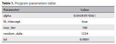
PURPOSE: We developed an artificial intelligence program for calculating intraocular lenses and analyzed its accuracy rate via ultrasonic biometry. This endeavor is aimed at enhancing precision and efficacy in the selection of intraocular lenses, particularly in cases where optical biometry is unavailable.
METHODS: Data was collected from the Hospital de Clínicas de Porto Alegre, which included cases of phacoemulsification with intraocular lens implantation, in which the lens selection was based on ultrasonic biometry. The program, implemented in Python, Java, and PHP, employs the ridge regression method. Two design options were developed: a basic model, which uses only keratometry variables (K1 and K2), axial size and final target refraction in the spherical equivalent, and an advanced model, which incorporates preoperative refraction and the patient's age. The Universal Barrett II formula was used to compare both models.
RESULTS: The sample consisted of 486 eyes from 313 patients, with 350 eyes used for program training and 136 for program validation. The spherical equivalent hit rates, with a variation of ±0.5 D, were 86% and 87.5% for the basic and advanced models, respectively, with no statistically significant difference between them. With the Barret Universal II formula, the success rate was 69%, which was significantly different from the values of the two aforementioned models (p<0.0001). The system was better for medium and long eyes but worse for short eyes (<=22.00 mm).
CONCLUSION: The developed artificial intelligence program was superior to the Barrett formula in terms of performance, in the general context and within the subgroup of patients with longer eyes. This innovation can considerably contribute to the selection of intraocular lenses, particularly in cases where optical biometry is unavailable.
Keywords: Biometry; Intraocular lens; Cataract; Artificial intelligence
Abstract
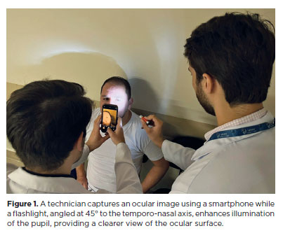
PURPOSE: This pilot study evaluated the diagnostic accuracy of a deep learning model for detecting pterygium in anterior segment photographs taken using smartphones in the Brazilian Amazon. The model’s performance was benchmarked against assessments made by experienced ophthalmologists, considered the clinical gold standard.
METHODS: In this cross-sectional study, 38 participants (76 eyes) from Barcelos, Brazil, were enrolled. Trained nonmedical health workers captured high-resolution anterior segment images using smartphones. These images were analyzed using a deep learning model based on the MobileNet-V2 convolutional neural network. Diagnostic metrics–including sensitivity, specificity, accuracy, positive predictive value, negative predictive value, and area under the receiver operating characteristic curve–were calculated and compared with the ophthalmologists’ evaluations.
RESULTS: The deep learning model achieved a sensitivity of 91.43%, specificity of 90.24%, positive predictive value of 88.46%, negative predictive value of 92.79%, and an area under the curve of 0.91. Logistic regression revealed no statistically significant association between pterygium and demographic variables such as age or gender.
CONCLUSIONS: The deep learning model demonstrated high diagnostic performance in identifying pterygium in a remote Amazonian population. These preliminary findings support the potential use of artificial intelligence–based tools to facilitate early detection and screening in underserved regions, thereby enhancing access to ophthalmic care.
Keywords: Pterygium/diagnostic imaging; Smartphone; Diagnostic techniques, ophthalmological; Deep learning; Telemedicine; Artificial intelligence; Cross-sectional studies; Brazil/epidemiology
Abstract
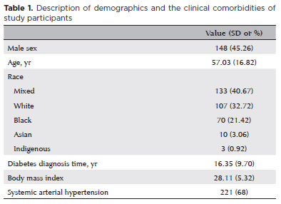
PURPOSE: Diabetic retinopathy screening in low- and middle-income countries is limited by restricted access to specialized care. Portable retinal cameras offer a practical alternative; however, image quality – affected by mydriasis – directly influences the performance of artificial intelligence models. This study evaluated the effect of mydriasis on image gradability and AI-based diabetic retinopathy detection in real-world, resource-limited settings.
METHODS: The proportions of gradable images were compared between mydriatic and non-mydriatic groups. Generalized estimating equations were used to identify factors associated with image gradability, including age, sex, race, diabetes duration, and systemic hypertension. A ResNet-200d model was trained on the mobile Brazilian Ophthalmological dataset and externally validated on both mydriatic and non-mydriatic images. Model performance was evaluated using accuracy, F1 score, area under the curve, and confusion matrix metrics. Sensitivity differences were assessed using the McNemar test, and area under the curves were compared using DeLong's test. The Youden index was used to determine optimal classification thresholds. Agreement between macula- and disc-centered images was analyzed using Cohen's κ.
RESULTS: The mydriatic group demonstrated a higher proportion of gradable images compared with the non-mydriatic group (82.1% vs. 55.6%; p<0.001). In non-mydriatic images, lower gradability was associated with systemic hypertension, older age, male sex, and longer diabetes duration. The AI model achieved better performance in mydriatic images (accuracy, 85.15%; area under the curve, 0.94) than in non-mydriatic images (accuracy, 79.68%; area under the curve, 0.93). The McNemar test showed a significant difference in sensitivity (p=0.0001), whereas DeLong's test revealed no significant difference in area under the curve (p=0.4666). The Youden index indicated that optimal classification thresholds differed based on mydriasis status. Agreement between image fields was moderate to substantial and improved with mydriasis.
CONCLUSION: Mydriasis significantly improves image gradability and enhances AI performance in diabetic retinopathy screening. Nonetheless, in low- and middle-income countries where pharmacologic dilation may be impractical, optimizing model calibration and thresholding for non-mydriatic images is essential to ensure effective AI implementation in real-world clinical environments.
Keywords: Artificial intelligence; Bias; Diabetic retinopathy; Portable camera; Retina
Abstract
PURPOSE: Using advanced imaging techniques, this study aimed to evaluate corneal stability, epithelial remodeling, and tear film changes over a one-year period in first-time soft-contact lens wearers.
METHODS: A retrospective study was conducted on 100 eyes of 50 first-time daily soft-contact lens users aged 21–65 years with no prior rigid gas-permeable lens wear. The Sirius Scheimpflug imaging system was used to assess corneal topography, epithelial thickness, and non-invasive tear break-up time at baseline, 3, 6, and 12 months. Corneal warpage was evaluated using symmetry indices and Baiocchi Calossi Versaci indices. We performed statistical analysis using repeated-measures analyses of variance with Greenhouse-Geisser correction.
RESULTS: The mean baseline central corneal thickness was 537.83 (±7.92) µm, with no significant thinning after one year. The average simulated keratometry values remained stable, indicating no progressive corneal steepening or flattening. There were no significant changes in warpage indices over time, suggesting corneal shape preservation. Higher-order aberrations (coma, trefoil, and spherical aberrations) and non-invasive tear break-up time remained unchanged throughout the study period.
CONCLUSIONS: Modern silicone hydrogel soft-contact lenses do not induce significant corneal warpage, epithelial remodeling, or optical aberrations over a one-year period. We found that corneal morphology and tear film stability were preserved, supporting the safety of soft-contact lens use. These findings provide clinically relevant insights into the long-term impact of contact lens wear. They may facilitate improved lens fitting strategies and preoperative refractive surgery assessments.
Keywords: Contact lenses, hydrophilic; Cornea/surgery; Corneal diseases; Corneal topography; Adaptation, ocular/physiology; Endothelium, corneal/pathology; Refractive errors; Tears/metabolism.
Abstract
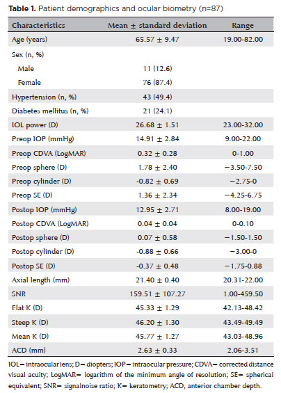
PURPOSE: To compare the refractive prediction error of Hill-radial basis function 3.0 with those of 3 conventional formulas and 11 combination methods in eyes with short axial lengths.
METHODS: The refractive prediction error was calculated using 4 formulas (Hoffer Q, SRK-T, Haigis, and Hill-RBF) and 11 combination methods (average of two or more methods). The absolute error was determined, and the proportion of eyes within 0.25-diopter (D) increments of absolute error was analyzed. Furthermore, the intraclass correlation coefficients of each method were computed to evaluate the agreement between target refractive error and postoperative spherical equivalent.
RESULTS: This study included 87 eyes. Based on the refractive prediction error findings, Hoffer Q formula exhibited the highest myopic errors, followed by SRK-T, Hill-RBF, and Haigis. Among all the methods, the Haigis and Hill-RBF combination yielded a mean refractive prediction error closest to zero. The SRK-T and Hill-RBF combination showed the lowest mean absolute error, whereas the Hoffer Q, SRK-T, and Haigis combination had the lowest median absolute error. Hill-radial basis function exhibited the highest intraclass correlation coefficient, whereas SRK-T showed the lowest. Haigis and Hill-RBF, as well as the combination of both, demonstrated the lowest proportion of refractive surprises (absolute error >1.00 D). Among the individual formulas, Hill-RBF had the highest success rate (absolute error ≤0.50 D). Moreover, among all the methods, the SRK-T and Hill-RBF combination exhibited the highest success rate.
CONCLUSIONS: Hill-radial basis function showed accuracy comparable to or surpassing that of conventional formulas in eyes with short axial lengths. The use and integration of various formulas in cataract surgery for eyes with short axial lengths may help reduce the incidence of refractive surprises.
Keywords: Cataract; Lenses, intraocular; Axial length, eye; Refractive errors; Artificial intelligence
Abstract
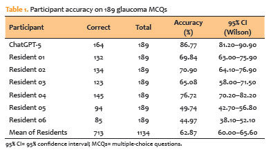
PURPOSE: To assess the performance of a contemporary large language model (ChatGPT-5) against ophthalmology residents on a standardized set of glaucoma multiple-choice questions.
METHODS: We conducted a cross-sectional comparative study with 189 text-only glaucoma multiple-choice questions from the Cybersight question bank. ChatGPT-5 was tested under standardized conditions, with each item placed in a new chat and limited to letter-only outputs. Six ophthalmology residents from a Brazilian training program (two Postgraduate Year 1, two Postgraduate Year 2, and two Postgraduate Year 3) answered the same questions under supervision. Accuracy was calculated using the official key. McNemar’s exact test was used to compare items between ChatGPT-5 and residents, and matched odds ratios and 95% confidence intervals (95% CIs) were calculated using the Haldane–Anscombe correction.
RESULTS: ChatGPT-5 received 164 of 189 correct responses (86.8%; 95% CI, 81.2–90.9). Residents’ overall accuracy was 62.9% (713/1,134; 95% CI, 60.0–65.6). The top-performing resident earned 76.7%. ChatGPT-5 outperformed all residents in head-to-head comparisons, with odds ratios ranging from 1.84 (95% CI, 1.10–3.08) to 13.15 (95% CI, 5.93–29.20), all p≤0.023. ChatGPT-5 correctly answered 17/189 items (9.0%), but fewer than half of residents were correct (“large language model-only wins”), whereas residents were more successful on items that ChatGPT-5 overlooked.
CONCLUSIONS: ChatGPT-5 outperformed ophthalmology residents on text-based glaucoma multiple-choice questions, indicating its potential as a subspecialty education and assessment tool. Generalizability is limited by the single question bank, text-only items, a small resident cohort, and the evaluation of one large language model version at a single time point. Before incorporating these findings into clinical decision-making, larger, multimodal, and longitudinal studies are required.
Keywords: Glaucoma; Artificial intelligence; Large language models; Education, medical; Medical staff, hospital
Abstract
PURPOSE: Standard automated perimetry has been the standard method for measuring visual field changes for several years. It can measure an individual’s ability to detect a light stimulus from a uniformly illuminated background. In the management of glaucoma, the primary objective of perimetry is the identification and quantification of visual field abnormalities. It also serves as a longitudinal evaluation for the detection of disease progression. The development of artificial intelligence-based models capable of interpreting tests could combine technological development with improved access to healthcare.
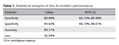
METHODS: In this observational, cross-sectional, descriptive study, we used an artificial intelligence-based model [Inception V3] to interpret gray-scale crops from standard automated perimetry that were performed in an ophthalmology clinic in the Brazilian Amazon rainforest between January 2018 and December 2022.
RESULTS: The study included 1,519 standard automated perimetry test results that were performed using Humphrey HFA-II-i-750 (Zeiss Meditech). The Subsequently, 70%, 10%, and 20% of the dataset were used for training, validation, and testing, respectively. The model achieved 80% (68.23%–88.9%) sensitivity and 94.64% (88.8%–98%) specificity for detecting altered perimetry results. Furthermore, the area under the receiver operating characteristic curve was 0.93.
CONCLUSIONS: The integration of artificial intelligence in the diagnosis, screening, and monitoring of pathologies represents a paradigm shift in ophthalmology, enabling significant improvements in safety, efficiency, availability, and accessibility of treatment.
Keywords: Glaucoma; Disease progression; Perimetry; Visual Fields; Visual field tests; Artificial intelligence; Neural networks, computers; Machine learning
09-fig01.jpg)
Abstract
Objetivo: Relatar um experimento projetado para determinar alterações anatômicas em córneas porcinas após a colocação de um novo implante depolímero na córnea.
Métodos: Foi utilizado olho de porco ex vivo. Um novo agente modelador biocompatível, de colágeno tipo 1, com 6mm de diâmetro foi moldado com excimer laser em sua face posterior, para criar três formatos planocôncavos. Os implantes foram inseridos dentro de um bolsão, dissecado manualmente, a 200 micrômetros (µm). Foram definidos três grupos de tratamento: grupo A (n=3), teve a profundidade máxima de ablação de70 µm; o grupo B (n=3), profundidade máxima de ablação de 64 µm; e o grupo C (n=3), profundidade máxima de ablação de 104 µm, com buraco central. O grupo controle, D (n=3), foi incluído, com a criação do bolsão estromal, porém sem inserir o material. A avaliação desses olhos foi realizada por tomografia de coerência óptica (OCT) e por tomografia corneana.
Resultados: A tomografia corneana mostrou uma tendência para diminuição da ceratometria média em todos os 4 grupos. A tomografia de coerência óptica mostrou córneas com implantes localizados no estroma anterior e aplanamento visível, enquanto as córneas não mudaram qualitativamente o formato no grupo controle.
Conclusões: O novo implante de biomaterial planocôncavo descrito aqui foi capaz de remodelar a córnea em modelo de animal ex vivo, resultando no aplanamento corneano. Novos estudos são necessários usando modelos animais in vivo para confirmar tais achados.
Keywords: Córnea; Cirurgia da córnea a laser; Substância própria; Proteses e implantes; Lasers de excimer; Materiais biocompatíveis; Animais; Suínos

Abstract
PURPOSE: To assess the outcomes of deep anterior lamellar keratoplasty or penetrating keratoplasty at the scar and the edema stages.
METHODS: Forty-five patients (45 eyes) with keratoconus scar stage (scar group, n=26; penetrating keratoplasty a subgroup, n=7; deep anterior lamellar keratoplasty b subgroup, n=19) and keratoconus edema stage (edema group, n=19; penetrating keratoplasty c subgroup, n=12; deep anterior lamellar keratoplasty d group, n=7) who received penetrating keratoplasty or deep anterior lamellar keratoplasty from 2000 to 2022 were retrospectively studied. At 1, 6, and 12 months after surgery, the best-corrected visual acuity, astigmatism, spherical equivalent, corneal endothelial cell density, and complications were analyzed.
RESULTS: The best-corrected visual acuity and average corneal endothelial cell loss rate were not significantly different between the scar and edema groups (p>0.05). At 6 and 12 months after surgery, the astigmatism and spherical equivalent in the scar group were significantly lower than those in the edema group (p<0.05). The spherical equivalent of the deep anterior lamellar keratoplasty b subgroup was lower than that of the penetrating keratoplasty a subgroup in the scar group 6 months after surgery (p<0.05). In the edema group, there was no significant difference in spherical equivalent between subgroups (p>0.05). There were no significant differences in best-corrected visual acuity and astigmatism between subgroups within the two groups (p>0.05). In comparison to the scar group, the edema group experienced more complications. According to a survival analysis, there was no statistically significant difference between the scar group and the edema group regarding the progression of vision.
CONCLUSIONS: In terms of the outcomes and prognosis for vision after keratoplasty with edema stage and scar stage, deep anterior lamellar keratoplasty may be as effective as penetrating keratoplasty.
Keywords: keratoconus; Edema; Cicatrix; keratoplasty, penetrating; Corneal transplantation; Astigmatism; Corneal endothelial cell loss; Endothelial cells
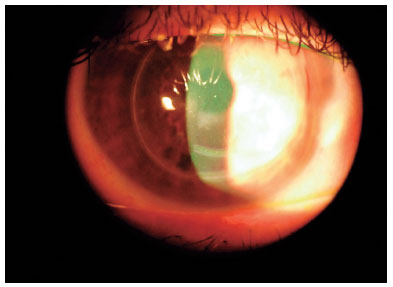
04-fig01.jpg)
Abstract
O ceratocone é uma doença progressiva que se manifesta como uma elevação semelhante a um cone da córnea central ou paracentral inferior e é associada a uma redução irregular da espessura do estroma. Há uma diminuição gradual da acuidade visual devido à assimetria da córnea, ao astigmatismo irregular e a um aumento das aberrações ópticas, o que prejudica a qualidade de vida. Foram desenvolvidos vários procedimentos para tentar interromper ou mesmo reverter a evolução da doença. Um deles é o chamado procedimento de Bader, que inclui um padrão de incisões em volta da circunferência da córnea e na base do cone protuberante. Essas incisões penetram até 70%-90% da profundidade da córnea e têm o objetivo de achatar a topografia e diminuir a assimetria da córnea e o astigmatismo irregular. Embora essa técnica seja muito promissora, segundo um estudo anterior, aqui se apresenta o caso de um paciente no qual esses objetivos não foram atingidos. Esse paciente recebeu lentes de contato para restaurar e manter sua visão, enquanto sua ectasia corneana e a redução da espessura progrediram ao longo da década seguinte.
Keywords: Ceratocone; Astigmatismo; Córnea; Topografia da córnea; Procedimentos cirúrgicos oftalmológicos; Lentes de contato; Dilatação patológica; Acuidade visua; Qualidade de vida.
Abstract
O ceratocone é uma doença progressiva que se manifesta como uma elevação semelhante a um cone da córnea central ou paracentral inferior e é associada a uma redução irregular da espessura do estroma. Há uma diminuição gradual da acuidade visual devido à assimetria da córnea, ao astigmatismo irregular e a um aumento das aberrações ópticas, o que prejudica a qualidade de vida. Foram desenvolvidos vários procedimentos para tentar interromper ou mesmo reverter a evolução da doença. Um deles é o chamado procedimento de Bader, que inclui um padrão de incisões em volta da circunferência da córnea e na base do cone protuberante. Essas incisões penetram até 70%-90% da profundidade da córnea e têm o objetivo de achatar a topografia e diminuir a assimetria da córnea e o astigmatismo irregular. Embora essa técnica seja muito promissora, segundo um estudo anterior, aqui se apresenta o caso de um paciente no qual esses objetivos não foram atingidos. Esse paciente recebeu lentes de contato para restaurar e manter sua visão, enquanto sua ectasia corneana e a redução da espessura progrediram ao longo da década seguinte.
Keywords: Ceratocone; Astigmatismo; Córnea; Topografia da córnea; Procedimentos cirúrgicos oftalmológicos; Lentes de contato; Dilatação patológica; Acuidade visua; Qualidade de vida
 ABO is licensed under a Creative Commons Attribution-NonComercial 4.0 Internacional.
About
Issues
Editorial Board
Submission
Arquivos Brasileiros de Oftalmologia
Official publication of Brazilian Council of Ophthalmology - Conselho Brasileiro de Oftalmologia (CBO)
Rua Casa do Ator, 1.117 - 2nd floor - Zip Code: 04546-004
São Paulo - SP, Brazil
TEL: +55 11 3266-4000
E-mail: [email protected]