Arq. Bras. Oftalmol. 2022;85 (6 )
:558-564
| DOI: 10.5935/0004-2749.20220073
Abstract
Objetivo: Analisar o perfil epidemiológico dos casos de evisceração e enucleação no pronto-socorro oftalmológico de um hospital terciário brasileiro.
Métodos: Análise retrospectiva dos casos tratados no pronto-socorro oftalmológico do Hospital São Paulo (Universidade Federal de São Paulo) entre os anos de 2013 a 2018. Os casos urgentes de evisceração e enucleação foram incluídos e os casos eletivos foram excluídos. A análise dos prontuários médicos foi baseada em: dados demográficos, causas imediatas e associadas ao procedimento, acuidade visual informada, duração dos sintomas antes do atendimento oftalmológico, complicações, distância da residência até o hospital e tempo de hospitalização.
Resultados: 61 enucleações e 121 eviscerações foram incluídas no estudo. Os pacientes tinham uma média de idade de 63,27 ± 18,68 anos; 99 eram do sexo masculino (54,50%) e 83 do sexo feminino (45,60%). As indicações de evisceração e enucleação foram: perfuração corneana com (44,50%) e sem (23,63%) sinais infecciosos, endoftalmite (15,38%), trauma ocular (14,29%), neoplasia (0,55%), queimadura (1,10%) e phthisis bulbi (0,55%). A acuidade visual informada foi de ausência de percepção luminosa (87,36%), percepção luminosa (1.10%), ausência de colaboração (3,30%) e sem dados informados (8,24%). A média de tempo até a busca pelo serviço oftalmológico foi de 18,32 dias. Houve 2 casos de oftalmia simpática após evisceração.
Conclusões: Eviscerações foram predominantemente realizadas em comparação a enucleações em todo o período de estudo. As características demográficas mais comuns foram idade >60 anos e sexo masculino. As principais indicações para procedimentos urgentes de evisceração e enucleação foram perfuração corneana com e sem infecção, endoftalmite e trauma ocular. Este estudo poderia guiar medidas preventivas para evitar procedimentos oculares destrutivos.
Keywords: Evisceração do olho; Enucleação ocular; Úlcera da córnea/epidemiologia; Endoftalmite; Traumatismos oculares; Serviços médicos de emergência; Serviços de saúde ocular.
Arq. Bras. Oftalmol. 2023;86 (5 )
:1-6
| DOI: 10.5935/0004-2749.20230064
Abstract
Objetivo: Avaliar a resposta tecidual e clínica a um implante orbitário de polimetilmetacrilato, oco e multiperfurado em sua porção posterior em modelo animal após evisceração.
Métodos: Dezesseis coelhos da raça Nova Zelândia foram submetidos à evisceração do globo ocular direito. Todos receberam implante oco de polimetilmetacrilato de 12 mm de diâmetro, multiperfurado em sua semiesfera posterior. O estudo foi dividido em avaliação clínica e histopatológica. A avaliação clínica foi diária até 14 dias pós-evisceração e, a cada sete dias, até completar 180 dias. Os animais foram divididos em grupos de quatro animais e cada um foi submetido à exenteração com 07, 30, 90 e 180 dias e depois à eutanásia. A análise histopatológica teve por fim caracterizar o padrão inflamatório, a estrutura do colágeno e o grau de neovascularização. Para isso, além da tradicional coloração pela hematoxilina-eosina, utilizou-se o corante Picrosirius Red (PSR) e imuno-histoquímica com o marcador CD 34.
Resultados: Não houve sinais de infecção, afinamento conjuntival ou escleral, exposição ou extrusão do implante em nenhum animal durante o estudo. Já no sétimo dia, o tecido neoformado migrou para dentro do implante formando uma rede fibrovascular através dos canais posteriores. A resposta inflamatória diminuiu ao longo do tempo avaliado e não foram encontradas células gigantes multinucleadas.
Conclusão: O implante analisado permite a sua integração aos tecidos orbitários pelo crescimento fibrovascular em seu interior. Os autores acreditam que este modelo de implante orbital pode fazer parte de testes com humanos.
Keywords: Implantes orbitários; Polimetilmetacrilato; Evisceração ocular; Anoftalmia; Procedimentos cirúrgicos oftalmológicos; Coelhos.
Arq. Bras. Oftalmol. 2026;89 (1 )
:1-6
| DOI: 10.5935/0004-2749.2025-0049
Abstract
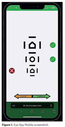
PURPOSE: This cross-sectional study compared best-corrected visual acuity obtained using Cloudscaper symbols, a novel optotype developed according to ETDRS specifications for children's virtual screening, with that obtained using LEA symbols.
METHODS: A total of 560 children aged 3-16 yr underwent visual acuity test with both Cloudscaper symbols and LS. The test application was standardized using the EyeSpy algorithm. Additionally, 147 participants were tested with the standard Snellen E paper chart. Paired t tests were performed to assess the clinical significance of logMAR visual acuity differences.
RESULTS: The mean logMAR visual acuity with LEA symbols was 0.12 (standard deviation [SD]=0.18; range, -0.10 to 0.80), while with Cloudscaper symbols it was 0.18 (SD=0.19; range, -0.10 to 0.80). The mean difference between Cloudscaper symbols and LEA symbols was 0.099 logMAR (approximately 0.5 optotypes; SD=0.08; range, 0.0-0.14; p<0.0001). Cloudscaper symbols slightly underestimated visual acuity compared to LEA symbols. Visual acuity measured by both methods was highly correlated (Spearman's r=0.74, p<0.0001). The mean visual acuity difference between Cloudscaper symbols and the Snellen E chart was 0.0045 (p=0.805; 95% confidence interval [95% CI]), whereas the difference between LEA symbols and Snellen E was 0.0883 (p<0.001; 95% CI).
CONCLUSIONS: Cloudscaper symbols provide a reliable tool for visual screening in children. Although they slightly underestimate visual acuity compared to LEA symbols – a finding also reported when comparing ETDRS letters with LEA symbols – Cloudscaper symbols show strong agreement with Snellen E chart measurements. This suggests that Cloudscaper symbols allow precise visual acuity assessment comparable to the gold standard.
Keywords: Vision screening; Vision tests; Visual acuity; Mobile applications; Eye health; Child health; Diagnostic techniques, Ophthalmological; Child; Preschool child; Adolescent
Arq. Bras. Oftalmol. 2025;88 (6 )
:1-7
| DOI: 10.5935/0004-2749.2025-0006
Abstract
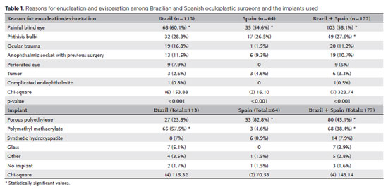
PURPOSE: This study aimed to evaluate the practices employed by oculoplastic surgeons in the assessment and management of anophthalmic sockets and external ocular prostheses.
METHODS: Oculoplastic surgeons from two countries, who specialized in the management of anophthalmic sockets, participated in a web-based survey. Data collected included demographics, types of surgery, implant use, external ocular prostheses management (including fabrication and cleaning), complications encountered, and follow-up times. The frequencies and distributions of the responses were statistically analyzed.
RESULTS: A total of 177 oculoplastic surgeons participated, 113 (63.8%) from Brazil, the remainder from Spain. Evisceration was the preferred surgical procedure of 149 (84.2%) surgeons. The most commonly reported indication for enucleation was a painful blind eye (n=103, 58.1%; both Brazil and Spain, p<0.001). Brazilian surgeons preferred polymethyl methacrylate implants (n=65, 57.5%), while Spanish surgeons favored porous polyethylene implants (n=53, 82.8%; p<0.001). Discharge was the most frequently observed clinical feature during socket evaluation (n=164, 92.6%; p<0.001). Brazilian surgeons recommended daily (n=53, 46.9%) or weekly (n=41, 36.2%) cleaning of external ocular prostheses, while Spanish surgeons more commonly recommended monthly cleaning (n=31, 48.4%; p<0.001). The majority of Brazilian surgeons (n=83, 73.4%) advised patients to remove their external ocular prostheses at night. Only a small number of Spanish surgeons (n=3, 4.6%) suggested this practice (p<0.001). Overall, the follow-up recommendations varied, with 70 (39.5%) surgeons recommending follow-up based on individual case needs, and 59 (33.3%) suggesting annual visits (p<0.001). The primary indications for external ocular prostheses replacement were edge damage (n=75, 42.3%) and loss of volume (n=68, 38.4%). The replacement intervals given typically ranged from 1 to 5 years (n=92, 51.9%; p<0.001).
CONCLUSION: Oculoplastic surgeons in Brazil and Spain demonstrated similar practices in the management of anophthalmic sockets. However, notable differences were observed in the choice of implant materials, cleaning protocols, and recommendations regarding external ocular prostheses removal during sleep.
Keywords: Anophthalmos; Eye, artificial; Polymethyl methacrylate; Polyethylene; Surgeons; Surveys and questionnaires; Brazil; Spain.
Arq. Bras. Oftalmol. 2025;88 (6 )
:1-6
| DOI: 10.5935/0004-2749.2025-0153
Abstract
PURPOSE: This clinical study aimed to assess the effectiveness of microemulsion artificial tears containing povidone and propylene glycol in the management of dry eye disease. Secondary objectives included evaluating improvements in tear-film stability, measured by tear break-up time and corneal staining scores, along with the tolerability and safety of the formulation.
METHODS: This was a prospective, single-arm interventional study involving 30 participants (52 eyes) diagnosed with dry eye disease. Participants self-administered the investigational eye drops twice daily for 28 consecutive days. Primary and secondary outcomes included changes in the Ocular Surface Disease Index, tear break-up time, and corneal staining scores. Adverse events were documented throughout the study period.
RESULTS: Significant improvements in Ocular Surface Disease Index scores were observed, reflecting a reduction in dry eye disease symptoms. Tear break-up time increased notably between baseline and follow-up assessments, with the proportion of eyes exhibiting tear break-up time ≥10 srising from 25.0% to 63.5%. Additionally, the percentage of eyes with a corneal staining score of zero improved from 23.1% to 69.2%. Conjunctival staining also decreased, with the proportion of eyes with scores of 2 and 3 dropping from 11.5% to 3.8% and 5.8% to 0%, respectively.
CONCLUSIONS: The findings suggest that povidone and propylene glycol-based artificial tears significantly enhance tear-film stability and alleviate symptoms in patients with mild to moderate dry eye disease, with minimal adverse effects. This formulation represents a safe and effective short-term treatment option for dry eye disease management.
Keywords: Artificial tears; Dry eye disease; Tear-film stability; Propylene glycol; Povidone; Visual acuity; Surveys and questionnaires
Arq. Bras. Oftalmol. 2025;88 (2 )
:1-7
| DOI: 10.5935/0004-2749.2023-0215
Abstract
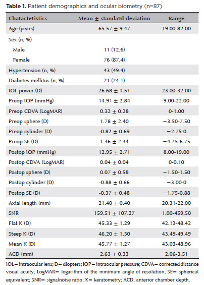
PURPOSE: To compare the refractive prediction error of Hill-radial basis function 3.0 with those of 3 conventional formulas and 11 combination methods in eyes with short axial lengths.
METHODS: The refractive prediction error was calculated using 4 formulas (Hoffer Q, SRK-T, Haigis, and Hill-RBF) and 11 combination methods (average of two or more methods). The absolute error was determined, and the proportion of eyes within 0.25-diopter (D) increments of absolute error was analyzed. Furthermore, the intraclass correlation coefficients of each method were computed to evaluate the agreement between target refractive error and postoperative spherical equivalent.
RESULTS: This study included 87 eyes. Based on the refractive prediction error findings, Hoffer Q formula exhibited the highest myopic errors, followed by SRK-T, Hill-RBF, and Haigis. Among all the methods, the Haigis and Hill-RBF combination yielded a mean refractive prediction error closest to zero. The SRK-T and Hill-RBF combination showed the lowest mean absolute error, whereas the Hoffer Q, SRK-T, and Haigis combination had the lowest median absolute error. Hill-radial basis function exhibited the highest intraclass correlation coefficient, whereas SRK-T showed the lowest. Haigis and Hill-RBF, as well as the combination of both, demonstrated the lowest proportion of refractive surprises (absolute error >1.00 D). Among the individual formulas, Hill-RBF had the highest success rate (absolute error ≤0.50 D). Moreover, among all the methods, the SRK-T and Hill-RBF combination exhibited the highest success rate.
CONCLUSIONS: Hill-radial basis function showed accuracy comparable to or surpassing that of conventional formulas in eyes with short axial lengths. The use and integration of various formulas in cataract surgery for eyes with short axial lengths may help reduce the incidence of refractive surprises.
Keywords: Cataract; Lenses, intraocular; Axial length, eye; Refractive errors; Artificial intelligence
Arq. Bras. Oftalmol. 2025;88 (5 )
:1-8
| DOI: 10.5935/0004-2749.2024-0312
Abstract
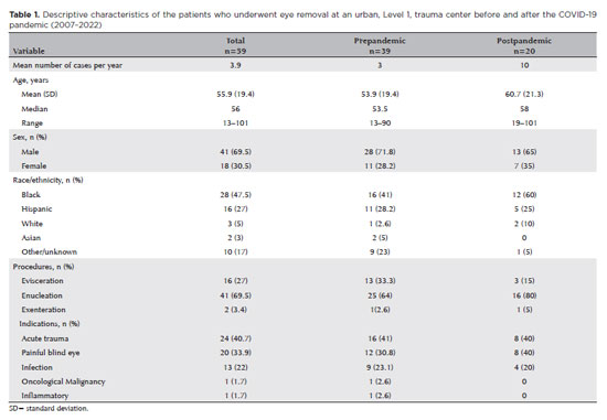
PURPOSE: To evaluate the changes in the rates and indications of eye removal procedures during the recent COVID-19 pandemic.
METHODS: The medical records of all patients who underwent eye removal from 2007 to 2022 were retrospectively reviewed. The patient demographic data and indications for surgery were collected. Data from two groups of patients (prepandemic surgery and postpandemic surgery) were compared. Statistical significance was set at p<0.05.
RESULTS: Fifty-nine patients underwent enucleation (69%), evisceration (27%), or exenteration (3%). The mean (SD) age of the patients was 55.9 (19.4) years, and most (69%) of the patients were males. Most (47%) of the study population were Black. The common indications for eye removal were trauma (41%), painful blind eye (34%), and infection/inflammation (24%). The types of trauma were assault (55%), accidental (39%), and self-inflicted (6%). The mean (SD) monthly rates of eye removal increased from 0.25 (0.50) in the prepandemic period to 0.77 (0.91) during the pandemic (p<0.001). These increases were noted in both males (p=0.003) and females (p=0.001) and were the highest among Black patients [0.42 (0.76); p<0.001]. Among the indications of eye removal, painful blind eyes [0.35 (0.75); p<0.001] and ocular trauma [0.31 (0.47); p=0.051] exhibited the greatest increases following the pandemic.
CONCLUSION: The rate of eye removal procedures increased during the recent pandemic. Although delayed care of chronic eye conditions may have contributed to the increased rates of painful blind eyes, the increased trauma-related eye removals may be attributed to the simultaneous spike in violent assaults in New York City.
Keywords: Eye injuries; Eye enucleation; COVID-19; Pandemics; Ethinicity; Inflammation, Trauma centers
Arq. Bras. Oftalmol. 2026;89 (3 )
:1-6
| DOI: 10.5935/0004-2749.2025-0332
Abstract
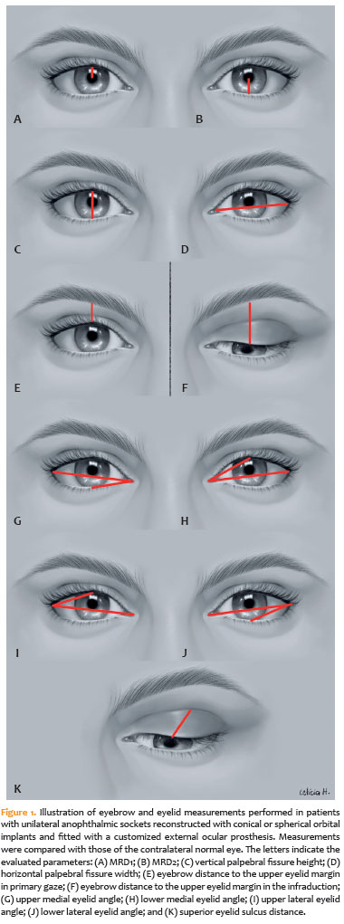
PURPOSE: To quantitatively compare eyebrow and eyelid positions in anophthalmic sockets reconstructed with conical or spherical orbital implants combined with customized external ocular prostheses.
METHODS: This cross-sectional observational study included 38 patients with unilateral anophthalmic sockets, of whom 21 received conical implants, and 17 received spherical implants. Eyelid and eyebrow parameters—including margin reflex distance 1 and 2, vertical and horizontal palpebral fissure dimensions, eyebrow-to-upper-eyelid margin distance in primary gaze and infraduction, medial and lateral eyelid angles in primary gaze, and superior eyelid sulcus depth —were quantitatively assessed using standardized digital photographs analyzed with Image J software. The contralateral healthy eye served as the control. Statistical analyses were performed to compare measurements between groups.
RESULTS: In the primary gaze position, conical and spherical implants showed comparable margin-reflex distance1, margin-reflex distance2, vertical palpebral fissure height, eyelid margin position, and medial and lateral eyelid angles. During infraduction, the upper eyelid margin was significantly lower in sockets reconstructed with conical implants. Compared with contralateral normal eyes, anophthalmic sockets exhibited a reduced horizontal palpebral fissure and a deeper superior eyelid sulcus, irrespective of implant shape.
CONCLUSION: Anophthalmic sockets reconstructed with conical or spherical implants demonstrate similar eyebrow and eyelid positioning in primary gaze. However, conical implants are associated with a lower eyelid margin during infraduction. Independent of implant format, anophthalmic sockets show a narrower horizontal palpebral fissure and increased superior sulcus depth compared with normal eyes.
Keywords: Anophthalmos; Prosthesis implantation; Anophthalmic socket; Conical implants; Spherical implants; Orbital implants; Eyelid measurements
Arq. Bras. Oftalmol. 2025;88 (5 )
:1-7
| DOI: 10.5935/0004-2749.2024-0074
Abstract
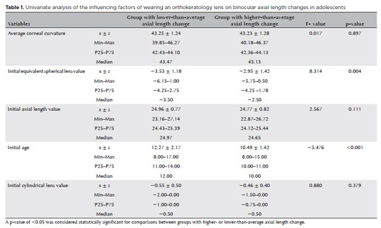
PURPOSE: This study aimed to identify factors influencing axial length changes in adolescents wearing orthokeratology lenses.
METHODS: A retrospective analysis was conducted on 84 adolescents (aged 9-17 yr) who wore orthokeratology lenses at our hospital. Axial length changes were calculated as the difference between the first and last visits. Patients were categorized into two groups based on axial length change: lower-than-average and higher-than-average. Data on sex, age at orthokeratology lens initiation, family history, initial equivalent spherical lens value, initial cylindrical lens value, initial average K value, and initial axial length were collected. Univariate and mixed-effects model analyses were performed to assess their influence on axial length changes.
RESULTS: Age (p<0.05) and initial equivalent spherical value (p<0.05) were significant predictors of axial length changes in both eyes and the left eye. For the right eye, only age was a significant factor (p<0.05). The mixed-effects model revealed that the difference between the left and right eyes, duration of orthokeratology lens use, age, initial equivalent spherical lens value, and initial axial length significantly influenced axial length changes in adolescents (p<0.05).
CONCLUSION: The factors influencing axial length changes in adolescents wearing orthokeratology lenses differ between the left and right eyes. These changes depend on the duration of lens wear, age, initial equivalent spherical lens value, and initial axial length. This study provides a theoretical basis for evaluating the clinical efficacy of orthokeratology lenses in managing myopia progression in adolescents.
Keywords: Orthokeratology; Contact lens; Myopia; Adolescent; Axial length, eye
Arq. Bras. Oftalmol. 2024;87 (2 )
:1-9
| DOI: 10.5935/0004-2749.2021-0456
Abstract
OBJETIVO: Este estudo visou avaliar os mecanismos da lesão e os tipos de fraturas orbitárias e sua relação com commotio retinae simultânea.
MÉTODOS: Este estudo retrospectivo avaliou registros de pacientes com fraturas orbitárias cujos diagnósticos foram confirmados por tomografia computadorizada entre julho de 2017 e setembro de 2019. Foram registrados os dados demográficos, circunstâncias da lesão, os resultados do exame oftalmológico e achados radiológicos. A análise estatística dos dados usou os testes de t-Student bicaudal, qui-quadrado e cálculos de odds ratio. O significado estatístico foi fixada em p<0,05.
RESULTADOS: Dos 204 pacientes com fraturas orbitárias incluídos neste estudo, 154 (75,5%) eram sexo masculino (75,5%). A média de idade foi de 42,1 anos. As fraturas orbitárias envolvendo uma parede orbital (58,8%) foram mais comuns do que as que acometeram várias paredes (41,2%). A maioria das fraturas acometeu a parede inferior (60,3%), sendo as paredes mediais as próximas mais frequentemente afetadas (19,6%). A causda mais comum de lesão foi agressão (59,3%), e a segunda mais comum foi queda (24%). A commotio retinae foi observada em 20,1% dos casos de fratura orbital e foi mais associada a lesões causadas por agressão (OR=5,22, p<0,001) e menos associada com aquelas causadas por quedas (OR=0,06, p<0,001). As restrições de movimentos oculares eram mais comuns na comoção central do que na periférica (OR=3,79, p=0,015) e com fraturas da parede medial do que com fraturas de outras paredes orbitais (OR=7,16, p<0,001). As chances de comoção não foram maiores em pacientes com fraturas orbitais de paredes múltiplas do que naqueles com fraturas de parede simples (p=0,967).
CONCLUSÕES: Na população do estudo, a agressão foi a causa mais comum de fraturas orbitais e resultou em commotio retinae mais grave do que qualquer outra causa. Os oftalmologistas devem estar cientes da probabilidade de commotio retinae em pacientes com fraturas orbitais resultantes de agressão, independentemente da extensão das lesões do paciente.
Keywords: Fraturas orbitárias; Movimentos oculares; Retina; Ferimentos e lesões
Arq. Bras. Oftalmol. 2024;87 (2 )
:1-8
| DOI: 10.5935/0004-2749.2022-0241
Abstract
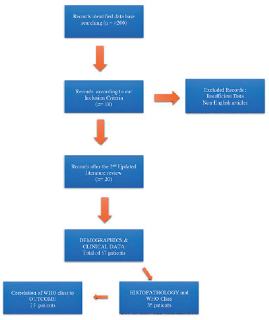
PURPOSE: We aimed to study reported cases of nasopharyngeal carcinoma presenting with ophthalmic manifestations with and without a prior diagnosis of nasopharyngeal carcinoma.
METHODS: We conducted a systematic review following the Preferred Reporting Items for Systematic Reviews and Meta-Analyses (PRISMA). A literature search was conducted using the MEDLINE database in PubMed and Google Scholar. We included patients with a previous diagnosis of nasopharyngeal carcinoma in Group I and those without a prior diagnosis of nasopharyngeal carcinoma in Group II. Data included demographics, clinical presentation, history of nasopharyngeal carcinoma, treatment, histopathological description, World Health Organization classification, and outcome.
RESULTS: Fifty-eight patients (26 in Group I and 32 in Group II) were included. The male-to-female ratio was 3:1. The mean age of the patients (53.3 ± 11.7 years and 54.8 ± 16.2 years, respectively) and gender did not differ significantly between the two groups. The most common ocular presentations were diplopia and proptosis in the first group (each in 34.6%), whereas visual disturbance was most common in the second group (46.9%). Treatment options and World Health Organization grading were comparable. The outcome in 38 patients (after a comparable follow-up period) was significantly better in group II (p=0.003). There was no statistically significant difference in the outcome of 23 patients in correlation with World Health Organization grades II versus III irrespective of group (p=0.094).
CONCLUSIONS: The demographics of patients with nasopharyngeal carcinoma presenting with ophthalmic manifestations were similar between the two study groups, with a wide age range and male predominance. Patients presenting initially to ophthalmologists with no history of nasopharyngeal carcinoma have a more favorable outcome. World Health Organization grading may have less value as a prognostic indicator.
Keywords: Nasopharyngeal carcinoma; Carcinoma; Eye manifestations; Exophthalmos; Diplopia; Systematic review
 ABO is licensed under a Creative Commons Attribution-NonComercial 4.0 Internacional.
09-fig01.jpg)
06-fig01.jpg)
15-tab01tb.jpg)
13-fig01.jpg)