Showing of 1 until 15 from 177 result(s)
Search for: Electroretinography; Cones (retina); Colour perception; Visual acuity; Low vision
03-tab01.jpg)
Abstract
Objetivo: A Escala Bayley de Desenvolvimento Infantil (Bayley-III) é uma ferramenta que avalia o desenvolvimento de crianças nos 3 primeiros anos de vida, incluindo os domínios cognitivo e motor. Este estudo tem como objetivo correlacionar a acuidade visual de grades e a funcionalidade visual em crianças saudáveis usando a Bayley-III.
Métodos: A acuidade visual binocular de grades foi medida usando o teste dos Cartões de Acuidade de Teller seguido pela Bayley-III em crianças saudáveis com idade entre 1-42 meses. Os escores da acuidade visual (logMAR) e da Bayley-III para habilidades cognitivas e motoras (grossa e fina) foram comparados.
Resultados: Um grupo de 40 crianças (20 meninos) com idades entre 1,2-42,1 meses foi testado e a média da acuidade visual foi de 0,39 ± 0,27 logMAR, sendo que todas estavam dentro dos limites normais para a idade. Houve uma forte correlação negativa e significante entre acuidade visual e idade (r=-0,83; p<0,001). A média do escore cognitivo foi de 49,92 ± 18,93 pontos, com forte correlação positiva e significante entre o escore cognitivo e a idade (r=0,81; p<0,001). A média do escore motor grosso foi de 41,72 ± 16,23 pontos, com forte correlação positiva e significante entre o escore motor grosso e a idade (r=0,75; p<0,001). A média do escore motor fino foi de 39,75 ± 14,63 pontos, com uma forte correlação positiva e significante entre o escore motor fino e a idade (r=0,77; p<0,001). A regressão linear múltipla mostrou que maior idade e melhor acuidade visual foram significantemente associadas à escores cognitivo e motor mais altos.
Conclusões: Neste estudo foi encontrada alta correlação entre a acuidade visual de grades medida pelos cartões de acuidade de Teller e os escores cogninitivo e motor medidos pela Bayley-III em crianças saudáveis. A Bayley-III pode ser uma ferramenta útil para avaliar a repercussão da deficiência visual no desenvolvimento cognitivo e motor de crianças.
Keywords: Desenvolvimento infantil; Acuidade visual; Cognição; Destreza motora; Transtornos da visão; Testes neuropsicológicos; Criança
04-tab01tb.jpg)
Abstract
Objetivo: Verificar se pacientes com dislexia do desenvolvimento (DD) apresentam déficits coerentes com uma disfunção magnocelular visual.
Métodos: Participantes com diagnóstico confirmado de dislexia do desenvolvimento (n=62; faixa etária=8 a 25 anos; Média da idade=13.8 anos, desvio padrão=3.9; 77% homens) foram comparados a um grupo controle com desenvolvimento típico, pareado por idade, sexo, dominância ocular, acuidade visual e compreensão de texto. A perimetria Frequency-Doubling Technology avaliou o limiar de sensibilidade ao contraste do campo visual periférico. O rastreador ocular Visagraph-III registrou os movimentos dos olhos durante leitura de texto.
Resultados: O grupo com dislexia do desenvolvimento apresentou piores limiares de sensibilidade no Frequency-Doubling Technology, com tamanho de efeito forte, do que o grupo controle. O grupo com dislexia do desenvolvimento apresentou mais olhos classificados com déficits na sensibilidade à ilusão de frequência duplicada do que o grupo controle. O grupo com dislexia do desenvolvimento apresentou pior habilidade motora ocular e no desempenho de leitura, revelado pela diferença entre os grupos em relação às fixações oculares, regressões, alcance de reconhecimento, taxa de leitura e eficiência relativa. Foi encontrada correlação significativa entre a sensibilidade ao contraste e as habilidades motoras oculares. Os participantes com boa eficiência relativa apresentaram uma sensibilidade ao contraste significativamente melhor do que os participantes com baixa eficiência relativa.
Conclusões: O grupo com dislexia do desenvolvimento apresentou desempenho inferior nas variáveis visuais relacionadas à função visual magnocelular (i.e., perimetria de frequência duplicada e habilidades motoras oculares), quando comparado ao grupo controle pareado. Os profissionais precisam estar cientes da importância de investigar a visão dos pacientes com dislexia do desenvolvimento além da acuidade visual e incluir nos seus procedimentos diagnósticos instrumentos para avaliar o processamento temporal, com limiar de sensibilidade ao contraste.
Keywords: Dislexia; Leitura; Percepção visual; Transtornos da visão; Músculos oculomotores; Movimentos oculares
07-fig01.jpg)
Abstract
Objetivo: Avaliar a acuidade visual através de potenciais evocados visuais de varredura em crianças saudáveis e ambliópicas, comparando-a com a acuidade visual pelo teste de Snellen.
Métodos: Foram incluídas no estudo 160 crianças com idades entre 6 e 17 anos. Desse total, 104 crianças (65%) estavam entre 7 e 17 anos de idade, eram capazes de comunicação verbal e não tinham nenhuma patologia ocular ou sistêmica (Grupo 1). O grupo 2 incluiu 56 crianças verbais (35%) com idades entre 6 e 17 anos e portadoras de estrabismo ou ambliopia anisometrópica, com a melhor acuidade visual corrigida entre 0,1 e 0,8. Todos os pacientes foram submetidos a um exame oftalmológico detalhado e a uma medição do potencial evocado visual por varredura. Registraram-se as características demográficas, os achados oculares, a melhor acuidade visual corrigida e os resultados do potencial evocado visual por varredura.
Resultados: No Grupo 1, os valores médios e máximos da acuidade visual pelo potencial evocado visual por varredura mostraram-se menores que a melhor acuidade visual corrigida medida através do teste de Snellen (p<0,001 para ambas as medições). Uma análise de Bland-Altman revelou que no grupo 1, a distribuição das diferenças entre a melhor acuidade visual corrigida pelo teste de Snellen e a média do potencial evocado visual por varredura foi de ± 0,11 logMAR, enquanto a distribuição das diferenças entre a melhor acuidade visual corrigida pelo teste de Snellen e o valor máximo do potencial evocado visual por varredura foi de ± 0,023 logMAR. No Grupo 2, os valores médio e máximo do potencial evocado visual por varredura mostraram-se menores que a melhor acuidade visual corrigida pelo teste de Snellen (respectivamente, p<0,001 e p=0,009). A análise de Bland-Altman revelou que a distribuição das diferenças entre a melhor acuidade visual corrigida pelo teste de Snellen e a média do potencial evocado visual por varredura foi de ± 0,16 logMAR, enquanto a distribuição das diferenças entre a melhor acuidade visual corrigida pelo teste de Snellen e o valor máximo do potencial evocado visual por varredura foi de ± 0,19 logMAR.
Conclusões: As medidas da acuidade visual através do potencial evocado visual por varredura mostram resultados comparáveis às medidas da acuidade visual pelo teste de Snellen. Essa técnica é um método objetivo e confiável de se avaliar a acuidade visual em crianças.
Keywords: Ambliopia; Acuidade visual; Potenciais evocados visuais; Testes visuais; Humanos; Criança; Adolescente.
07-tab01.jpg)
Abstract
Objetivo: Avaliar a eficácia das lentes de contato gelatinosas HydroCone, de hidrogel com silicone, em pacientes com microftalmia posterior.
Métodos: Foram revisados retrospectivamente 26 olhos com microftalmia posterior, a partir dos prontuários de 13 pacientes que receberam lentes de contato gelatinosas HydroCone, de hidrogel com silicone. Todos os pacientes foram submetidos ao exame de acuidade visual não corrigida e com melhor correção por óculos e com refração cicloplégica. Todos os pacientes receberam lentes de contato de acordo com os parâmetros obtidos na análise topográfica e foi obtida a melhor acuidade visual corrigida com lentes de contato.
Resultados: O equivalente esférico do olho direito variou de 10,00 a 19,25 dioptrias, e o do olho esquerdo de 11,00 a 21,5 dioptrias. Os comprimentos médios axiais e das câmaras posteriores foram menores do que para a população de mesma idade. No entanto, os valores médios dos parâmetros do segmento anterior, como o diâmetro horizontal visível da íris, a profundidade da câmara anterior central, a espessura da lente e a espessura central da córnea estavam dentro da faixa normal. Os valores médios da ceratometria revelaram curvatura corneana aumentada em relação à população normal. A média da melhor acuidade visual corrigida com lentes de contato foi significativamente maior que a média da melhor acuidade visual corrigida com óculos em ambos os olhos (p=0,045).
Conclusão: As lentes de contato gelatinosas de silicone HydroCone proporcionam melhor acuidade visual que óculos em pacientes com microftalmia posterior.
Keywords: Microftalmia; Lentes de contato hidrofílicas; Silicones; Transtornos da visão/reabilitação; Acuidade visual.
05-tab01.jpg)
Abstract
OBJETIVO: Avaliar, depois de 30 meses, a função visual e as alterações na espessura macular central de pacientes com degeneração macular relacionada à idade sem resposta terapêutica ao ranibizumabe (Lucentis®) que mudaram seu tratamento para o aflibercepte (Eylea®).
MÉTODOS: Realizou-se um estudo retrospectivo de pacientes com degeneração macular neovascular relacionada à idade que mudaram o tratamento para o aflibercepte após 6 ou mais injeções intravítreas de ranibizumabe a intervalos de 4-8 semanas. Todos os pacientes mudaram para o aflibercepte intravítreo (2,0 mg) e depois de 3 injeções consecutivas, seguidas de um regime de dosagem pro re nata, foram avaliados após 30 meses de tratamento. A melhor acuidade visual corrigida, o exame biomicroscópico, a pressão intraocular, a fundoscopia e a espessura macular central foram registrados no início do tratamento, antes da transição para o tratamento com aflibercepte intravítreo e aos 6, 12, 18, 24 e 30 meses de tratamento com o aflibercepte intravítreo.
RESULTADOS: Satisfizeram aos critérios de inclusão 33 olhos. A mediana da idade dos pacientes foi de 73,57 ± 7,98 anos. Dos pacientes, 21 (61,8%) eram homens e 12 (35,3%) eram mulheres. Antes da transição para o tratamento com o aflibercepte intravítreo, os pacientes receberam em média 16,8 ± 8,8 injeções de ranibizumabe (faixa 6-38).Depois da transição, o número médio de injeções de aflibercepte foi de 9,09 ± 3,94. Não houve diferenças significativas na melhor acuidade visual corrigida depois da mudança para o aflibercepte em qualquer das avaliações. Houve diminuição significativa da espessura macular central aos 6, 12, 18 e 30 meses (respectivamente, p=0,01, p=0,03, p=0,05, p=0,05 e p<0,001).
CONCLUSÃO: Pacientes com degeneração macular neovascular relacionada à idade que mudaram seu tratamento para o aflibercepte intravítreo devido à falta de resposta ao ranibizumabe intravítreo, tiveram melhora anatômica significativa da retina; mas embora esse estado tenha persistido, não foi observado nenhum ganho funcional significativo.
Keywords: Degeneração macular; Ranibizumab/uso terapêutico; Inibidores de angiogênese /uso terapêutico; Injeções intravítreas; Retina/patologia; Acuidade visual
Abstract
PURPOSE: This study aimed to assess grating visual acuity and functional vision in children with congenital Zika syndrome.
METHODS: Initial and final grating visual acuity was measured using Teller acuity cards. Cerebral vision impairment standardized tests were used to assess functional vision. Patients were referred to the early visual intervention program for visually disabled children. Neuroimaging was performed.
RESULTS: In this study, 10 children were included with an age range of 1–37 months. Eight patients presented with macular atrophic scars. Neuroimaging revealed microcephaly and cerebral abnormalities in all patients. Low vision and cerebral vision impairment characteristics were observed in all children. The final grating visual acuity in this group varied from 3.00 to 0.81 logMAR.
CONCLUSIONS: The grating visual acuity test revealed low vision in all children with congenital Zika syndrome. Functional vision evaluation revealed cerebral vision impairment characteristics in all patients, who were referred to the early visual intervention program. Visual acuity improved in six children.
Keywords: Zika virus infection/congenital; Low vision; Vision disorders; Atrophy, Microcephaly; Visual acuity; Child
Abstract
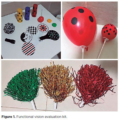
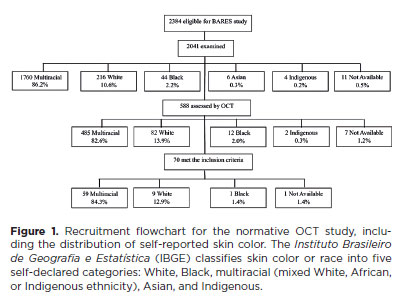
PURPOSE: Glaucoma is one of the leading causes of irreversible blindness worldwide. When topical hypotensive agents or laser trabeculoplasty fail to adequately control the disease, escalation of therapy becomes necessary, with transscleral cyclophotocoagulation being one of the available options. Several variations of transscleral cyclophotocoagulation exist, including traditional continuous wave, MicroPulse, and slow-coagulation techniques. We propose a novel variation – custom slow-coagulation transscleral cyclophotocoagulation – which combines elements of both continuous wave and slow-coagulation approaches. This study aimed to evaluate the outcomes of this technique in patients with refractory glaucoma.
METHODS: This retrospective, interventional study included 104 eyes of 83 patients with refractory glaucoma who underwent custom slow-coagulation transscleral cyclophotocoagulation. Changes in intraocular pressure, visual acuity, the number of glaucoma medications, and postoperative complications were analyzed. A paired t test was used to compare changes in intraocular pressure and visual acuity, while the Wilcoxon signed-rank test was applied to categorical variables. Success rates following custom slow-coagulation transscleral cyclophotocoagulation were estimated using Kaplan–Meier survival analysis.
RESULTS: Mean intraocular pressure decreased significantly from 38.9 ± 15.8 mmHg at baseline to 16.3 ± 9.9 mmHg at Month 12 (p<0.001). The mean number of glaucoma medications also decreased significantly from 3.6 ± 0.6 to 1.8 ± 1.4 (p<0.001). No significant reduction in mean visual acuity was observed during follow-up.
CONCLUSIONS: Custom slow-coagulation transscleral cyclophotocoagulation effectively reduced baseline intraocular pressure and the number of glaucoma medications, with a low rate of complications and no decline in visual acuity over a 12-month follow-up period. This novel technique demonstrated a high safety profile in a Hispanic population and represents a low-cost, minimally invasive procedure with rapid recovery and promising efficacy in intraocular pressure control.
Keywords: Glaucoma/surgery; Sclera; Filtering surgery; Laser coagulation/methods; Lasers, semiconductor/therapeutic use; Intraocular pressure; Blindness/prevention & control; Vision, low/epidemiology; Visual acuity
Abstract
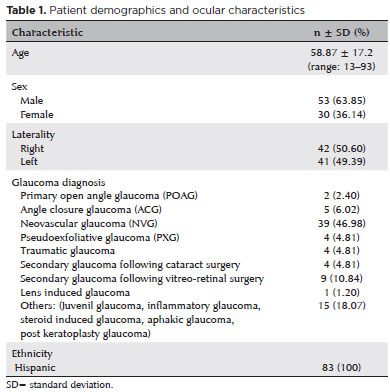
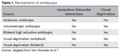
PURPOSE: Amblyopia is a cortical neurological disorder caused by abnormal visual experiences during the critical period for visual development. Recent works have shown that, in addition to the well-known visual alterations, such as changes in visual acuity, several perceptual aspects of vision are affected. This study aims to analyze and compare the effects of different types of amblyopia on visual color processing and determine whether these effects are correlated with visual acuity.
METHODS: Our study sample comprised 42 amblyopic individuals, aged 7-40 years, (strabismus, n=16; anisometropia, n=18; and mixed-cause, n=8) and 33 age-matched controls. Color vision was tested by measuring the chromaticity threshold of each patient on the protan, deutan, and tritan axes using version 02 of the Cambridge Color Test. Spatial stimulation cues were eliminated using spatial noise and luminance.
RESULTS: The color discrimination thresholds on the protan, deutan, and tritan axes were similar between control participants and amblyopic patients (p>0.05). There was no correlation between VA values and color thresholds (p>0.05).
CONCLUSION: Patients with amblyopia have normal color vision in contexts that include luminance and spatial noise. Our results may be indicative of independent neural pathways for spatial and chromatic visual processing.
Keywords: Amblyopia; Anisometropia; Color vision; Strabismus; Vision disorders; Visual acuity
Abstract
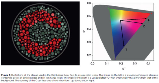
PURPOSE: This study evaluated macular thickness using spectral-domain optical coherence tomography in healthy participants from a population-based eye survey.
METHODS: The Brazilian Amazon Region Eye Survey was a population-based study assessing the prevalence and causes of visual impairment, blindness, and ocular diseases in adults aged ≥45 years from urban and rural areas of Parintins. A subgroup was selected based on inclusion criteria for both eyes: best-corrected visual acuity ≥20/32, normal eye examination results, and no prior ocular surgery. Scans were performed using the iVue optical coherence tomography device. Measurements were taken from the nine subfields defined by the Early Treatment Diabetic Retinopathy Study, examining the full retina as well as the inner and outer retinal layers. Associations of retinal thickness with age and sex were also analyzed. Statistical significance was set at p≤0.05.
RESULTS: In total, 70 healthy participants (25 males), aged 45–65 years (mean=52 ± 5), were included. Mean central foveal thickness was 248.71 ± 18.73 μm. A significant age-related reduction in macular thickness was observed, particularly in the inner superior parafovea (p=0.036), nasal perifovea (p=0.001), superior perifovea (p=0.028), outer layer of inferior parafovea (p=0.049), and the inferior perifovea of the full retina (p=0.029). Males showed significantly greater thickness in the outer layer, especially in the outer parafovea (p=0.004) and perifovea (p<0.0001).
CONCLUSIONS: This study established normative macular thickness values for healthy older adults in the Brazilian Amazon region using spectral-domain optical coherence tomography. Age and sex were found to significantly influence macular thickness and should be considered when interpreting measurements. These data will support future studies of retinal diseases in this population.
Keywords: Retinal diseases/diagnosis; Macula lutea/pathology; Macular degeneration/diagnosis; Diabetic retinopathy/diagnosis; Vision, low; Vision tests; Tomography, optical coherence/methods; Young adult; Cross-sectional studies; Brazil/epidemiology
Abstract
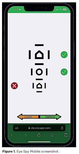
PURPOSE: This cross-sectional study compared best-corrected visual acuity obtained using Cloudscaper symbols, a novel optotype developed according to ETDRS specifications for children's virtual screening, with that obtained using LEA symbols.
METHODS: A total of 560 children aged 3-16 yr underwent visual acuity test with both Cloudscaper symbols and LS. The test application was standardized using the EyeSpy algorithm. Additionally, 147 participants were tested with the standard Snellen E paper chart. Paired t tests were performed to assess the clinical significance of logMAR visual acuity differences.
RESULTS: The mean logMAR visual acuity with LEA symbols was 0.12 (standard deviation [SD]=0.18; range, -0.10 to 0.80), while with Cloudscaper symbols it was 0.18 (SD=0.19; range, -0.10 to 0.80). The mean difference between Cloudscaper symbols and LEA symbols was 0.099 logMAR (approximately 0.5 optotypes; SD=0.08; range, 0.0-0.14; p<0.0001). Cloudscaper symbols slightly underestimated visual acuity compared to LEA symbols. Visual acuity measured by both methods was highly correlated (Spearman's r=0.74, p<0.0001). The mean visual acuity difference between Cloudscaper symbols and the Snellen E chart was 0.0045 (p=0.805; 95% confidence interval [95% CI]), whereas the difference between LEA symbols and Snellen E was 0.0883 (p<0.001; 95% CI).
CONCLUSIONS: Cloudscaper symbols provide a reliable tool for visual screening in children. Although they slightly underestimate visual acuity compared to LEA symbols – a finding also reported when comparing ETDRS letters with LEA symbols – Cloudscaper symbols show strong agreement with Snellen E chart measurements. This suggests that Cloudscaper symbols allow precise visual acuity assessment comparable to the gold standard.
Keywords: Vision screening; Vision tests; Visual acuity; Mobile applications; Eye health; Child health; Diagnostic techniques, Ophthalmological; Child; Preschool child; Adolescent
Abstract
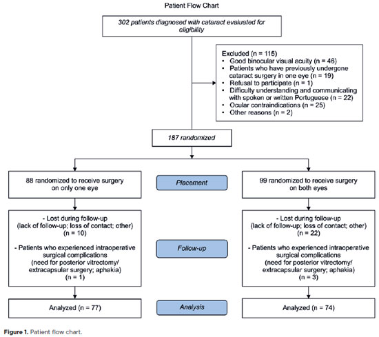
PURPOSE: This prospective, randomized, unmasked, clinical trial aimed to report the visual outcomes of cataract surgery on both eyes versus cataract surgery on one eye in Brazilian patients.
METHODS: This study included patients with bilateral cataracts and binocular visual acuity worse than or equal to 0.3 logarithm of the minimum angle of resolution. The patients were randomly assigned to undergo surgery on one (Control Group) or both eyes (one eye at a time; Intervention Group). Postoperatively, self-reported visual function using Catquest-9SF (primary outcome measure), binocular visual acuity, stereopsis, and ocular dominance (secondary outcome measures) were compared.
RESULTS: A total of 151 patients (77 and 148 eyes in the Control and Intervention Groups, respectively) completed the follow-up. Patients who underwent surgery on both eyes exhibited significantly better self-reported visual function (p=0.036) and stereopsis (p=0.026) than those who underwent surgery on one eye. Binocular visual acuity and ocular dominance did not affect the group comparisons.
CONCLUSIONS: Surgery on both eyes resulted in significantly better self-reported visual function and stereopsis than surgery on one eye.
Keywords: Cataract; Cataract extraction; Quality of life; Treatment outcome; Visual acuity; Binocular vision; Stereopsis
Abstract
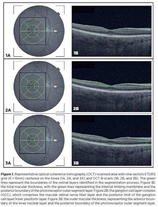
PURPOSE: This study aimed to evaluate the total macular thickness as well as the thickness of the inner and outer retinal layers in patients with Parkinson's disease. It also aimed to verify the correlation of these parameters with motor symptoms and cognitive function.
METHODS: A total of 46 eyes of 23 patients with Parkinson's disease and 40 eyes of 20 healthy controls were included in the study. The patients' cognitive, functional, and nonmotor symptoms were evaluated using the Katz Index of Independence and Pfeffer's Activities of Daily Living, Mini-Mental State Examination, Frontal Assessment Battery, Schwab and England Staging Scales, and Movement Disorders Society Nonmotor Symptoms Scale. The macular thickness measurements obtained via total, inner, and outer optical coherence tomography were recorded. Furthermore, the correlation of the parameters of optical coherence tomography with cognitive, functional, and nonmotor symptoms was assessed.
RESULTS: The scores of the Katz Index of Independence and Pfeffer's Activities of Daily Living as well as the Movement Disorders Society Nonmotor Symptoms Scale were significantly lower in patients with Parkinson's disease than in healthy controls. Moreover, the former had greater total macular thickness. The temporal and inferior outer sectors were significantly greater for the ganglion cell complex thickness in patients. A significant correlation was observed between the total macular thickness and the Movement Disorder Society-Unified Parkinson's Disease Rating Scale, Parte III (MDS-UPDRS-III) values. Contrarily, there was a negative correlation between the outer macular thickness and the MDS-UPDRS-III values. Meanwhile, the total macular thickness and ganglion cell complex thickness were significantly correlated with the scores of the Mini-Mental State Examination, Schwab and England Staging Scale, Frontal Assessment Battery, and Katz Index of Independence and Pfeffer's Activities of Daily Living. In addition, the Schwab and England scale was correlated with the outer macular thickness.
CONCLUSION: The total and inner macular thicknesses at the temporal and inferior outer sectors were greater in patients with Parkinson's disease than in the control group. These findings indicate that macular thickness may be greater in those with Parkinson's disease, particularly when associated with mild motor symptoms. In addition, the parameters of the total, inner, and outer optical coherence tomography were significantly associated with motor and nonmotor symptoms as well as cognitive function impairment.
Keywords: Parkinson's disease; Tomography, optical coherence; Neurodegenerative diseases; Cognitive dysfunction; Cognition; Motor perception; Visual acuity; Retina
13-fig01.jpg)
Abstract
O treinamento de biofeedback por microperimetria é um método de reabilitação da visão que envolve treinamento de atenção, controle oculomotor e reabilitação do locus preferencial de fixação da retina. Esse treinamento pode melhorar significativamente a acuidade visual para longe e perto na degeneração macular relacionada à idade. Estudos anteriores mostraram que o treinamento de biofeedback usando a nistagmografia elétrica pode reduzir a amplitude do nistagmo e aumentar o período de foveação. Entretanto, os resultados não se mantiveram após o término das sessões. Aqui é relatado um caso de tratamento com biofeedback por microperimetria para melhorar a acuidade visual e a estabilidade de fixação em uma criança de 11 anos de idade. O treinamento teve impacto benéfico e afetou positivamente a estabilidade da fixação e a visão para longe, para perto e de leitura. Subjetivamente, foi relatada melhoria da qualidade de vida. Em contraste com estudos anteriores, os efeitos positivos foram mantidos até 12 meses após a terapia. Até onde sabemos, este é o primeiro caso na literatura que relata benefícios de longo prazo.
Keywords: Nistagmo patológico/reabilitação; locus retiniano preferencial; Baixa visão; Testes de campo visual
16-fig01.jpg)
Abstract
O uso de preenchedores dérmicos é uma prática bem estabelecida de rejuvenescimento facial. Embora seja um procedimento minimamente invasivo, pode levar a complicações graves como cegueira. Uma revisão de casos de perda visual pós preenchimento facial estético foi conduzida para descrever os mecanismos, considerações anatômicas, quadro oftalmológico, atuais estratégias de prevenção e manejo, e tendências ao longo dos anos. Foram identificados 233 casos, e 172 pacientes tiveram ao menos um olho com baixa visão ao final do seguimento. O paciente típico é uma mulher jovem submetida a preenchimento de ácido hialurônico na glabela ou nariz, apresentando dor ocular súbita, ptose e oftalmoplegia devido à oclusão vascular. Este estudo também destaca um possível aumento de profissionais não habilitados realizando este procedimento. Apesar do contínuo desenvolvimento dos preenchedores dérmicos e aprimoramento das opções de tratamento disponíveis, mais estudos e estratégias são necessários para reduzir a incidência de complicações e minimizar suas consequências.
Keywords: Preenchedor dérmico, Injeção; Técnica cosmética/efeito adverso; Oclusão da artéria retiniana; Baixa visão/etiologia
Abstract
This study aimed to propose a guideline for amblyopia treatment and follow-up. Studies show that amblyopia leads to a series of perceptual deficits, including loss of visual acuity, stereoacuity, and contrast sensitivity. Perceptual changes are also found in the sound eye, such as those involving the types of motion perception. The gold standard of treatment remains the prescription of eyeglasses, when indicated, and patching of the dominant eye. The treatment is mostly effective in patients aged <7 years and must be discontinued gradually, tapering off patching for at least 5 weeks. Atropine may be performed for penalization in hyperopic children whose amblyopic eye has better visual acuity under cycloplegia than the fellow eye. The discovery of significant neural plasticity in the amblyopic brain after the critical period opens possibilities for new treatment modalities even after childhood.
Keywords: Amblyopia; Atropine; Contrast sensitivity; Motion perception; Eyeglasses; Visual acuity; Prescriptions
 ABO is licensed under a Creative Commons Attribution-NonComercial 4.0 Internacional.
About
Issues
Editorial Board
Submission
Arquivos Brasileiros de Oftalmologia
Official publication of Brazilian Council of Ophthalmology - Conselho Brasileiro de Oftalmologia (CBO)
Rua Casa do Ator, 1.117 - 2nd floor - Zip Code: 04546-004
São Paulo - SP, Brazil
TEL: +55 11 3266-4000
E-mail: [email protected]