Arq. Bras. Oftalmol. 2021;84 (3 )
:209-213
| DOI: 10.5935/0004-2749.20210035
Abstract
OBJETIVOS: Identificar vasos linfáticos em espécimes orbitários de cadáveres humanos através de microscopia óptica e análise imunohistoquímica.
MÉTODOS: Um estudo postmortem incluiu dez espécimes orbitários provenientes de dez cadáveres humanos. Todos os espécimes orbitários foram obtidos até 12 horas após a morte com uma técnica cirúrgica de exenteração orbitária e dissecados em glândula lacrimal, nervo óptico, gordura órbitária e músculos extraoculares. Para classificar como um vaso linfático, os critérios histológicos incluíram vasos endoteliais de parede única sem membrana basal bem desenvolvida, irregulares e lúmen sem hemácias, e os critérios imunohistoquímicos incluíram vasos endoteliais de parede única, com formato irregular e lúmen sem hemácias e reagentes a podoplanina D2-40.
RESULTADOS: As lâminas histológicas de glândula lacrimal, nervo óptico, tecido adiposo e músculos extraoculares reagiram positivamente a podoplanina D2-40.
CONCLUSÃO: Este estudo demonstrou vasos linfáticos na órbita humana, mais exatamente, na glândula lacrimal, no nervo óptico, na gordura orbitária e nos músculos extrínsecos extraoculares via microscopia óptica e imunohistoquímica.
Keywords: Vasos linfáticos; Órbita; Nervo óptico; Aparelho lacrimal; Músculos oculomotores; Tecido adiposo; Microscopia
Arq. Bras. Oftalmol. 2025;88 (6 )
:1-9
| DOI: 10.5935/0004-2749.2024-0411
Abstract
PURPOSE: This study evaluated macular thickness using spectral-domain optical coherence tomography in healthy participants from a population-based eye survey.
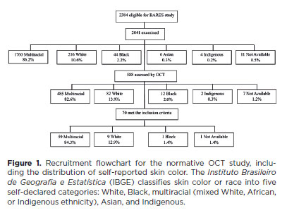
METHODS: The Brazilian Amazon Region Eye Survey was a population-based study assessing the prevalence and causes of visual impairment, blindness, and ocular diseases in adults aged ≥45 years from urban and rural areas of Parintins. A subgroup was selected based on inclusion criteria for both eyes: best-corrected visual acuity ≥20/32, normal eye examination results, and no prior ocular surgery. Scans were performed using the iVue optical coherence tomography device. Measurements were taken from the nine subfields defined by the Early Treatment Diabetic Retinopathy Study, examining the full retina as well as the inner and outer retinal layers. Associations of retinal thickness with age and sex were also analyzed. Statistical significance was set at p≤0.05.
RESULTS: In total, 70 healthy participants (25 males), aged 45–65 years (mean=52 ± 5), were included. Mean central foveal thickness was 248.71 ± 18.73 μm. A significant age-related reduction in macular thickness was observed, particularly in the inner superior parafovea (p=0.036), nasal perifovea (p=0.001), superior perifovea (p=0.028), outer layer of inferior parafovea (p=0.049), and the inferior perifovea of the full retina (p=0.029). Males showed significantly greater thickness in the outer layer, especially in the outer parafovea (p=0.004) and perifovea (p<0.0001).
CONCLUSIONS: This study established normative macular thickness values for healthy older adults in the Brazilian Amazon region using spectral-domain optical coherence tomography. Age and sex were found to significantly influence macular thickness and should be considered when interpreting measurements. These data will support future studies of retinal diseases in this population.
Keywords: Retinal diseases/diagnosis; Macula lutea/pathology; Macular degeneration/diagnosis; Diabetic retinopathy/diagnosis; Vision, low; Vision tests; Tomography, optical coherence/methods; Young adult; Cross-sectional studies; Brazil/epidemiology
Arq. Bras. Oftalmol. 2025;88 (4 )
:1-6
| DOI: 10.5935/0004-2749.2024-0278
Abstract
PURPOSE: This study aimed to evaluate the prevalence of orbital conditions in a tertiary ophthalmic outpatient hospital in Sao Paulo, Brazil, with a focus on the main diagnoses and their distribution.
METHODS: A retrospective chart review was conducted involving patients registered and admitted to the orbital disease unit at the Department of Ophthalmology, University of São Paulo Medical School, from January 2004 to March 2018. A total of 838 medical charts were analyzed, of which 37 were excluded due to incomplete data. The remaining charts were categorized into eight diagnostic groups: Graves’ orbitopathy , inflammatory disorders, tumors, vascular lesions, acquired structural abnormalities, congenital structural abnormalities, infectious diseases, and others.
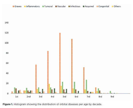
RESULTS: Of the 837,300 ophthalmological appointments, 3,372 (0.4%) were related to orbital diseases. The study included 801 patients, of whom 63.45% were women. The patients’ mean age was 42.86 years. Graves’ orbitopathy was the most common (55%), followed by tumor (17%), inflammatory disorders (9%), vascular lesions (7%), acquired structural abnormalities (5%), congenital structural abnormalities (4%), others (2%), and infectious diseases (1%). The study found significant differences in the incidence and types of orbital diseases, indicating the specialized nature of tertiary care and referral biases.
CONCLUSION: Published data on epidemiological orbital diseases is scarce. Therefore, this study focused on the diverse nature of orbital diseases and their low incidence among ophthalmology appointments. The major trends align with other epidemiological studies, demonstrating a preponderance of Graves’ orbitopathy in middle-aged adults and a bimodal distribution of tumors. These findings are essential in shaping resident training programs and healthcare policies, particularly in tertiary settings. Understanding the epidemiology of orbital diseases can improve diagnostic accuracy, treatment approaches, and patient outcomes as well as support future systemic prospective studies.
Keywords: Orbital diseases; Orbital tumors; Neoplasms; Inflammation; Graves’ ophthalmopathy; Outpatients
Arq. Bras. Oftalmol. 2024;87 (4 )
:1-8
| DOI: 10.5935/0004-2749.2021-0415
Abstract
Objetivo: Fenótipos Stargardt-like já foram associados a variantes patogênicas no gene ABCA4. O propósito desse estudo é descrever quatro pacientes com achados retinianos semelhantes a doença de Stargardt com resultados moleculares diferentes do esperado.
Métodos: Esse relato fez a revisão de prontuários médicos de quatro pacientes com distrofia macular e achados clínicos sugestivos de doença de Stargardt. Foram realizados avaliação oftalmológica, exames de imagens e testes usando next generation sequencing para avaliar variantes patogênicas associadas aos fenótipos dos pacientes.
Resultados: Os pacientes apresentavam atrofia macular e alterações pigmentares sugerindo achados clínicos de doença de Stargardt. Dois pacientes foram associados a genes com herança autossômica dominante (RIMS1 e CRX) e dois pacientes foram associados a genes com herança autossômica recessiva (CRB1 e RDH12) com variantes preditoras de serem patogênicas.
Conclusão: Distrofias maculares podem ter similaridades fenotípicas com fenótipo de Stargardt-like associados a outros genes além dos classicamente já descritos.
Keywords: Doença de Stargardt; Estudos de associação genética; Fenótipo; Padrões de herança; Sequenciamento de nucleotídeos em larga escala; Degeneração macular; Distrofias retinianas; Doenças genéticas
Arq. Bras. Oftalmol. 2024;87 (2 )
:1-6
| DOI: 10.5935/0004-2749.2021-0527
Abstract
OBJETIVO: Os níveis séricos de N-óxido de trimetilamina têm sido associados ao diabetes mellitus tipo 2 e suas complicações. O presente estudo tem como objetivo responder a duas questões, entre elas: O nível plasmático de N-óxido de trimetilamina poderia ser um novo marcador no diagnóstico de retinopatia diabética? e Ele poderia ser utilizado no diagnóstico diferencial de retinopatia diabética e não diabética?
MÉTODOS: Trinta pacientes com retinopatia diabética, 30 pacientes com retinopatia não diabética, 30 pacientes com diabetes mellitus tipo 2 sem retinopatia e 30 participantes saudáveis do grupo controle foram incluídos no estudo. Parâmetros bioquímicos, níveis séricos de IL-6, de TNF-α e de N-óxido de trimetilamina foram medidos em todos os participantes.
RESULTADOS: O nível de N-óxido de trimetilamina foi significativamente maior na retinopatia diabética do que nos outros grupos (p<0,001). Não houve diferença significativa no nível de N-óxido de trimetilamina entre o grupo de retinopatia não diabética, do grupo controle ou do grupo de diabetes mellitus tipo 2. Houve uma correlação positiva significativa entre o nível de N-óxido de trimetilamina e os níveis elevados de FPG, IMC, HOMA-IR, BUN, IL-6 e TNF-α.
CONCLUSÃO: O estudo atual mostrou que o nível de N-óxido de trimetilamina encontra-se elevado na retinopatia diabética. Esses achados sugerem que o nível sérico de N-óxido de trimetilamina pode ser um novo marcador na retinopatia diabética, podendo ser usado no diagnóstico diferencial de retinopatia diabética e não diabética.
Keywords: Biomarcadores; Retinopatia diabética; Diagnóstico diferencial; Óxidos; Diabetes mellitus tipo 2; N-óxido de trimetilamina; Trimetilamina


 ABO is licensed under a Creative Commons Attribution-NonComercial 4.0 Internacional.


02-fig01.jpg)
05-tab01tb.jpg)
10-tab01tb.jpg)
13-fig01.jpg)
11-fig01.jpg)
14-fig01.jpg)
03-fig01.jpg)
07-tab01tb.jpg)