Arq. Bras. Oftalmol. 2023;86 (3 )
:1-4
| DOI: 10.5935/0004-2749.20230041
Abstract
Objetivos: Avaliar o perfil das consultas de emergência oftalmológicas durante os primeiros meses de quarentena por pandemia de covid-19 em um hospital oftalmológico de referência em São Paulo e compará-lo com o mesmo período do ano anterior.
Métodos: Os dados foram extraídos do serviço de emergência do Hospital Ophthal Especializado, São Paulo, Brasil. Todos os casos registrados entre 23 de março e 19 de maio de 2020 foram incluídos no estudo como Grupo 2020. Os casos registrados entre 23 de março e 19 de maio de 2019 foram incluídos no estudo como Grupo de 2019. Tabelas de frequência foram utilizadas para análises descritivas. Os testes Qui-quadrado e Exato de Fisher foram aplicados para comparar variáveis categóricas entre os grupos.
Resultados: Observamos uma diminuição de 46,15% no número de casos durante a pandemia Covid-19 em 2020 quando comparado ao mesmo período sem pandemia em 2019. Foi observado um aumento significativo nos achados de pálpebra (+12,3%), córnea (+97,1%), retina (+173,1%), refração (+62,9%), glaucoma (+43,9%), esclera (+68,8%), trauma (+39,3%), herpes (+54,7%) e catarata (+549,9%) em 2020 quando comparado para 2019. Por outro lado, houve redução nos casos de conjuntivite (-33,4%), transtornos do aparelho lacrimal (-81,0%), iridociclite (-39,9%) e consultas pós-operatórias (-80,1%).
Conclusão: durante a fase inicial da pandemia Covid-19, foi observada uma redução drástica no número de pacientes que procuram o serviço de emergência ocular. Houve também uma mudança nos principais motivos para visitas com frequências mais altas de casos de alta gravidade como distúrbios da retina, distúrbios da córnea, glaucoma e trauma, e frequências mais baixas de condições transmissíveis como conjuntivite.
Keywords: COVID-19; Emergências; Isolamento Social; Oftalmologia; Quarentena.
Arq. Bras. Oftalmol. 2021;84 (3 )
:214-219
| DOI: 10.5935/0004-2749.20210029
Abstract
OBJETIVO: Avaliar a influência da dinâmica pupilar na curva de desfoco de olhos implantados com lente intraoculares multifocais difrativas.
MÉTODOS: Estudo prospectivo e randomizado realizado na Faculdade de Medicina de Ribeirão Preto - Universidade de São Paulo - Departamento de Oftalmologia. Trinta e oito pacientes foram aleatoriamente designados para receber bilateralmente lentes intraoculares SN6AD1 (n=20) (mfIOL) ou SN60WF (n=18) (aIOL). Além da acuidade visual para longe e perto, corrigida e não corrigida, e curva de desfoco, foi ainda realizada pupilometria dinâmica. A área sob a curva de desfoco foi calculada usando um modelo polinomial empírico.
RESULTADOS: Um total de 16 e 17 pacientes (n=32 e 34 olhos) completaram 1 ano de seguimento nos grupos mfIOL e aIOL, respectivamente. Não houve diferenças significativas entre grupos para as acuidades visuais seja para longe ou perto. As curvas de desfoco do grupo mfIOL mostraram um pico duplo; enquanto o SN60WF mostrou apenas um pico, típico para uma lente intraoculares monofocal. A média da área sob a curva de desfoco do grupo aIOL foi (4,66 ± 1,51 logMAR.dp), e essa é estatisticamente significante diferente da métrica do grupo mfIOL (1,99 ± 1,31 logMAR.dp). A pupila na contração máxima após a exposição a um flash de 30 cd/m2 por 1 segundo foi significativamente correlacionada com uma melhor área de foco no grupo mfIOL (r=0,54; p=0,0017), essa relação não foi observada para o grupo aIOL.
CONCLUSÃO: Estes dados indicam que quanto menor a pupila durante contração, melhor é a área sob a curva de desfoco e, portanto, o desempenho visual dos olhos implantados com essa mfIOL. Esta correlação não foi encontrada para lentes intraoculares monofocais.
Keywords: Lentes intraoculares multifocais; Pupila/fisiologia, Catarata; Facoemulsificacão
Arq. Bras. Oftalmol. 2021;84 (1 )
:17-21
| DOI: 10.5935/0004-2749.20210003
Abstract
OBJETIVO: Comparar a gravidade e a lateralidade do ceratocone de acordo com a rinite alérgica, os hábitos de coçar e dormir e a destreza manual.
MÉTODOS: Foram realizadas questões objetivas sobre rinite alérgica, prurido ocular e posição do sono em pacientes com ceratocone, diagnosticados com base na tomografia corneana. Esses exames foram analisados e classificados de acordo com a classificação de Amsler-Krumeich.
RESULTADOS: O prurido ocular foi referido por 29 (85,29%) dos 34 voluntários. Dezoito sujeitos (62,07%) relataram coçar ambos os olhos igualmente, 6 (20,69%) mais no olho direito e 5 (17,24%) mais no olho esquerdo. Comparando-se a posição de dormir principal e o olhos com apresentação mais grave da doença, foi encontrada alguma relação baseada no teste exato de Fisher (0,567 no olho direito e 0,568 no olho esquerdo), embora nenhuma comparação parecesse estatisticamente significante.
CONCLUSÕES: A associação entre maiores valores de ceratometria e posição do sono parece ser mais importante do que entre ceratometria e prurido ou destreza manual.
Keywords: Ceratocone; Hipersensibilidade; Sono/fisiologia; Rinite alérgica; Córnea; Tomografia
Arq. Bras. Oftalmol. 2021;84 (6 )
:561-568
| DOI: 10.5935/0004-2749.20210093
Abstract
Objetivo: Fornecer orientações sobre a frequência e os componentes dos exames oftalmológicos para crianças saudáveis de 0 a 5 anos.
Métodos: Essas diretrizes foram desenvolvidas com base em revisão bibliográfica e experiência clínica de um comitê de especialistas. Foram realizadas buscas PubMed/Medline; documentos selecionados não se restringiram a revisões sistemáticas, ensaios clínicos randomizados e estudos observacionais. Quando adequado, o perfil GRADE foi aplicado para graduá-los e o consenso de especialistas foi usado nos tópicos sem evidência científica. Também foram revisadas as recomendações pela Academia Americana de Pediatria, Associação Americana de Oftalmologia Pediátrica e Estrabismo, Academia Americana de Oftalmologia, Royal College of Ophthalmologist e Sociedade Canadense de Oftalmologia. O documento final foi aprovado pela Sociedade Brasileira de Oftalmologia Pediátrica e Sociedade Brasileira de Pediatria.
Resultados: Os recém-nascidos devem ser submetidos ao teste do reflexo vermelho e inspeção dos olhos e anexos pelo pediatra dentro de 72 horas de vida ou antes da alta da maternidade. O teste do reflexo vermelho deve ser repetido pelo pediatra durante as consultas de puericultura pelo menos três vezes ao ano durante os primeiros 3 anos de vida. Se factível, um exame oftalmológico completo pode ser feito entre 6 a 12 meses de vida. Até os 36 meses de idade, os marcos visuais, função visual apropriada para a idade, fixação e alinhamento ocular também devem ser avaliados pelo pediatra ou médico da família. Pelo menos um exame oftalmológico completo deve ser realizados entre 3 e 5 anos de idade. O exame deve conter pelo menos inspeção dos olhos e anexos, avaliação da função visual apropriada para a idade, avaliação da motilidade e alinhamento ocular (testes de cobertura), refração sob cicloplegia e avaliação do fundo de olho dilatado.
Conclusões: As diretrizes sobre a frequência da avaliação oftalmológica são ferramentas importantes para orientar os médicos sobre a melhor prática a fim de evitar problemas visuais tratáveis na infância, que poderiam comprometer seu desenvolvimento social, escolar e global, além de causar perda permanente da visão.
Keywords: Técnicas de diagnóstico oftalmológico, Triagem visual; Testes visuais; Criança, Lactente
Arq. Bras. Oftalmol. 2026;89 (2 )
:1-6
| DOI: 10.5935/0004-2749.2025-0244
Abstract
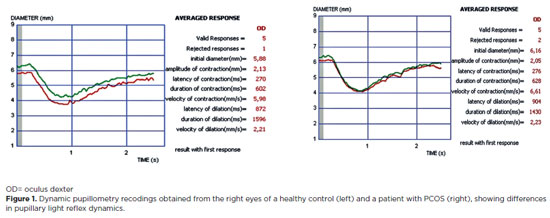
PURPOSE: Polycystic ovary syndrome is frequently associated with autonomic nervous system dysfunction, even in the absence of obesity or overt metabolic abnormalities. Alterations in pupillary responses may reflect early autonomic involvement and serve as a potential tool for early diagnosis, risk stratification, and disease monitoring. This study aimed to investigate pupillary reflex parameters using dynamic pupillometry in newly diagnosed non-obese women with polycystic ovary syndrome and to compare the findings with those of healthy controls. Methods: This prospective cross-sectional study included 48 newly diagnosed women with polycystic ovary syndrome and 44 age- and sex-matched healthy controls. Pupillary function parameters were measured using dynamic pupillometry (MonPackOne; Metrovision, France). Results: The mean age did not differ significantly between the groups (p=0.870). Initial pupil diameter, pupil contraction amplitude, and contraction velocity were significantly lower in the PCOS group than in the control group, whereas pupillary dilation duration was significantly longer (p<0.001, p<0.001, p=0.007, and p=0.032, respectively). No significant differences were observed between the groups regarding contraction latency, contraction duration, dilation latency, or dilation velocity (p=0.749, p=0.925, p=0.653, and p=0.310, respectively). Conclusion: Newly diagnosed non-obese women with polycystic ovary syndrome exhibit significant alterations in pupillary dynamics, suggesting a generalized reduction in both sympathetic and parasympathetic activity. Dynamic pupillometry may represent a practical, noninvasive tool for detecting early autonomic hypoactivity and identifying patients at risk for future metabolic or cardiovascular complications.
Keywords: Autonomic nervous system; IPupil/physiology; Reflex, Pupillary/physiology; Polycystic ovary syndrome/diagnosis; Menstruation disturbances; Ideal body weight
Arq. Bras. Oftalmol. 2025;88 (1 )
:1-4
| DOI: 10.5935/0004-2749.2022-0375
Abstract
PURPOSE: This study aimed to assess grating visual acuity and functional vision in children with congenital Zika syndrome.
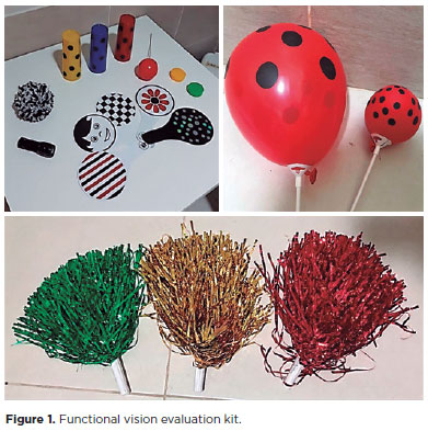
METHODS: Initial and final grating visual acuity was measured using Teller acuity cards. Cerebral vision impairment standardized tests were used to assess functional vision. Patients were referred to the early visual intervention program for visually disabled children. Neuroimaging was performed.
RESULTS: In this study, 10 children were included with an age range of 1–37 months. Eight patients presented with macular atrophic scars. Neuroimaging revealed microcephaly and cerebral abnormalities in all patients. Low vision and cerebral vision impairment characteristics were observed in all children. The final grating visual acuity in this group varied from 3.00 to 0.81 logMAR.
CONCLUSIONS: The grating visual acuity test revealed low vision in all children with congenital Zika syndrome. Functional vision evaluation revealed cerebral vision impairment characteristics in all patients, who were referred to the early visual intervention program. Visual acuity improved in six children.
Keywords: Zika virus infection/congenital; Low vision; Vision disorders; Atrophy, Microcephaly; Visual acuity; Child
Arq. Bras. Oftalmol. 2025;88 (4 )
:1-12
| DOI: 10.5935/0004-2749.2023-0263
Abstract
PURPOSE: Amblyopia is a cortical neurological disorder caused by abnormal visual experiences during the critical period for visual development. Recent works have shown that, in addition to the well-known visual alterations, such as changes in visual acuity, several perceptual aspects of vision are affected. This study aims to analyze and compare the effects of different types of amblyopia on visual color processing and determine whether these effects are correlated with visual acuity.
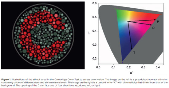
METHODS: Our study sample comprised 42 amblyopic individuals, aged 7-40 years, (strabismus, n=16; anisometropia, n=18; and mixed-cause, n=8) and 33 age-matched controls. Color vision was tested by measuring the chromaticity threshold of each patient on the protan, deutan, and tritan axes using version 02 of the Cambridge Color Test. Spatial stimulation cues were eliminated using spatial noise and luminance.
RESULTS: The color discrimination thresholds on the protan, deutan, and tritan axes were similar between control participants and amblyopic patients (p>0.05). There was no correlation between VA values and color thresholds (p>0.05).
CONCLUSION: Patients with amblyopia have normal color vision in contexts that include luminance and spatial noise. Our results may be indicative of independent neural pathways for spatial and chromatic visual processing.
Keywords: Amblyopia; Anisometropia; Color vision; Strabismus; Vision disorders; Visual acuity
Arq. Bras. Oftalmol. 2025;88 (6 )
:1-5
| DOI: 10.5935/0004-2749.2024-0340
Abstract
PURPOSE: This study aimed to report the surgical outcomes and success predictors of micropulse transscleral cyclophotocoagulation in eyes with refractory glaucoma.
METHODS: This was a noncomparative, interventional case series. Patients with refractory glaucomas, defined as eyes with prior incisional glaucoma surgery failure and uncontrolled intraocular pressure, who underwent micropulse transscleral cyclophotocoagulation between March 2017 and June 2021 were enrolled. A minimum follow-up period of 6 months was required. Preoperative and postoperative intraocular pressure, number of hypotensive medications, surgical complications, and any subsequent related events were recorded. Success criteria were as follows: 1) intraocular pressure reduction ≥20% and intraocular pressure ≤18 mmHg; 2) intraocular pressure reduction ≥30% and intraocular pressure ≤15 mmHg. The need for topical hypotensive medications was not considered a failure.
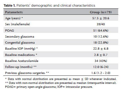
RESULTS: Seventy-nine (79) eyes (79 patients; mean age, 57.5 ± 20.6 years) were included. Overall, the median follow-up duration was 12.0 (interquartile interval, 6–24) months, and the mean intraocular pressure was reduced from 22.8 ± 6.8 mmHg to 15.5 ± 5.6 mmHg at the last follow-up visit (p<0.001). The mean number of medications was reduced from 2.8 ± 0.7 to 2.0 ± 1.0 (p<0.01). At 12 months postoperatively, the success rates for criteria 1 and 2 were 54.9% and 49.7%, respectively. Aside from one case of corneal ulcer, which fully resolved with clinical treatment, and two cases of persistent hypotony (with no visual acuity loss during follow-up), no other vision-threatening complications were observed during the postoperative period. The magnitude of intraocular pressure reduction at 1 month (adjusted to preoperative intraocular pressure; HR=1.01; p=0.002).
CONCLUSION: Our findings suggest that micropulse transscleral cyclophotocoagulation is a relatively effective alternative for managing refractory glaucomas, with minor postoperative complications. In addition, the initial intraocular pressure reduction was a statistically significant predictor of 1-year success in patients undergoing micropulse transscleral cyclophotocoagulation.
Keywords: Intraocular pressure/physiology; Glaucoma, open-angle/surgery; Trabeculectomy; Laser coagulation/methods; Tonometry, ocular/methods; Postoperative complications; Antihypertensive agents/therapeutic use.
Arq. Bras. Oftalmol. 2024;87 (6 )
:1-5
| DOI: 10.5935/0004-2749.2021-0482
Abstract
OBJETIVO: Nos últimos 20 anos, o número de escolas médicas no Brasil aumentou, mas as vagas para especialização em Oftalmologia não acompanharam a demanda crescente. Este estudo quer estimar a demanda por especialização e avaliar a oferta de oportunidades de aprendizado em Oftalmologia.
MÉTODOS: Estudo epidemiológico com pesquisa em banco de dados provenientes do Ministério da Educação e Conselho Brasileiro de Oftalmologia. Estes dados foram checados através de 120 editais publicados pelos serviços de Residência em 2021.
RESULTADOS: De 2002 a 2021, o número de vagas em faculdades de Medicina aumentou 370%, enquanto o número de vagas certificadas de especialização em Oftalmologia aumentou 64%. Houve um desalinhamento de 11.4% entre os dados do Conselho Brasileiro de Oftalmologia e do Ministério da Educação.
CONCLUSÃO: A proporção de graduados em Medicina aumentou muito mais do que a oferta de oportunidades de especialização em Oftalmologia, o impacto disto na busca por vagas de especialização não acreditadas é desconhecido, políticas de monitoramento das vagas de especialização em Oftalmologia devem ser estabelecidas.
Keywords: Oftalmologia; Ensino; Educação médica; Especialização
Arq. Bras. Oftalmol. 2026;89 (3 )
:1-9
| DOI: 10.5935/0004-2749.2025-0312
Abstract
PURPOSE: To quantitatively assess changes in central corneal sensitivity after phacoemulsification and to characterize recovery patterns up to 90 days using standardized esthesiometry.
METHODS: This prospective observational study included 44 patients (88 eyes) undergoing uncomplicated phacoemulsification with intraocular lens implantation. Central corneal sensitivity was measured using a Cochet-Bonnet® esthesiometer preoperatively and at 30 and 90 days postoperatively. Repeated-measures data were analyzed using Friedman and Wilcoxon signed-rank tests (p<0.05). Inter-eye differences were assessed with a paired Wilcoxon test. Individual changes from baseline (Δ30, Δ90) were calculated, and 90-day recovery was categorized according to thresholds aligned with the 5-mm device resolution. Spearman correlation was used to explore associations between age and Δ90.
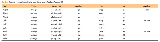
RESULTS: Corneal sensitivity decreased after surgery. In right eyes, mean sensitivity declined from 41.14 ± 7.77 mm at baseline to 36.82 ± 9.03 mm at 30 days and partially recovered to 38.64 ± 7.73 mm at 90 days. In left eyes, sensitivity decreased from 44.11 ± 6.29 mm to 37.39 ± 9.05 mm at 30 days and recovered to 41.82 ± 7.63 mm at 90 days. Left eyes showed higher sensitivity than right eyes at baseline (p=0.023) and at 90 days (p=0.018). At 90 days, complete or near-complete recovery (within ± 5 mm of baseline) occurred in 73.2% of right eyes and 78.0% of left eyes, while improvement above baseline (≥ +5 mm) occurred in 7.3% and 4.9%, respectively. Age showed weak, nonsignificant correlations with Δ90 (p=−0.14 to −0.19; p>0.2).
CONCLUSION: Phacoemulsification with a 2.75-mm clear corneal incision leads to a temporary reduction in central corneal sensitivity, with partial recovery by 90 days. Recovery patterns vary among individuals, highlighting the value of postoperative sensitivity monitoring to identify atypical trajectories and guide ocular surface care during visual rehabilitation.
Keywords: Phacoemulsification; Cornea/innervation; Ophthalmic nerve/physiology; Optometry/instrumentation; Diagnostic techniques, ophthalmological; Neural regeneration; Visual rehabilitation.
Arq. Bras. Oftalmol. 2026;89 (5 )
:1-6
| DOI: 10.5935/0004-2749.2026-0019
Abstract
PURPOSE:To evaluate ocular findings in children born to mothers who were infected with severe acute respiratory syndrome coronavirus 2 (SARS-CoV-2) during pregnancy.
METHODS: This prospective observational study was conducted in Brasília, Brazil, between October 2020 and 2021. Children born to mothers with laboratory-confirmed SARS-CoV-2 infection during pregnancy were included and compared with a control group of nonexposed children. All participants underwent a comprehensive ophthalmological examination performed by pediatric ophthalmologists, including assessment of ocular motility, slit-lamp biomicroscopy, cycloplegic refraction, intraocular pressure measurement, central corneal pachymetry, indirect ophthalmoscopy, and retinal imaging. Maternal and neonatal clinical data were also collected. Ocular parameters were compared between the exposed and control groups.
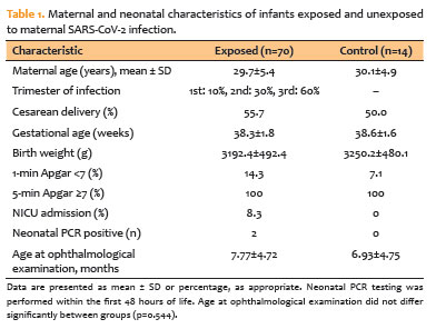
RESULTS: A total of 84 children were evaluated, including 70 exposed to maternal SARS-CoV-2 infection and 14 nonexposed controls. The mean gestational age was 38.3±1.8 weeks, and the mean birth weight was 3,192±492 g. No significant differences were observed between the groups in terms of ocular motility, anterior or posterior segment findings, intraocular pressure, central corneal thickness, or refractive status. Isolated findings, including epicanthus and congenital ptosis, were observed in a small number of children but were not significantly associated with maternal SARS-CoV-2 infection.
CONCLUSIONS: In this prospective cohort, no significant ocular abnormalities were identified in children exposed in utero to maternal SARS-CoV-2 infection compared with nonexposed controls. However, the relatively small and unbalanced control group limits the strength of these findings, which should not be interpreted as evidence that maternal SARS-CoV-2 infection does not affect ocular development. Larger prospective studies with adequately sized and balanced comparison groups are warranted to further investigate the potential ocular effects of prenatal SARS-CoV-2 exposure.
Keywords: COVID-19; SARS-CoV-2; Pregnancy; Infants; Ocular findings; Vertical transmission; Ophthalmology
Arq. Bras. Oftalmol. 2026;89 (5 )
:1-9
| DOI: 10.5935/0004-2749.2026-0074
Abstract
PURPOSE:To determine the frequency and clinical characteristics of ophthalmologic manifestations in children aged ≤16 years with autism spectrum disorder who were evaluated at a private academic healthcare network and a public tertiary hospital.
METHODS: This retrospective descriptive study was conducted through a review of the medical records of children aged <16 years with autism spectrum disorder who were examined between 2023 and 2024. Demographic characteristics, visual function testability, refractive errors, strabismus, and prescribed treatments were analyzed. Dynamic retinoscopy was used to objectively assess the binocular accommodative response. Statistical analyses included 95% confidence intervals (Wilson score method) and exploratory multivariate logistic regression.
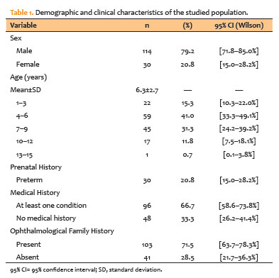
RESULTS: A total of 144 children were included; 79.2% were male, and the mean age was 6.3±2.7 years. Refractive errors were identified in 81.9% of children, of whom 63.2% had clinically significant errors requiring optical correction. Strabismus was diagnosed in 32.6% of cases, with exotropia being the most prevalent type. Stereopsis was successfully assessed in 72.2% of children. Notably, dynamic retinoscopy demonstrated perfect agreement (κ=1.000, p<0.001) with the presence of clinically significant refractive errors. Multivariate regression showed that increasing age was strongly associated with successful visual acuity testing, whereas neurological comorbidities and greater autism spectrum disorder severity significantly increased the odds of non-testability. Among children who continued follow-up, 63.9% experienced significant improvement in visual acuity. Although optical correction was generally well tolerated, adherence to occlusion therapy was notably low (6.8%), and 42.4% of the cohort was lost to follow-up.
CONCLUSIONS: Ophthalmologic abnormalities, particularly refractive errors and strabismus, are highly prevalent among children with autism spectrum disorder. Given the limited reliability of subjective visual assessment in this population, objective measures, particularly dynamic retinoscopy and cycloplegic refraction, are essential for accurate diagnosis. The substantial loss to follow-up further highlights the need for adapted clinical strategies to ensure continuity of ophthalmologic care in this population.
Keywords: Autism spectrum disorder; Pediatric ophthalmology; Refractive errors; Strabismus


 ABO is licensed under a Creative Commons Attribution-NonComercial 4.0 Internacional.


07-tab01.jpg)
03-fig01.jpg)
03-tab01.jpg)
06-tab01tb.jpg)
12-tab01.jpg)
11-fig01tb.jpg)
06-fig01.jpg)