

Arq. Bras. Oftalmol. 2026; 89 (5): 10.5935/0004-2749.2026-0074
Total: 427
Lucía Pereira1; Ivana Ormazábal1; Geraldine Kohn1; Marcela Dillems1; Rodolfo Garretón1; César Simonovis2
DOI: 10.5935/0004-2749.2026-0074
ABSTRACT
PURPOSE:To determine the frequency and clinical characteristics of ophthalmologic manifestations in children aged ≤16 years with autism spectrum disorder who were evaluated at a private academic healthcare network and a public tertiary hospital.
METHODS: This retrospective descriptive study was conducted through a review of the medical records of children aged <16 years with autism spectrum disorder who were examined between 2023 and 2024. Demographic characteristics, visual function testability, refractive errors, strabismus, and prescribed treatments were analyzed. Dynamic retinoscopy was used to objectively assess the binocular accommodative response. Statistical analyses included 95% confidence intervals (Wilson score method) and exploratory multivariate logistic regression.
RESULTS: A total of 144 children were included; 79.2% were male, and the mean age was 6.3±2.7 years. Refractive errors were identified in 81.9% of children, of whom 63.2% had clinically significant errors requiring optical correction. Strabismus was diagnosed in 32.6% of cases, with exotropia being the most prevalent type. Stereopsis was successfully assessed in 72.2% of children. Notably, dynamic retinoscopy demonstrated perfect agreement (κ=1.000, p<0.001) with the presence of clinically significant refractive errors. Multivariate regression showed that increasing age was strongly associated with successful visual acuity testing, whereas neurological comorbidities and greater autism spectrum disorder severity significantly increased the odds of non-testability. Among children who continued follow-up, 63.9% experienced significant improvement in visual acuity. Although optical correction was generally well tolerated, adherence to occlusion therapy was notably low (6.8%), and 42.4% of the cohort was lost to follow-up.
CONCLUSIONS: Ophthalmologic abnormalities, particularly refractive errors and strabismus, are highly prevalent among children with autism spectrum disorder. Given the limited reliability of subjective visual assessment in this population, objective measures, particularly dynamic retinoscopy and cycloplegic refraction, are essential for accurate diagnosis. The substantial loss to follow-up further highlights the need for adapted clinical strategies to ensure continuity of ophthalmologic care in this population.
Keywords: Autism spectrum disorder; Pediatric ophthalmology; Refractive errors; Strabismus
INTRODUCTION
Autism spectrum disorder (ASD) is defined by the Centers for Disease Control and Prevention (CDC) as a group of developmental disabilities that can cause difficulties with social interaction, communication, and behavior. It is characterized by persistent deficits in communication and social interaction as well as restricted and repetitive patterns of behavior, activities, and interests(1).
Its diagnosis has increased significantly in recent decades, and it is now considered a public health issue with an estimated global prevalence of 1%. In Chile, studies estimate a prevalence of 1.96% and a male-to-female ratio of 4:1(2,3).
Current management of ASD necessitates a multidisciplinary approach that includes audiological, genetic, metabolic, and neurological evaluations as well as input from occupational therapists and speech pathologists. In this context, ophthalmological evaluation is critical, as up to 40% of these children have pathologies such as clinically significant refractive errors, strabismus, and amblyopia, which can impair cognitive and social development(4).
The purpose of this study was to determine the frequency and characteristics of ophthalmological manifestations in pediatric children (aged up to 16 years) diagnosed with ASD who attended the Pediatric Ophthalmology Service at a private academic healthcare network and a public tertiary hospital, with a focus on the pediatric ophthalmologist’s role in the multidisciplinary team.
METHODS
The Institutional Ethics Committee approved a retrospective, descriptive study that followed the principles of the Declaration of Helsinki. Children under the age of 16 diagnosed with ASD and treated by pediatric ophthalmologists at a private academic healthcare network and a public tertiary hospital from 2023 to 2024 were included. Children with severe systemic diseases were excluded.
All children received a formal diagnosis of ASD from a pediatric neurologist based on the DSM-5 criteria. The Autism Diagnostic Observation Schedule, Second Edition (ADOS-2) and the Childhood Autism Rating Scale, Second Edition (CARS-2) were used to assist with diagnostic assessment. Trained psychologists assessed cognitive functioning using the Wechsler Intelligence Scale for Children, Fifth Edition (WISC-V), as well as the Conners rating scales to assess attention-deficit/hyperactivity disorder (ADHD) symptoms and related behavioral/emotional conditions (1). Children underwent multidisciplinary follow-up, which included child psychiatry, occupational therapy, and speech therapy.
Medical records were reviewed, including demographic information, perinatal, morbid, and family history. Visual acuity, refractive error, ocular motility and sensory abnormalities, dynamic refraction, strabismus, biomicroscopy findings, color vision, and a fundus examination were all recorded. Hunter(5) described a dynamic retinoscopy technique that was used to assess binocular accommodative response and capacity. This procedure was carried out in a dimly lit room without cycloplegia (wearing distance correction when applicable) with a detailed near-fixation target placed next to the retinoscope. The retinoscopic reflex was tested during distance fixation (“with” motion) and after shifting focus to near. A normal response was defined as rapid, complete, and continuous bilateral neutralization. Responses were considered abnormal if they were incomplete, sluggish, transient, or exhibited accommodative lag. Cohen’s kappa coefficient (k) was used to assess the diagnostic agreement between dynamic retinoscopy results and the presence or absence of clinically significant refractive errors, with corresponding 95% confidence intervals (95% CIs). A k value of 1.000 indicated perfect agreement, and statistical significance was set at p<0.05. In this study, clinically significant refractive errors were determined using the American Association for Pediatric Ophthalmology and Strabismus (AAPOS) guidelines for amblyopia risk factors(6). For children aged 12 to 30 months, the following threshold criteria were used to determine clinical significance and indicate optical correction: astigmatism >2.00 D, hyperopia >+4.50 D, anisometropia >2.50 D, or myopia >−3.50 D. D. For children aged 31–48 months, astigmatism >2.00 D, hyperopia >+4.00 D, anisometropia >2.00 D, or myopia >−3.00 D; for children over 48 months: astigmatism >1.50 D, hyperopia >+3.50 D, anisometropia >1.50 D, or myopia >−1.50 D.
Treatment responses and follow-ups were also documented. Because objective measurement tools were unavailable, treatment adherence for both spectacles and occlusion therapy was assessed retrospectively using caregiver reports documented in the clinical follow-up notes. Adherence was classified qualitatively as follows: “Adequate adherence” (consistent daily use as prescribed), “partial adherence” (intermittent or inconsistent use), and “poor or low adherence” (rare or no use, or complete rejection of the treatment).
Python was used for statistical analysis (SciPy library, version 1.10). The normality of continuous variables was examined. Age was not normally distributed, so it was expressed as median and interquartile range and compared between groups using the non-parametric Mann–Whitney U test; continuous variables were presented as mean±standard deviation (SD). Categorical variables were summarized as absolute frequencies and percentages. Point estimates of prevalence and testability rates were reported, alongside their respective 95% CIs, calculated using the Wilson score method for binomial proportions. Bivariate comparisons between clinical subgroups were conducted. When expected cell counts were <5, categorical variables were compared using Pearson’s chi-square (χ2) test or Fisher’s exact test. Additionally, an exploratory multivariate logistic regression analysis was carried out. A two-tailed p-value <0.05 was deemed statistically significant for all analyses.
RESULTS
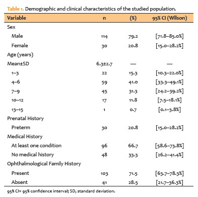
A total of 144 children were included, the majority from a private academic healthcare network (n=130, 90.3%) and the remainder from a public tertiary hospital (n=14, 9.7%). According to the DSM-5, the majority of children (74.3%, n=107) required Level 1 support, followed by Levels 2 (14.6%, n=21) and 3 (11.1%, n=16). Table 1 shows demographic and clinical characteristics in detail. The sample primarily consisted of school-aged males with a mean age of 6.3±2.7 years. In terms of perinatal history, nearly one-fifth of the children were born preterm, with moderate to late prematurity accounting for the majority (73.3%). Although a family history of ophthalmological conditions was common (71.5%), refractive errors were responsible for the vast majority of these cases (87.4%). Within this group, a clinically relevant subset of 60 children (41.7% of the total sample) had both a family history of refractive errors and needed glasses, accounting for 66.7% of those with a positive family history. Table 2 summarizes the children’s medical history, demonstrating a high prevalence of otorhinolaryngological, neurological, and respiratory conditions.
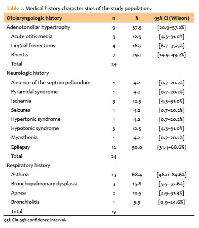
The most common reason for consultation was preventive ophthalmological check-up (39.6%), followed by strabismus (22.9%) and refractive errors (21.5%). Other reasons for consultation included red eye (6.3%), referral from another center (5.6%), chalazion (3.5%), and epiphora (0.7%). In the preventive check-up group, 73.7% had an ophthalmological abnormality, with refractive errors being the most common finding (76.2%), followed by strabismus (38.1%). Exotropia was the most common finding (48.5%), but other ocular motility abnormalities were also found. In the nine cases of red eye consultation, inflammatory pathologies of the ocular surface predominated, with allergic conjunctivitis and blepharitis being the most common.
Table 3 presents the data from the ophthalmological evaluation.
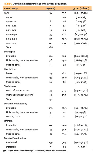
When testability was analyzed by age, both quantitative visual acuity measurements and stereopsis evaluation showed a gradual, age-dependent increase. Specifically, the success rate for quantitative visual acuity was lowest in the youngest cohort (1–3 years) at 18.2%, but increased significantly to 64.4% in children aged 4–6 years, 82.2% in the 7–9 years group, and 94.1% in children aged 10–12 years. Similarly, 45.5% of children aged 1–3 years had successful stereopsis evaluation, which increased to 71.2% and 80.0% in the 4–6 and 7–9 years cohorts, respectively, and peaked at 88.2% in the 10–12 years group. The single patient aged 13 to 15 years category completed both assessments (100.0%).
In terms of functional visual examination testability, stereopsis was successfully evaluated in 72.2% of children (n=104/144), while 25.0% (n=36) were unable to be tested due to lack of cooperation or inability to complete the task. 2.8% of cases (n=4) had no data recorded or were missing. Fine stereopsis (60–40 seconds of arc) was achieved by 18.3% of the assessable children (n=19), moderate stereopsis (140–80 seconds of arc) by 34.6% (n=36), and coarse stereopsis (800–200 seconds of arc) by 26.0% (n=27). A stereopsis level of 3552 seconds of arc was observed in 20.2% (n=21), while 1.0% (n=1) showed no stereopsis (nil). Binocular fusion was observed in 16.0% of the children.
Dynamic retinoscopy accurately identified clinically significant refractive errors (k=1.000, p<0.001). Among the 139 testable children, 25.9% (n=36/139; 95% CI, 19.3–33.8) had an abnormal accommodative response, which corresponded solely to children with clinically significant refractive status. In contrast, normal responses accounted for 74.1% (n=103/139; 95% CI, 66.2–80.7), which corresponded to cases with no clinically significant refractive errors.
Lower testability rates were found for color vision evaluation using Ishihara plates (34.0%; 95% CI, 26.8–42.1; n=49/144).
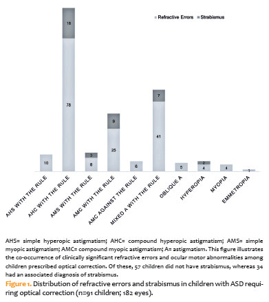
Astigmatism was the most common refractive diagnosis, with compound hyperopia accounting for 42.9%, followed by mixed hyperopia (22.5%) and compound myopia (13.7%). Clinically significant refractive errors requiring optical prescription were found in 63.2% of children (95% CI, 55.1%–70.6; n=91/144), while physiological refractive states accounted for 18.8% (95% CI, 13.2–25.9; n=27/144).
The most common range for positive spherical equivalent (n=117) was +1.25 to +2.25 diopters (38.5%), followed by values between +2.50 and +3.50 diopters (20.5%). The vast majority of children (94.0%) had positive spherical equivalents of less than +5.00 diopters. For the negative spherical equivalent (n=65), the most common range was less than −1.00 diopters (41.5%), followed by −1.25 to −2.25 diopters (30.8%).
The overall prevalence of strabismus was 32.6% (95% CI, 25.5−40.7; n=47/144). Strabismus associated with refractive errors was identified in 23.6% (95% CI, 17.4–31.2; n=34/144), while primary or isolated strabismus occurred in 9.0% (95% CI, 5.4–14.8; n=13/144; Figure 1). The most common type of strabismus in children with refractive errors was exotropia (61.5%), followed by esotropia (17.9%). We also observed associated ocular motility changes, including hyperfunction of the oblique muscles (12.8%) and nystagmus (7.7%). Exotropia strabismus was the most common in children with strabismus without associated refractive error, accounting for 92.3% of cases, while esotropia occurred in only 7.7%. Furthermore, in children with strabismus (n=47), stereopsis could be assessed in 28 cases (59.6%), whereas it could not be assessed in 19 children (40.4%) or the result was not recorded. Among the children with assessable stereopsis, coarse and moderate stereopsis predominated, with 3552 arc seconds in 39.3% and 800–200 arc seconds in 32.1%. No cases of fine stereopsis (≤60 arc seconds) were observed.
A fundus examination was performed on 139 children (96.5%), with abnormalities in five cases (3.6%). The findings included optic disk and retinal myelination in two children, peripapillary retinal pigment epithelium abnormality in one patient, retinal dystrophy in another, and retinopathy of prematurity in one.
The treatment and follow-up results are summarized in Table 4. 91.6% of children (n=132) required treatment, which included optical correction (69.7%), pharmacological (15.2%), surgery (6.8%), and occlusion therapy (6.8%). Strabismus surgery was the most common procedure (88.9%), with one (11.1%) being lacrimal duct probing.
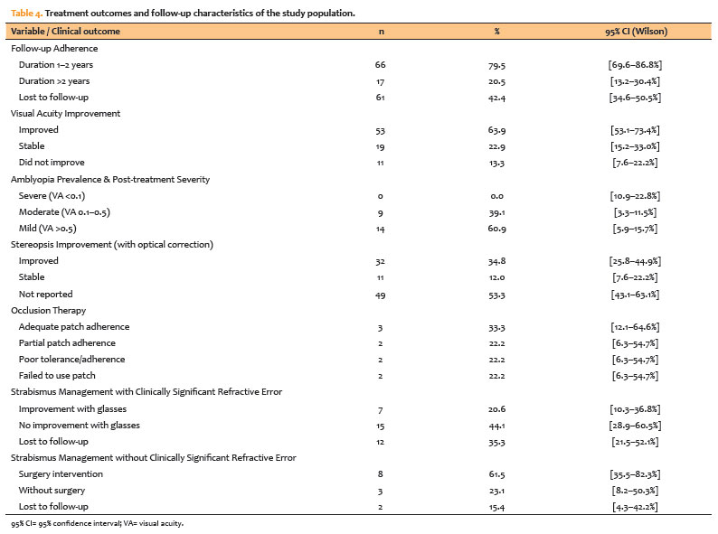
Of the total number of children evaluated, 83 (57.6%) continued ophthalmological follow-up, while 61 (42.4%) did not attend subsequent check-ups until the time of data collection. Of the 83 children who continued to receive ophthalmological care, the majority (54.2%) had a follow-up period of 1 to 2 years.
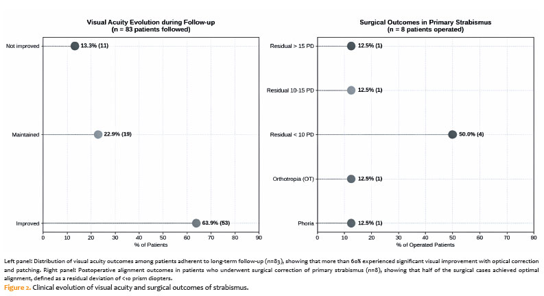
Visual acuity increased in 53 cases (63.9%), remained stable in 19 children (22.9%), and did not improve in 11 children (13.2%). It is worth noting that 13 children in the group that improved did so without the use of optical correction, which could be attributed to visual maturation, improved cooperation during check-ups, and progressive adaptation to the ophthalmological examination in children with ASD. The degree of improvement was 2 lines in 19 cases (35.8%), 3 lines in 14 children (26.4%), and 4 or more lines in 9 children (17.0%). 20.8% showed an improvement of one line.
Amblyopia was diagnosed during follow-up after appropriate optical correction was prescribed and visual acuity reassessed. According to this definition, amblyopia was detected in 16.0% of the total cohort (95% CI, 10.9–22.8; n=23/144), with 60.9% (n=14/23) presenting mild amblyopia and 39.1% (n=9/23) presenting moderate amblyopia; no cases of severe amblyopia were found. Among children who attended follow-up (n=83), visual acuity improved in 63.9% (95% CI, 53.1–73.4; n=53/83), with 79.2% (n=42/53) improving by two or more lines on optotype charts.
Stereopsis improved in 32 cases (34.8%) of the 92 children with refractive error who received spectacles.
In 20.6% of children with strabismus and associated refractive error, optical correction improved their vision. Postoperative results were favorable in 75.0% of children with strabismus without associated refractive error who presented with phoria, orthotropia, or a residual deviation <10 prism diopters. 25% of children had residual deviations ≥10 prism diopters. Figure 2. Clinical evolution of visual acuity and surgical outcomes for strabismus.
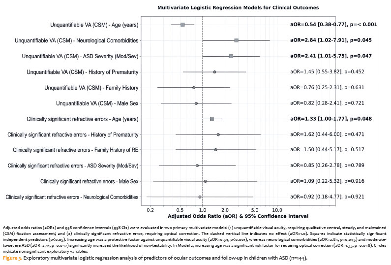
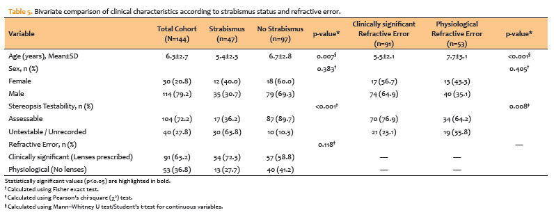
Bivariate analysis revealed significant relationships between demographics and ocular conditions. Children with strabismus are significantly younger than non-strabismic children (5.4±2.3 vs. 6.7±2.8 years; p=0.007). Similarly, children with pathological refractive errors requiring an optical prescription had a significantly lower mean age compared to those with physiological refractive states (5.5±2.1 vs. 7.7±3.1 years; p<0.001). Strabismus significantly reduced stereopsis testability, with only 36.2% of children completing the test compared to 89.7% of non-strabismic children (p<0.001). Stereopsis testability was also found to be significantly associated with refractive state (p=0.008). In addition, exploratory multivariate logistic regression models with demographic, clinical, and neurodevelopmental predictors revealed significant independent associations with visual evaluation capacity and refractive status (Figure 3 and Table 5).
DISCUSSION
This study emphasizes the high prevalence of ophthalmologic abnormalities in children with ASD, particularly refractive errors and strabismus(7,8). More than 80% of the children in our cohort, which included 79.2% males, a mean age of 6.3 years, and a high burden of medical comorbidities, had at least one ocular finding. Refractive errors were the most common abnormality (81.9%), especially astigmatism and moderate hyperopia. Strabismus was diagnosed in 32.6% of children, and it was frequently associated with refractive errors, the most common of which was exotropia. This refractive-strabismus association is consistent with previous research, which has linked some strabismus in ASD to a refractive and amblyogenic component(9-11). The observed prevalence is consistent with previous clinical studies, which reported figures ranging from 20% to 40%(4,9,12). Divergent strabismus is the most common type in children with ASD(4,12,13). Stereopsis could be assessed in 72.2% of cases, and amblyopia was typically mild to moderate. Correction of these conditions may improve visual function and enable interaction with the environment, though neurodevelopmental and functional outcomes were not directly assessed in this study.
These findings are consistent with multiple clinical studies that show a higher frequency of ophthalmological abnormalities in the pediatric ASD population than in the general population(7-12,14). The 81.9% prevalence of refractive errors is consistent with international cohorts that report refractive errors as the primary ocular manifestation, alongside a relatively uniform pattern of astigmatism and moderate hyperopia(15-17). The observed strabismus prevalence (32.6%) is within the previously reported range of 20% to 40%(4,8,13), with a similar prevalence of exotropia(4,13,14). Furthermore, our findings on binocular vision, such as the predominance of coarse and moderate stereopsis and the absence of fine stereopsis in children with strabismus, are consistent with previous studies(16,18). The presence of mild to moderate amblyopia is also comparable to the 11%–19% reported in other ASD studies(15,17,18).
These findings highlight the clinical significance of using individualized examination strategies and tailored follow-up for children with ASD. In this context, the pediatric ophthalmologist is an important member of the multidisciplinary team(19). Dynamic retinoscopy (accommodative response) showed perfect diagnostic agreement (k=1.000, p<0.001) for clinically significant refractive errors. This establishes dynamic retinoscopy as an objective and highly reliable tool in this population, allowing clinicians to accurately identify which children require cycloplegic refraction for definitive correction while overcoming the communication barriers inherent in subjective testing(20). While optical correction was generally well tolerated with progressive adaptation, we found very low adherence to occlusion therapy (6.8%), which was primarily due to tactile hypersensitivity and behavioral resistance. To address this, alternative strategies such as pharmacological penalization (e.g., atropine drops) or structured behavioral approaches, such as systematic desensitization and positive reinforcement therapy guided by behavioral specialists, should be actively considered and discussed during caregiver counseling(21,22). Furthermore, 42.4% of the cohort missed follow-up appointments. This high dropout rate is most likely caused by systemic and socioeconomic barriers such as transportation issues, the indirect financial costs of specialized care, a lack of sensory-friendly clinical environments, and the overwhelming cumulative medical burden placed on caregivers of children with complex phenotypes (23,24).
This study’s strengths include a clinically relevant sample size, the inclusion of children from both public and private facilities, and a comprehensive ophthalmological evaluation that allowed for the identification of refractive, motor, and sensory abnormalities. However, this study has some limitations that must be acknowledged. First, the retrospective and descriptive design makes it impossible to determine causality or the long-term functional impact of the ophthalmologic findings. Second, there was no standardized confirmation of the ASD diagnosis, as well as no classification of ASD severity or cognitive level, both of which can have a significant impact on a child’s cooperation during an examination. Third, visual function testing was occasionally incomplete; to address this, we made a systematic distinction between tests that could not be evaluated due to poor cooperation and data that were simply missing or unrecorded in the clinical charts. Fourth, treatment adherence was not measured, but rather based on subjective caregiver reports, so these specific outcomes should be interpreted with caution. Furthermore, using a clinical cohort from a referral center may introduce referral bias and a higher burden of comorbidities, while the lack of a control group limits direct comparisons to the general pediatric population. Finally, the significant loss to follow-up (42.4%) limits the comprehensive assessment of long-term therapeutic and surgical interventions in these children.
In conclusion, this study found a high frequency of ophthalmologic abnormalities, primarily refractive errors and strabismus, in a clinical cohort of children with ASD. These findings highlight the importance of conducting systematic ophthalmological evaluations in this population. Given the inherent difficulties and unreliability of subjective visual testing, objective measures, particularly dynamic retinoscopy and cycloplegic refraction, are required for accurate diagnosis. Finally, the observed significant loss to follow-up emphasizes the critical need for tailored follow-up strategies to ensure these children receive effective and continuous visual care.
ACKNOWLEDGMENTS
The authors thank the children and their families for their willingness and collaboration as well as the staff of the UC Christus Health Network and La Florida Hospital Dr. Eloísa Díaz for their support during clinical care and data collection. The collaboration of the healthcare teams involved in the multidisciplinary care of children with ASD is also acknowledged.
The authors would like to thank Enago (www.enago.br) for the English language review.
AUTHORS’ CONTRIBUTIONS:
Significant contribution to conception and design: Pereira Lucía, Kohn Geraldine, Dillems Marcela, Ormazábal Ivana, Garretón Rodolfo. Data acquisition: Pereira Lucía, Kohn Geraldine, Dillems Marcela, Ormazábal Ivana, Garretón Rodolfo. Data analysis and interpretation: Pereira Lucía, Kohn Geraldine, Dillems Marcela, Ormazábal Ivana, Garretón Rodolfo, Simonovis César. Manuscript drafting: Pereira Lucía, Kohn Geraldine, Dillems Marcela, Ormazábal Ivana, Garretón Rodolfo, Simonovis César. Significant intellectual content revision of the manuscript: Pereira Lucía, Kohn Geraldine, Dillems Marcela, Ormazábal Ivana, Garretón Rodolfo, Simonovis César. Final approval of the submitted manuscript: Pereira Lucía, Kohn Geraldine, Dillems Marcela, Ormazábal Ivana, Garretón Rodolfo, Simonovis César. Statistical analysis: Pereira Lucía, Kohn Geraldine, Dillems Marcela, Ormazábal Ivana, Garretón Rodolfo, Simonovis César. Obtaining funding: not applicable. Supervision of administrative, technical, or material support: Pereira Lucía, Kohn Geraldine, Dillems Marcela, Ormazábal Ivana, Garretón Rodolfo, Simonovis César. Research group leadership: Pereira Lucía.
REFERENCES
1. Maenner MJ, Warren Z, Williams AR, Amoakohene E, Bakian AV, Bilder DA, et al. Prevalence and characteristics of autism spectrum disorder among children aged 8 years—Autism and Developmental Disabilities Monitoring Network, 11 sites, United States, 2020. MMWR Surveill Summ. 2023;72(2):1–14.
2. Biblioteca del Congreso Nacional de Chile. Políticas de apoyo al espectro autista. Santiago: BCN; 2018.
3. Yáñez C, Maira P, Elgueta C, Brito M, Crockett MA, Troncoso L, et al. Estimación de la prevalencia de trastorno del espectro autista en población urbana chilena. Andes Pediatr. 2021;92(4):519–25.
4. Ikeda J, Davitt BV, Ultmann M, Maxim R, Cruz OA. Brief report: incidence of ophthalmologic disorders in children with autism. J Autism Dev Disord. 2013;43(6):1447–51.
5. Hunter DG. Dynamic retinoscopy: the missing data. Surv Ophthalmol. 2001;46(3):269–74.
6. Donahue SP, Arthur B, Neely DE, Arnold RW, Silbert D, Ruben JB; POS Vision Screening Committee. Guidelines for automated preschool vision screening: a 10-year, evidence-based update. J AAPOS. 2013;17(1):4–8.
7. Lord C, Elsabbagh M, Baird G, Veenstra-Vanderweele J. Autism spectrum disorder. Lancet. 2018;392(10146):508–20.
8. Khanna RK, Kovarski K, Arsene S, Siwiaszczyk M, Pisella PJ, Bonnet-Brilhault F, et al. Ophthalmological findings in children with autism spectrum disorder. Graefes Arch Clin Exp Ophthalmol. 2020;258(4):909r–16.
9. Denis D, Burillon C, Livet MO, Burguière O. Signes ophtalmologiques chez l’enfant autiste. J Fr Ophtalmol. 1997;20(2):103-10. French.
10. Gogate P, Soneji FR, Khandekar R, et al. Ophthalmologic disorders and refractive errors in children with autism spectrum disorder. J AAPOS. 2019;23(1):46.e1–46.
11. Chang MY, Gandhi N, O’Hara M. Ophthalmologic disorders and risk factors in children with autism spectrum disorder. J AAPOS. 2019;23(6):337.e1–336.e6.
12. Milne E, Griffiths H, Buckley D, Scope A. Vision in children and adolescents with autistic spectrum disorder: evidence for reduced convergence. J Autism Dev Disord. 2009;39(7):965–75.
13. Reynolds M, Culican SM. Visual Autism. Children (Basel). 2023 Mar 23;10(4):606.
14. Scharre JE, Creedon MP. Assessment of visual function in autistic children. Optom Vis Sci. 1992;69(6):433–9.
15. Ohya T, Morita K, Yamashita Y, Egami C, Ishii Y, Nagamitsu S, et al. Impaired exploratory eye movements in children with Asperger’s syndrome. Brain Dev. 2014;36(3):241-7.
16. Granet DB, Gomi CF, Ventura R, Miller-Scholte A. The relationship between convergence insufficiency and ADHD. Strabismus. 2005;13(4):163–8.
17. Stevenson M, Tedone E. Assessment of autism spectrum disorders in children with visual impairment and blindness: a scoping review. J Autism Dev Disord. 2025;55(4):1411-23.
18. Wang J, Ding G, Li Y, Hua N, Wei N, Qi X, et al. Refractive status and amblyopia risk factors in Chinese children with autism spectrum disorder. J Autism Dev Disord. 2018;48(5):1530–6.
19. Kabataş EU, Ozer PA, Ertugrul GT, Kurtul BE, Bodur S, Alan BE. Initial ophthalmic findings in Turkish children with autism spectrum disorder. J Autism Dev Disord. 2015;45(8):2578–81.
20. Anketell PM, Saunders KJ, Gallagher SM, Bailey C, Little JA. Accommodative Function in Individuals with Autism Spectrum Disorder. Optom Vis Sci. 2018;95(3):193–201.
21. Tomchek SD, Dunn W. Sensory processing in children with and without autism: a comparative study using the short sensory profile. Am J Occup Ther. 2007;61(2):190–200.
22. Kupzyk S, Allen KD. A review of strategies to increase comfort and compliance with medical/dental routines in persons with intellectual and developmental disabilities. J Dev Phys Disabil. 2019;31(2):231–49.
23. Simmons DR, Robertson AE, McKay LS, Toal E, McAleer P, Pollick FE. Vision in autism spectrum disorders. Vision Res. 2009;49(22):2705–39.
24. Perna J, Bellato A, Ganapathy PS, Solmi M, Zampieri A, Faraone SV, et al. Association between Autism Spectrum Disorder (ASD) and vision problems. A systematic review and meta-analysis. Mol Psychiatry. 2023;28(12):5011-23.
Submitted for publication:
March 13, 2026.
Accepted for publication:
August 12, 2026.
Approved by the following research ethics committee: Pontifícia Universidad Católica de Chile (Protocol ID: 230828005).
Data Availability Statement: The datasets generated and/or analyzed during the current study are included in the manuscript.
Edited by
Editor-in-Chief: Newton Kara-Júnior
Associate Editor: Luisa Moreira Hopker
Funding: This study received no specific financial support.
Disclosure of potential conflicts of interest: The authors declare no potential conflicts of interest.

How to cite this article:
 ABO is licensed under a Creative Commons Attribution-NonComercial 4.0 Internacional.

