Arq. Bras. Oftalmol. 2001;64 (2 )
:123-126
| DOI: 10.1590/S0004-27492001000200006
Abstract
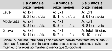
Objetivos: Ambliopia é o defeito visual mais comum em crianças e por mais de 250 anos a terapia oclusiva vem sendo o melhor tratamento. Sendo assim, propusemo-nos a determinar os fatores que influenciam no sucesso do tratamento da ambliopia por terapia oclusiva em nosso meio. Métodos: Foi realizado um estudo retrospectivo com 169 crianças amblíopes atendidas no Ambulatório de Ambliopia do Hospital de Clínicas da UNICAMP, Campinas (SP), entre janeiro de 1996 e maio de 1998. A população atendida foi classificada quanto ao sexo, idade de início do tratamento por faixa etária (3 grupos), olho afetado, tipo de ambliopia (estrabísmica, anisometrópica, por deprivação, associação de dois tipos), tempo de seguimento, gravidade da ambliopia (leve, moderada, grave), adesão ao tratamento (regular, irregular) e resposta obtida (cura, melhora, sem cura). Resultados: A adesão ao tratamento não diferiu entre as faixas etárias (p=0,68) e não foi influenciada pela gravidade da ambliopia (p=0,82). Dos pacientes estudados 52,67% curaram-se, 19,52% melhoraram e 27,81% não obtiveram cura. Os pacientes com adesão regular tiveram índice de cura significativamente maior do que os pacientes com adesão irregular (p=0,0009). O resultado do tratamento não dependeu da idade de início do mesmo (p=0,39) e da gravidade da ambliopia (p=0,30). Conclusão: Concluímos, assim, que, no nosso grupo de estudo, a adesão é o principal fator prognóstico no sucesso da terapia oclusiva.
Keywords: Ambliopia; Prognósticos; Estrabismo; Curativos oclusivos; Cooperação do paciente
 ABO is licensed under a Creative Commons Attribution-NonComercial 4.0 Internacional.
09-tab01tb.jpg)
09-tab01.jpg)
05-tab01tb.jpg)
09-fig01.jpg)
14-fig01.jpg)
10-fig01.jpg)
01-fig01tb.jpg)
07-tab01tb.jpg)