Showing of 1 until 15 from 123 result(s)
Search for: Cataract extraction; Lutein; Phacoemulsification; Capsulorhexis; Lenses, intraocular
01-ane01.jpg)
Abstract
OBJETIVO: Avaliar a relação entre a incidência de complicações e reações emocionais durante a cirurgia de catarata sob anestesia tópica em pacientes funcionalmente monoculares.
MÉTODOS: Estudo prospectivo, transversal, caso-controle de vinte e dois pacientes monoculares e dezenove controles pareados por idade e sexo . Dados demográficos foram analisados: idade, sexo e escolaridade. As cirurgias foram realizadas pelo mesmo cirurgião e durante o procedimento os sinais vitais dos pacientes (como pressão arterial sistêmica e frequência cardíaca) e eventos cirúrgicos (duração da cirurgia, movimentos corporais, sinais de aumento da pressão vítrea, dificuldade de realização da capsulorrexis e complicações) foram coletados. A acuidade visual pré e pós foi analisada. A distribuição normal dos dados foi confirmada com o teste de Shapiro-Wilk. Os dados foram expressos como média ± DP e porcentagem. A comparação dos diferentes testes clínicos entre os grupos foi realizada utilizando Student’s t-test e ANOVA com correção de Bonferroni. O qui-quadrado foi usado para comparar dados demográficos. Valor de p<0,05 foi considerado estatisticamente significante.
RESULTADOS: Este estudo incluiu vinte e dois olhos de 22 pacientes funcionalmente monoculares (6 homens e 13 mulheres) e dezenove olhos de 19 controles (11 homens e 11 mulheres). A média de idade foi de 73,05 ± 13,31 anos nos indivíduos monoculares e 69,74 ± 16,81 no controle. Considerando-se os sinais vitais não houve diferença significativa entre os grupos (p>0,05). Durante o procedimento, a percepção do cirurgião em relação aos movimentos excessivos de olho, pálpebra ou cabeça em ambos os grupos foi semelhante, assim como sinais de aumento da pressão vítrea (p=0,2 e p=0,1, respectivamente).
CONCLUSÃO: Este estudo sugere que é seguro realizar a extração de catarata com anestesia tópica em pacientes funcionalmente monoculares. Esses pacientes aparentemente se comportam de maneira semelhante aos pacientes binoculares.
Keywords: Facoemulsificação/psicologia; Capsulorrexe; Anestésicos locais; Acuidade visual
03-fig01.jpg)
Abstract
OBJETIVO: Avaliar a influência da dinâmica pupilar na curva de desfoco de olhos implantados com lente intraoculares multifocais difrativas.
MÉTODOS: Estudo prospectivo e randomizado realizado na Faculdade de Medicina de Ribeirão Preto - Universidade de São Paulo - Departamento de Oftalmologia. Trinta e oito pacientes foram aleatoriamente designados para receber bilateralmente lentes intraoculares SN6AD1 (n=20) (mfIOL) ou SN60WF (n=18) (aIOL). Além da acuidade visual para longe e perto, corrigida e não corrigida, e curva de desfoco, foi ainda realizada pupilometria dinâmica. A área sob a curva de desfoco foi calculada usando um modelo polinomial empírico.
RESULTADOS: Um total de 16 e 17 pacientes (n=32 e 34 olhos) completaram 1 ano de seguimento nos grupos mfIOL e aIOL, respectivamente. Não houve diferenças significativas entre grupos para as acuidades visuais seja para longe ou perto. As curvas de desfoco do grupo mfIOL mostraram um pico duplo; enquanto o SN60WF mostrou apenas um pico, típico para uma lente intraoculares monofocal. A média da área sob a curva de desfoco do grupo aIOL foi (4,66 ± 1,51 logMAR.dp), e essa é estatisticamente significante diferente da métrica do grupo mfIOL (1,99 ± 1,31 logMAR.dp). A pupila na contração máxima após a exposição a um flash de 30 cd/m2 por 1 segundo foi significativamente correlacionada com uma melhor área de foco no grupo mfIOL (r=0,54; p=0,0017), essa relação não foi observada para o grupo aIOL.
CONCLUSÃO: Estes dados indicam que quanto menor a pupila durante contração, melhor é a área sob a curva de desfoco e, portanto, o desempenho visual dos olhos implantados com essa mfIOL. Esta correlação não foi encontrada para lentes intraoculares monofocais.
Keywords: Lentes intraoculares multifocais; Pupila/fisiologia, Catarata; Facoemulsificacão
08-tab01.jpg)
Abstract
Objetivos: Avaliar a estabilidade e eficácia da técnica double-flanged com sutura de 5-0 polipropileno para fixação de cataratas subluxadas aos 18 meses e as possíveis complicações desta nova técnica.
Métodos: Esta técnica utiliza um monofilamento de polipropileno 5-0 para criar dois flanges com um termocautério para fixar um Segmento de Tensão Capsular na esclera a fim de estabilizar o saco capsular subluxado. Esta técnica foi implementada em 17 olhos que necessitavam do implante de lente intraocular em casos de diálise zonular devido a trauma, síndrome de Marfan, microesferofacia, subluxação idiopática ou pós-facoemulsificação que provocou subulxação do saco capsular intraoperatória.
Resultados: O seguimento dos pacientes foi de 18 meses. A acuidade visual corrigida melhorou significativamente de 0,85 para 0,39 (logMAR), enquanto os erros de refração esféricos e cilíndricos e a pressão intraocular permaneceram estáveis. Nenhuma fotodegradação de sutura ou pseudofacodonese foi encontrada.
Conclusão: A técnica double-flanged para fixação transescleral de saco capsular com sutura de 5-0 polipropileno mostrou resultados de estabilidade de longo prazo para o complexo lente/saco capsular. Então, aparenta ser uma opção segura para cirurgia de catarata, sem necessidade pontos, em olhos com fraqueza zonular ou diálise
Keywords: Catarata; Facoemulsificação; Lente intraocular; Técnica de sutura; Acuidade visual
03-fig01.jpg)
Abstract
Objetivo: Investigar os resultados pós-operatórios e avaliar os preditores de sucesso da facoemulsificação combinada à goniotomia com o Kahook Dual Blade para o tratamento da catarata e do glaucoma em olhos com glaucoma primário de ângulo aberto.
Métodos: Série de casos retrospectivos, não comparativos e intervencionistas, em que todos os pacientes com glaucoma primário de ângulo aberto submetidos ao procedimento de facoemulsificação combinada à goniotomia com o Kahook Dual Blade entre junho de 2018 e abril de 2019 foram inscritos. Todos os participantes tiveram um acompanhamento mínimo de 6 meses. Foram registrados os valores de pressão intraocular pré e pós-operatória (em 1, 3 e 6 meses), número de medicamentos antiglaucomatosos, melhor acuidade visual corrigida, complicações cirúrgicas e quaisquer eventos ou procedimentos subsequentes relacionados. A análise de regressão logística foi usada para investigar a associação entre diferentes variáveis e resultados cirúrgicos.
Resultados: Um total de 57 olhos de 47 pacientes foram incluídos (média de idade, 70,5 ± 7 anos). A pressão intraocular média reduziu de 15,5 ± 4,2 mmHg para 12,2 ± 2,4 mmHg na última visita de acompanhamento (p<0,001). O número médio de medicamentos antiglaucomatosos diminuiu significativamente de 1,9 ± 1,0 para 0,6 ± 1,0 durante o mesmo período (p<0,001). Com base no critério predefinido (redução da pressão intraocular ≥20% e/ou redução de ≥1 medicamento), a taxa de sucesso em 6 meses foi de 86%. Um valor de pressão intraocular pré-operatório mais alto (OR= 2,01; p=0,016) e maior porcentagem de redução da pressão intraocular inicial
(30 dias) (OR= 1,02; p=0,033) foram significativamente associados ao sucesso cirúrgico.
Conclusão: Nossos resultados sugerem que o procedimento de facoemulsificação combinada à goniotomia com o Kahook Dual Blade é uma alternativa eficaz e segura para o manejo da catarata em olhos com glaucoma primário de ângulo aberto, impactando positivamente no controle da pressão intraocular e no número de medicamentos. Olhos com pressão intraocular basal mais alta e resposta inicial mais pronunciada ao procedimento parecem apresentar melhores resultados em 6 meses. Mais estudos são necessários para avaliar a eficácia em longo prazo e o perfil de segurança.
Keywords: Glaucoma; Glaucoma de ângulo aberto; Catarata; Facoemulsificação; Pressão intraocular; Goniotomia
07-tab01tb.jpg)
Abstract
Objetivo: Criar modelos, em catarata pediátrica, para estimar valores futuros de ceratometria e comprimento axial, com base na ceratometria e no comprimento axial medidos na cirurgia, para previsão do poder da lente intraocular para emetropia em idades futuras.
Métodos: Olhos com catarata bilateral, ceratometria e comprimento axial medidos na cirurgia e pelo menos um exame pós-operatório com medidas de ceratometria e comprimento axial foram considerados para este estudo. Os modelos para estimar futuras ceratometrias e comprimentos axiais foram criados considerando (1) ceratometria e comprimento axial medidos na cirurgia, (2) a inclinação média da regressão logarítmica da ceratometria e comprimento axial criada para cada olho e (3) a idade na cirurgia. A lente intraocular para emetropia em idades futuras pode ser estimada usando esses valores em fórmulas de terceira geração. Os erros de estimativa da ceratometria, comprimento axial e poder da lente intraocular, usando os modelos, também foram calculados.
Resultados: 57 olhos de 29 pacientes preencheram os critérios de inclusão. A idade média na cirurgia e acompanhamento foram de 36,96 ± 32,04 meses e 2,39 ± 1,46 anos, respectivamente. A inclinação média da regressão logarítmica criada para cada olho foi de -3.286 para ceratometria e + 3.189 para o comprimento axial. Os erros médios de estimativa absoluta para ceratometria e comprimento axial foram respectivamente: 0,61 ± 0,54 D e 0,49 ± 0,55 mm, e para o poder da lente intraocular usando as fórmulas SRK-T, Hoffer-Q e Holladay I foram: 2,04 ± 1,73 D, 2,49 ± 2,10 D e 2,26 ± 1,87 D, respectivamente.
Conclusões: Os modelos apresentados podem ser utilizados para estimar o poder da lente intraocular que levaria a emetropia em idades futuras e orientar a escolha do poder da lente intraocular a ser implantada na catarata pediátrica.
Keywords: Catarata; Biometria/métodos; Emetropia; Comprimento axial do olho; Lentes intraoculares; Criança
Abstract
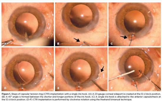
PURPOSE: To evaluate the effect of using a single iris retractor, affixed to the anterior capsulorhexis at the 12 o'clock position, on the ease of capsular tension ring implantation.
METHODS: This prospective comparative study comprised 37 patients with zonular weakness attributed to pseudoexfoliation syndrome who underwent capsular tension ring implantation during cataract surgery. In Group 1, a single iris retractor was inserted into the anterior capsulorhexis at the 12 o'clock position. Group 2 did not receive this intervention. Zonular weakness was graded on a scale of 1–5, and the subjective difficulty of capsular tension ring implantation was categorized as easy, medium, or difficult.
RESULTS: Group 1 and 2 comprised 20 and 17 patients, respectively. There were no significant differences between the groups in age, sex distribution, and presence of glaucoma (p=0.53, p=0.28, and p=1.00, respectively). The mean zonular weakness score was significantly higher in Group 1 (3.35 ± 0.45) than in Group 2 (2.71 ± 0.59; p=0.02). Capsular tension ring implantation was significantly easier in the iris retractor group (p<0.001).
CONCLUSIONS: Placement of a single iris retractor attached to the anterior capsulorhexis at the 12 o'clock position may facilitate easier capsular tension ring implantation, even in patients with greater zonular weakness. This technique could reduce the risk of capsular tension ring displacement into the iridocorneal angle or ciliary sulcus.
Keywords: Capsular tension ring; Cataract; Iris hook; Pseudoexfoliation syndrome; Zonular weakness; Cataract extraction; Phacoemulsification; Capsulorhexis.
Abstract
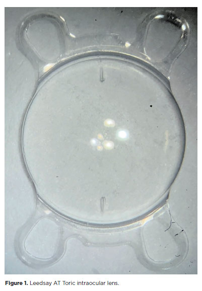
PURPOSE: The purpose of this study was to assess visual outcomes and patient satisfaction following cataract surgery involving the implantation of quad-loop intraocular lenses, including trifocal, bifocal, and toric variants.
METHODS: Information was obtained from both physical and electronic medical records of patients who underwent phacoemulsification cataract surgery with implantation of different intraocular lenses between January 1, 2022, and December 31, 2023. The study included individuals aged over 18 who received bilateral implantation of bifocal, trifocal, or monofocal toric intraocular lenses. Visual acuity was assessed at various postoperative time points using the logMAR scale. Quantitative variables were analyzed using mean and standard deviation.
RESULTS: A total of 92 eyes received premium intraocular lenses: 4 bifocal, 32 trifocal, 52 toric monofocal, and 4 trifocal toric lenses. The average preoperative corrected visual acuity was logMAR 0.478 ± 0.259. On the first postoperative day, the average uncorrected visual acuity was logMAR 0.301 ± 0.207. By day 30, 67.4% of eyes achieved uncorrected distance visual acuity of logMAR 0.2 or better. Patient satisfaction was high, with few reports of glare or halos.
CONCLUSION: Quad-loop intraocular lenses-including trifocal, bifocal, and toric models-demonstrated effective improvement in visual acuity and high levels of patient satisfaction. These lenses represent a suitable option for enhancing visual outcomes after cataract surgery. Additional studies with larger cohorts are recommended to confirm these results.
Keywords: Cataract extraction; Aberrometry/methods; Lenses, intraocular; Lens implantation, intraocular; Prosthesis design
Abstract
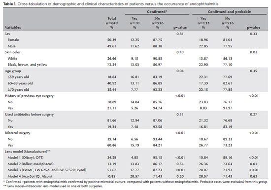
PURPOSE: Endophthalmitis is one of the most important adverse events after cataract surgery, as it can lead to total vision loss. This study aimed to describe the occurrence of endophthalmitis after phacoemulsification with intraocular lens implantation in patients treated in a community setting in Porto Velho, Rondônia, Brazil.
METHODS: This retrospective cohort study was conducted using a database of 649 medical records of patients who underwent surgery and were followed for three months. Poisson regression analysis was used to estimate relative risks and 95% confidence intervals (95% CIs).
RESULTS: The incidence of confirmed endophthalmitis was 11.94% (95% CI, 9.50-14.76), while the incidence of confirmed and probable cases was 20.50% (95% CI, 17.52-23.73). For confirmed cases, bilateral surgery and the use of lens model 3 were identified as risk factors for endophthalmitis, whereas age over 70 yr and preoperative antibiotic use were protective factors. For confirmed and probable cases, brown and yellow skin color, bilateral surgery, and the use of lens model 3 were also identified as risk factors. Gram-negative bacteria were the predominant etiological agents, and corneal edema was the main clinical manifestation. The mean duration of treatment was eight days, and 27.12% of patients used antibiotics.
CONCLUSION: The incidence observed was substantially higher than that reported in the literature, with a predominance of Gram-negative agents and an association with bilateral surgeries and the Eyeol intraocular lens model. These findings reinforce the need for continuous epidemiological surveillance and the implementation of specific biosafety and infection control protocols during cataract surgery campaigns.
Keywords: Endophthalmitis; Disease outbreaks; Phacoemulsification; Lens implantation, intraocular; Lenses, intraocular; Cataract; Risk factors; Anti-bacterial agents
Abstract
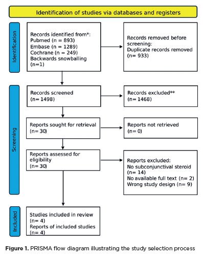
The advantages and disadvantages of using perioperative subconjunctival steroid injections in dropless cataract surgery continue to be debated. A systematic review of PubMed, EMBASE, and the Cochrane Central database identified five studies—two randomized controlled trials and three non-randomized studies—encompassing 70,751 eyes. Among these, 12,319 eyes (17.4%) received subconjunctival steroid injections, while 58,432 eyes (82.6%) were managed with topical steroids. The Cochrane Collaboration’s RoB 2 tool was applied for bias assessments in randomized controlled trials, and heterogeneity was assessed using the I² statistics. No statistically significant differences were found between the two groups regarding macular edema (p=0.249), visual acuity (p=0.73), or laser flare count (p=0.45). Both subconjunctival injections and topical steroids demonstrated comparable efficacy and safety in controlling postoperative inflammation after cataract surgery. Additional research is warranted to validate these conclusions.
Keywords: Cataract extraction; Phacoemulsification; Lens implantation, intraocular; Postoperative care; Intravitreal injections; Anti-inflammatory agents, non-steroidal/administration & dosage; Glucocorticoids; Triamcinolone acetonide; Research design; Randomiz
Abstract
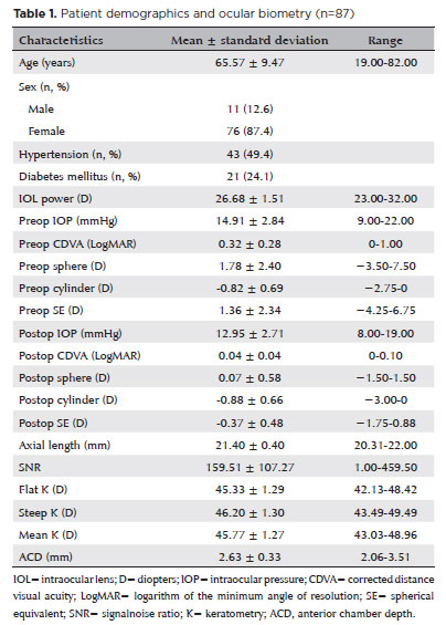
PURPOSE: To compare the refractive prediction error of Hill-radial basis function 3.0 with those of 3 conventional formulas and 11 combination methods in eyes with short axial lengths.
METHODS: The refractive prediction error was calculated using 4 formulas (Hoffer Q, SRK-T, Haigis, and Hill-RBF) and 11 combination methods (average of two or more methods). The absolute error was determined, and the proportion of eyes within 0.25-diopter (D) increments of absolute error was analyzed. Furthermore, the intraclass correlation coefficients of each method were computed to evaluate the agreement between target refractive error and postoperative spherical equivalent.
RESULTS: This study included 87 eyes. Based on the refractive prediction error findings, Hoffer Q formula exhibited the highest myopic errors, followed by SRK-T, Hill-RBF, and Haigis. Among all the methods, the Haigis and Hill-RBF combination yielded a mean refractive prediction error closest to zero. The SRK-T and Hill-RBF combination showed the lowest mean absolute error, whereas the Hoffer Q, SRK-T, and Haigis combination had the lowest median absolute error. Hill-radial basis function exhibited the highest intraclass correlation coefficient, whereas SRK-T showed the lowest. Haigis and Hill-RBF, as well as the combination of both, demonstrated the lowest proportion of refractive surprises (absolute error >1.00 D). Among the individual formulas, Hill-RBF had the highest success rate (absolute error ≤0.50 D). Moreover, among all the methods, the SRK-T and Hill-RBF combination exhibited the highest success rate.
CONCLUSIONS: Hill-radial basis function showed accuracy comparable to or surpassing that of conventional formulas in eyes with short axial lengths. The use and integration of various formulas in cataract surgery for eyes with short axial lengths may help reduce the incidence of refractive surprises.
Keywords: Cataract; Lenses, intraocular; Axial length, eye; Refractive errors; Artificial intelligence
Abstract
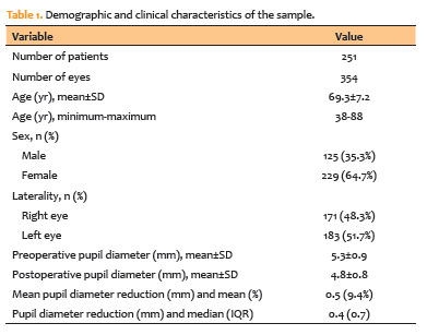
PURPOSE: To evaluate changes in scotopic pupil diameter before and after cataract surgery performed by phacoemulsification with intraocular lens implantation.
METHODS: This prospective longitudinal observational study included patients who underwent cataract surgery. Scotopic pupil diameter was measured preoperatively and 30-40 days postoperatively using an automated keratometer after a standardized dark-adaptation period under controlled ambient illumination. Each eye was considered an independent unit of observation. Because some participants contributed both eyes, intraindividual correlation was accounted for using a linear mixed-effects model with random patient intercepts. Time of assessment (preoperative versus postoperative), age, sex, and eye laterality were included as fixed effects.
RESULTS: A total of 354 eyes from 251 patients were analyzed. The mean patient age was 69.3±7.2 yr. Mean scotopic pupil diameter decreased from 5.3±0.9mm preoperatively to 4.8±0.8mm postoperatively, representing a mean reduction of 0.5mm (9.4%). In the linear mixed-effects model, cataract surgery was associated with a significant reduction in pupil diameter, with an adjusted mean difference of 0.45mm (95% confidence interval [95% CI], 0.39-0.51; p<0.001). Age (p=0.061), sex (p=0.920), and eye laterality (p=0.152) were not significantly associated with the magnitude of pupil diameter change.
CONCLUSION: Phacoemulsification with intraocular lens implantation was associated with a significant reduction in scotopic pupil diameter, independent of age, sex, and eye laterality. This finding should be considered during preoperative planning, particularly when selecting intraocular lenses whose optical performance depends on postoperative pupil size.
Keywords: Cataract; Pupil; Phacoemulsification; Lens implantation, intraocular; Lenses, intraocular; Pseudophakia
Abstract
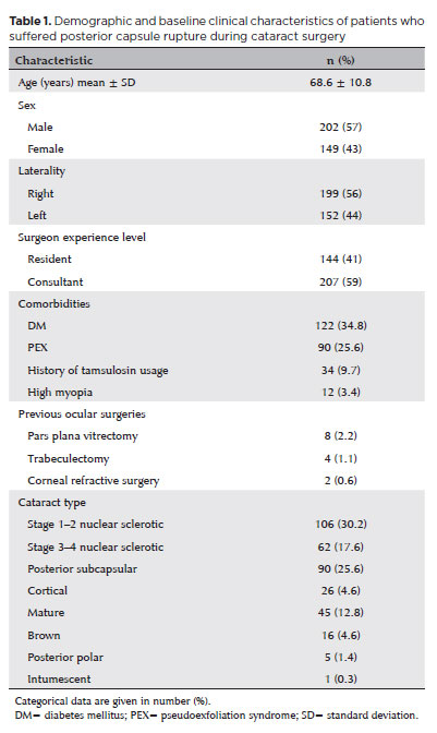
PURPOSE: Posterior capsule rupture is defined as an intraoperative posterior capsule tear resulting in vitreous loss. This study aimed to analyze the clinical characteristics, preoperative risk factors, intraoperative management strategies, and postoperative complications associated with posterior capsule rupture during phacoemulsification surgery.
METHODS: This was a retrospective observational cohort study of the medical records for 25,224 phacoemulsification surgeries performed at our tertiary eye care center between 2017 and 2022. We collected and collated the demographic characteristics and clinical findings of the patients in our cohort. Intraoperative management strategies and postoperative outcomes over a 1-year followup period were also recorded.
RESULTS: Posterior capsule rupture occurred in 351 eyes (351 patients), giving an overall posterior capsule rupture rate of 1.3%. The mean patient age was 68.6 ± 10.8 years. Pseudoexfoliation syndrome, mature cataracts, brown cataracts, and surgery performed by a resident were identified as risk factors for posterior capsule rupture (p<0.05 for each; the risk ratios were 2.70, 2.15, 2.44, 1.34, respectively). The most common intraoperative complications were dislocated lens fragments in the vitreous (8%) and iris damage (7.1%). The mean best-corrected visual acuity improved from 1.31 ± 0.84 (logMAR) postoperatively to 0.51 ± 0.56 at the end of the 1-year follow-up period (p<0.001). Corneal edema (55.6%) and elevated intraocular pressure (33.3%) were the most common early postoperative complications. Persistently elevated intraocular pressure (11.1%) and cystoid macular edema (5.1%) were the most common late postoperative complications.
CONCLUSION: Posterior capsule rupture is a common complication of phacoemulsification surgery that requires prolonged postoperative follow-up and a multidisciplinary approach. Despite the increased incidence of complications when rupture occurs, appropriate intraoperative and postoperative management can lead to satisfactory visual outcomes.
Keywords: Cataract extraction; Phacoemulsification; Posterior capsule rupture; Corneal edema; Risk factors; Postoperative complications; Intraoperative complications
Abstract
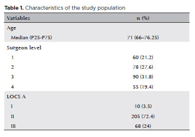
PURPOSE: To compare endothelial corneal cell changes following cataract surgery performed by phacoemulsification with intraocular lens implantation, conducted by surgeons with varying levels of experience.
METHODS: Two hundred and eighty-three eyes diagnosed with cataract were included. Lens opacity was classified into three categories (I, II, and III). Surgeons were categorized into four experience levels (1, 2, 3, and 4), based on years of practice and lifetime surgeries performed. Corneal endothelial characteristics were assessed using non-contact specular microscopy, with measurements taken before surgery and 30-60 days post-surgery.
RESULTS: Pre- and postoperative endothelial analysis showed no significant differences between surgeon levels regarding visual acuity achieved, corneal thickness, and endothelial hexagonality. However, the central endothelial cell density index showed a significantly greater reduction among level 1 surgeons (p=0.026). Grade II cataracts exhibited significant variations in the central endothelial cell density (p=0.011) and average cell size, with level 1 surgeons showing the largest increases (p=0.024).
CONCLUSIONS: The analysis revealed significant differences in visual acuity and endothelial indices between surgeon experience levels, with less experienced surgeons showing greater variations and poorer performance. Clinical protocols should consider these data to establish safer training protocols.
Keywords: Cataract extraction; Phacoemulsification; Endothelium; corneal; Lens implantation, intraocular; Visual acuity; Internship and residency; Surgeons
Abstract
We present a case report detailing the successful phacoemulsification surgery with artificial iris implantation for two individuals with oculocutaneous albinism. These women suffered from cataracts, resulting in reduced visual acuity and heightened photophobia due to iris pigmentary epithelium deficiency. The patients underwent phacoemulsification along with prosthetic artificial iris implantation into the posterior chamber. This intervention resulted in improved visual acuity, reduced photophobia and glare, and an overall enhanced quality of life. Our report highlights two cases of successful phacoemulsification and artificial iris implantation in patients with oculocutaneous albinism and cataracts, leading to improved visual acuity, reduced photophobia, and enhanced quality of life. Notably, there are no prior records in South American literature of cataract surgery combined with artificial iris implantation for oculocutaneous albinism patients up to the time of this publication.
Keywords: Cataract extraction; Albinism, oculocutaneous; Lens implantation, intraocular
01-fig01.jpg)
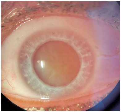
Abstract
Um homem de 59 anos apresentou embaçamento visual unilateral no olho esquerdo. Sua acuidade visual nesse olho era no nível de movimentos da mão. O paciente havia se submetido a uma cirurgia de facoemulsificação em que foi feita a implantação intraestromal de uma lente intraocular de câmara posterior. Foi feita a extração dessa lente intraestromal intraocular e uma nova lente intraocular foi implantada. A melhor acuidade visual corrigida final foi de 20/40 pela tabela de Snellen. Com este relato de caso, os autores desejam apontar que uma incisão de degrau único em córnea clara, quando combinada com a injeção de uma lente ocular através da incisão, pode levar a um direcionamento incorreto da lente intraocular para dentro do estroma corneano. Portanto, recomenda-se uma construção cuidadosa da incisão ao se remover uma lente intraocular direcionada incorretamente.
Keywords: Implante de lente intraocular; Lentes intraocular; Facoemulsificação; Cicatrização; Catarata; Acuidade visual
 ABO is licensed under a Creative Commons Attribution-NonComercial 4.0 Internacional.
About
Issues
Editorial Board
Submission
Arquivos Brasileiros de Oftalmologia
Official publication of Brazilian Council of Ophthalmology - Conselho Brasileiro de Oftalmologia (CBO)
Rua Casa do Ator, 1.117 - 2nd floor - Zip Code: 04546-004
São Paulo - SP, Brazil
TEL: +55 11 3266-4000
E-mail: [email protected]