Arq. Bras. Oftalmol. 2023;86 (5 )
:1-5
| DOI: 10.5935/0004-2749.20230063
Abstract
Objetivo: A injeção peribulbar de triancinolona é um tratamento alternativo para doenças oculares da tireoide; no entanto, a segurança desse procedimento continua controversa. O objetivo deste artigo é descrever os efeitos adversos locais e sistêmicos de injeções peribulbares de triancinolona em pacientes com doença ocular da tireoide.
Métodos: Estudo retrospectivo de uma série de casos. Foram analisados os prontuários médicos dos pacientes com doença ocular da tireoide tratados com injeções de triancinolona peribulbar em uma única instituição acadêmica entre 2007 e 2019. Foram documentadas as complicações locais e sistêmicas.
Resultados: Um total de 123 pacientes foram tratados. Apenas 11 (8,9%) pacientes apresentaram complicações locais, sendo a mais frequente a presença de equimoses palpebrais superficiais (7,3%), enquanto 2 (1,6%) pacientes apresentaram complicações sistêmicas (hiperglicemia e inibição da suprarrenal após a interrupção do tratamento). Todas estas complicações foram transitórias e nenhum paciente apresentou sequelas de longo prazo.
Conclusões: As injeções peribulbares de triancinolona nas doenças oculares da tireoide têm uma taxa muito baixa de complicações, tanto locais quanto sistêmicas. São necessários estudos prospectivos para aprofundar este tópico.
Keywords: Órbita/diagnóstico por imagem; Imageamento por ressonância magnética; Oftalmopatia de Graves; Triancinolona/efeitos adversos; Injeções.
Arq. Bras. Oftalmol. 2022;85 (6 )
:590-598
| DOI: 10.5935/0004-2749.20220081
Abstract
Objetivo: Identificar tendências no campo de pesquisa da orbitopatia de Graves nas últimas duas décadas e analisar os ramos de maior concentração de pesquisas nessa área.
Métodos: O banco de dados Web of Science foi usado para extrair artigos com “orbitopatia de Graves” ou seus sinônimos no título. Dados completos e referências foram exportados para o programa VOSviewer para serem analisados. Mapas e gráficos de visualização foram construídos a partir desses dados.
Resultados: Foram obtidos 1067 artigos sobre a orbitopatia de Graves a partir do banco de dados Web of Science. Os EUA ficaram em primeiro lugar em termos de número de publicações, seguidos pela Itália e pela República Popular da China. Dentre os autores, os artigos de Wiersinga WM tiveram o maior número de citações. Quanto às instituições, os artigos da Universidade de Amsterdã tiveram o maior número de citações, mas a Universidade de Pisa publicou o maior número de artigos. Dentre os periódicos, a revista Thyroid publicou o maior número de artigos. A análise de coautoria mostrou quatro agrupamentos de colaboração entre países. O primeiro agrupamento engloba países europeus; o segundo engloba os EUA, Brasil, Canadá, Coreia do Sul e Taiwan. A República Popular da China compreende um agrupamento por si só. O quarto agrupamento inclui Japão, Austrália e Polônia. A análise das palavras-chave revelou cinco agrupamentos de tópicos de palavras-chave: patogênese, gerenciamento, associação, qualidade de vida e cirurgia. A análise das referências citadas em conjunto revelou cinco agrupamentos: patogênese, manejo, fatores de risco, avaliação clínica e manejo cirúrgico.
Conclusão: A pesquisa no campo da orbitopatia de Graves cresceu nos últimos vinte anos. Os tópicos com a maior concentração de pesquisas são: patogênese, gerenciamento, fatores de risco, qualidade de vida e complicações. As tendências de pesquisa mudaram nas últimas duas décadas. Ficou evidente um aumento do interesse em explorar os mecanismos e associações da orbitopatia de Graves. Observou-se uma cooperação entre países europeus neste campo de pesquisa. Os EUA estabeleceram uma cooperação internacional mais ampla que outros países. Acreditamos que mais colaboração internacional envolvendo países em desenvolvimento seria recomendável.
Keywords: Oftalmopatia de Graves; Bibliometria; Oftalmopatia de Graves; Pesquisa
Arq. Bras. Oftalmol. 2023;86 (4 )
:359-364
| DOI: 10.5935/0004-2749.20230056
Abstract
Objetivo: Comparar as características radiológicas e clínicas do adenoma pleomórfico primário e do carcinoma adenoide cístico da glândula lacrimal.
Métodos: Este estudo revisou retrospectivamente os achados de imagem e os prontuários médicos de casos de adenoma pleomórfico e carcinoma adenoide cístico da glândula lacrimal.
Resultados: Foram avaliados 11 pacientes com adenoma pleomórfico e 16 pacientes com carcinoma adenoide cístico. Não houve diferenças estatisticamente significativas em relação à idade e sexo. Proptose foi o sintoma de apresentação mais comum em ambos os grupos. Os carcinomas adenoides císticos foram mais propensos que os adenomas pleomórficos a apresentarem massas palpáveis, diplopia, dor e perda sensorial, mas essa diferença entre os grupos não foi estatisticamente significativa. Não houve diferenças estatísticas em termos de homogeneidade e indentação do globo ocular entre os dois tipos de tumores em imagens de tomografia computadorizada (p>0,05). Também à tomografia computadorizada, a invasão óssea, a calcificação do tumor e o sinal em cunha foram mais frequentes nos carcinomas adenoides císticos, enquanto a remodelação óssea foi mais frequente nos adenomas pleomórficos, com significância estatística para todas essas manifestações (p<0,05). À ressonância magnética, os adenomas pleomórficos foram significativamente mais propensos a terem margens bem definidas, contornos lobulados, realce heterogêneo pelo contraste e hiperintensidade na ressonância magnética ponderada em T2 (p<0,05).
Conclusão: Ao se diferenciar o adenoma pleomórfico e o carcinoma adenoide cístico da glândula lacrimal, é muito importante avaliar as características radiológicas juntamente com as características clínicas. Os contornos lobulados podem ser uma característica radiológica significativamente distinta em favor do adenoma pleomórfico.
Keywords: Aparelho lacrimal/patologia; Adenoma pleomorfo; Carcinoma adenoide cístico; Tomografia computadorizada por raios x; Imagem por ressonância magnética.
Arq. Bras. Oftalmol. 2025;88 (5 )
:1-6
| DOI: 10.5935/0004-2749.2024-0103
Abstract
PURPOSE: This study aimed to evaluate abnormalities in the retinal nerve fiber layer and ganglion cell layer in patients with thyroid-associated orbitopathy using optical coherence tomography and to examine their relationship with disease severity.
METHODS: A cross-sectional study was conducted involving 74 participants, comprising 45 individuals with thyroid-associated orbitopathy and 29 healthy controls. All subjects underwent a comprehensive ophthalmological examination and optical coherence tomography using the Cirrus HD-OCT. The clinical activity score and the European Group on Graves’ Orbitopathy severity were also evaluated.
RESULTS: In the thyroid-associated orbitopathy group, the mean peripapillary retinal nerve fiber layer thickness was significantly reduced in the temporal quadrant (p<0.05). No significant differences were found in ganglion cell layer thickness across all sectors when compared with the control group. Besides, a significant correlation was observed between orbitopathy severity and decreased mean peripapillary retinal nerve fiber layer thickness (p<0.001).
CONCLUSION: Optical coherence tomography may serve as a useful tool for identifying changes in the retinal nerve fiber layer and ganglion cell layer in patients with thyroid-associated orbitopathy, including in the inactive phase and prior to the clinical manifestation of dysthyroid optic neuropathy. It may be a helpful adjunct in monitoring disease progression.
Keywords: Graves’ ophthalmopathy; Optic nerve disorders; Retinal nerve fiber layer; Retinal ganglion cells; Optical coherence tomography
Arq. Bras. Oftalmol. 2025;88 (2 )
:1-5
| DOI: 10.5935/0004-2749.2024-0113
Abstract
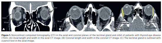
This study aimed to evaluate the morphometric and volumetric dimensions of the lacrimal gland in patients with inactive thyroid eye disease and compare them with the values reported in the literature. This case series evaluated consecutive patients with inactive thyroid eye disease treated at a tertiary eye hospital from 2015 to 2020. The patients' baseline demographics and clinical characteristics were obtained. The axial and coronal length, width, and volume of the lacrimal gland were measured on computed tomography scan images, and the results were statistically analyzed. A total of 21 patients (42 orbits) with inactive thyroid eye disease were evaluated. Their mean age was 49.0 ± 14.6 years, and 12 (57.1%) of them were men. The main complaint was dryness, and the majority of the patients had good vision and mild proptosis. The mean axial length and width of the lacrimal gland were 19.3 ± 3.9 mm and 7.5 ± 2.1 mm, respectively; coronal length and width, 20.4 ± 4.5 mm and 7.5 ± 2.1 mm, respectively; and lacrimal gland volume, 0.825 ± 0.326 mm3. Age, sex, or laterality were not found to be determinants of lacrimal gland enlargement. Patients with thyroid eye disease have enlarged lacrimal gland even in the nonactive phase of the disease multifactorial aspects influence the lacrimal gland in thyroid eye disease, making it difficult to establish a clear correlation with predisposing factors. Further studies are warranted to better understand the association between thyroid eye disease and the lacrimal gland.
Keywords: Graves' ophthalmology; Graves' disease; Lacrimal apparatus; Lacrimal apparatus diseases; X-ray computed tomography
Arq. Bras. Oftalmol. 2025;88 (5 )
:1-7
| DOI: 10.5935/0004-2749.2024-0319
Abstract
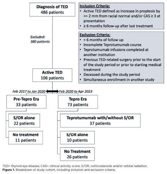
PURPOSE: This study evaluated rates of thyroid eye disease-related eyelid surgeries, strabismus surgeries, and orbital decompressions in active thyroid eye disease patients treated with teprotumumab compared to those who were not.
METHODS: In this single-center longitudinal study, we compared patients with active thyroid eye disease evaluated from 02/01/2017 to 01/31/2020 (pre-teprotumumab era) with those seen from 02/01/2020 to 04/30/2023 (teprotumumab era). Patients from the pre-teprotumumab era who received corticosteroids and/or orbital radiation were compared with those in the teprotumumab era treated with teprotumumab, with or without corticosteroids and/or orbital radiation. The primary outcomes were rates of orbital decompressions, strabismus surgery, and eyelid surgery among patients with at least 6 months of follow-up. Orbital decompressions involving two or more walls were classified as severe.
RESULTS: Of 486 records reviewed, 106 patients had active thyroid eye disease. Among them, 33 were from the pre-teprotumumab era; 22 received corticosteroids and/or orbital radiation, and 11 received no treatment. Seventy three patients were from the teprotumumab era; 37 received teprotumumab (with or without corticosteroids and/or orbital radiation), 10 received corticosteroids and/or orbital radiation alone, and 26 received no treatment. Demographics were comparable between groups. Orbital decompression was performed in 11 of 44 eyes (25.0%) in the pre-teprotumumab era treated with corticosteroids and/or orbital radiation (8 one-wall, 3 ≥two-wall), compared to 3 of 74 eyes (4.1%) in the teprotumumab era treated with teprotumumab with or without corticosteroids and/ or orbital radiation (all one-wall). The overall rate of orbital decompressions and the rate of ≥two-wall decompressions were significantly lower in the teprotumumab era (p=0.02 and p=0.0496, respectively). There was no significant difference in one-wall decompressions between era (p=0.07). Rates of strabismus surgeries (27.3% vs. 13.5%, p=0.19) and eyelid surgeries (22.7% vs. 21.6%, p=0.92) did not significantly differ between the era.
CONCLUSIONS: In patients with active thyroid eye disease, treatment with teprotumumab was associated with a significantly lower rate and severity of orbital decompressions compared to treatment with corticosteroids and/or orbital radiation alone. However, the rates of strabismus and eyelid surgeries remained similar between groups.
Keywords: Teprotumumab; Adrenal cortex hormone; Decompression; Graves ophthalmopathy; Strabismus
Arq. Bras. Oftalmol. 2024;87 (5 )
:1-7
| DOI: 10.5935/0004-2749.2023-0296
Abstract
PURPOSE: To compare inferomedial wall orbital decompression to balanced medial plus lateral wall orbital decompression in patients with Graves’ orbitopathy in the inactive phase with regard to exophthalmos reduction and the effects on quality of life.
METHODS: Forty-two patients with inactive Graves’ orbitopathy were randomly divided into two groups and submitted to one of two orbital decompression techniques: inferomedial wall orbital decompression or medial plus lateral wall orbital decompression. Preoperative and postoperative assessments included Hertel’s exophthalmometry and a validated Graves’ orbitopathy quality of life questionnaire. The results of the two groups were compared.
RESULTS: Compared to preoperative measurement, exophthalmos reduction was statistically significant in both groups (p<0.001) but more so in patients undergoing medial plus lateral wall orbital decompression (p=0.010). Neither orbital decompression techniques increased the visual functioning subscale score on the Graves’ orbitopathy quality of life questionnaire (inferomedial wall orbital decompression p=0.362 and medial plus lateral wall orbital decompression p=0.727), but a statistically significant difference was observed in the score of the appearance subscale in patients submitted to medial plus lateral wall orbital decompression (p=0.006).
CONCLUSIONS: Inferomedial wall orbital decompression is a good alternative for patients who do not require large exophthalmos reduction. However, medial plus lateral wall orbital decompression offers greater exophthalmos reduction and greater improvement in appearance (higher Graves’ orbitopathy quality of life questionnaire scores), making it a suitable option for esthetic-functional rehabilitation.
Keywords: Graves’ ophthalmopathy; Quality of life; Exophthalmos; Strabismus; Diplopia; Decompression, surgical
 ABO is licensed under a Creative Commons Attribution-NonComercial 4.0 Internacional.
05-tab01.jpg)
06-fig01.jpg)
11-tab01.jpg)

02-fig01.jpg)
03-fig01.jpg)
02-fig01.jpg)
04-fig01.jpg)
02-fig01.jpg)
14-fig01.jpg)
02-fig01.jpg)