Arq. Bras. Oftalmol. 2026;89 (2 )
:1-6
| DOI: 10.5935/0004-2749.2025-0244
Abstract
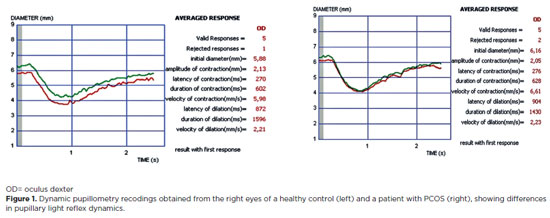
PURPOSE: Polycystic ovary syndrome is frequently associated with autonomic nervous system dysfunction, even in the absence of obesity or overt metabolic abnormalities. Alterations in pupillary responses may reflect early autonomic involvement and serve as a potential tool for early diagnosis, risk stratification, and disease monitoring. This study aimed to investigate pupillary reflex parameters using dynamic pupillometry in newly diagnosed non-obese women with polycystic ovary syndrome and to compare the findings with those of healthy controls. Methods: This prospective cross-sectional study included 48 newly diagnosed women with polycystic ovary syndrome and 44 age- and sex-matched healthy controls. Pupillary function parameters were measured using dynamic pupillometry (MonPackOne; Metrovision, France). Results: The mean age did not differ significantly between the groups (p=0.870). Initial pupil diameter, pupil contraction amplitude, and contraction velocity were significantly lower in the PCOS group than in the control group, whereas pupillary dilation duration was significantly longer (p<0.001, p<0.001, p=0.007, and p=0.032, respectively). No significant differences were observed between the groups regarding contraction latency, contraction duration, dilation latency, or dilation velocity (p=0.749, p=0.925, p=0.653, and p=0.310, respectively). Conclusion: Newly diagnosed non-obese women with polycystic ovary syndrome exhibit significant alterations in pupillary dynamics, suggesting a generalized reduction in both sympathetic and parasympathetic activity. Dynamic pupillometry may represent a practical, noninvasive tool for detecting early autonomic hypoactivity and identifying patients at risk for future metabolic or cardiovascular complications.
Keywords: Autonomic nervous system; IPupil/physiology; Reflex, Pupillary/physiology; Polycystic ovary syndrome/diagnosis; Menstruation disturbances; Ideal body weight
Arq. Bras. Oftalmol. 2026;89 (1 )
:1-10
| DOI: 10.5935/0004-2749.2025-0184
Abstract
PURPOSE: To investigate choroidal structural and vascular changes in patients with mild autonomous cortisol secretion using enhanced depth imaging optical coherence tomography and optical coherence tomography angiography.
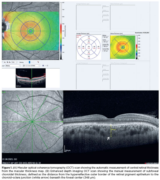
METHODS: This cross-sectional study included 60 eyes of 30 patients with mild autonomous cortisol secretion and 60 eyes of 30 subjects with nonfunctional adenoma (controls) between February 2023 and January 2024. Subfoveal choroidal thickness, pachychoroid spectrum disease and choroidal vascularity index were evaluated using spectral-domain optical coherence tomography. Group comparisons were performed, and correlations between subfoveal choroidal thickness and clinical features were analyzed.
RESULTS: Pachyvessels were more common in patients with mild autonomous cortisol secretion than in controls (71.4% vs. 42.9%, p=0.002). The frequency of pachychoroidal spectrum disease was significantly higher in the mild autonomous cortisol secretion Group (68.3% vs. 31.7%; p<0.001). Median subfoveal choroidal thickness was 355 μm (range, 150–535) in the mild autonomous cortisol secretion Group and 297 μm (range, 162–597) in controls (p=0.014). Choroidal vascularity index was comparable between groups (p=0.072). Subfoveal choroidal thickness correlated significantly with axial length, spherical equivalent, post-1-mg dexamethasone suppression test cortisol level, and disease duration.
CONCLUSION: Patients with mild autonomous cortisol secretion exhibited greater subfoveal choroidal thickness and a higher frequency of pachychoroidal spectrum disease compared with controls, whereas stromal and vascular structural alterations were proportionally similar between groups.
Keywords: Adrenal gland neoplasms; Central serous chorioretinopathy; Choroid; Cushing syndrome; Hydrocortisone; Optical coherence tomography
Arq. Bras. Oftalmol. 2024;87 (4 )
:1-8
| DOI: 10.5935/0004-2749.2021-0401
Abstract
Objetivos: Avaliar a eficácia do uso de toxina botulínica tipo A no tratamento do estrabismo em pacientes com comprometimento neurológico e avaliar os fatores associados ao sucesso do tratamento.
Métodos: Cinquenta pacientes com estrabismo e comprometimento neurológico foram incluídos no estudo. Em todas as crianças, a toxina botulínica tipo A foi injetada no músculo extraocular apropriado. A relação entre características demográficas, características clínicas e o sucesso do tratamento foram analisadas.
Resultados: No grupo de estudo, 34 pacientes tiveram esotropia e 16 pacientes tiveram exotropia, sendo trinta e seis pacientes com paralisia cerebral e 14 pacientes com hidrocefalia. O tempo médio de acompanhamento foi de 15,3 ± 7,3 meses. O número médio de aplicações foi de 1,4 ± 0,6. O ângulo de desvio médio foi de 42,5 ± 13,2 DP antes do tratamento e diminuiu para 12,8 ± 11,9 DP após o tratamento. Alinhamento motor bem sucedido (ortotropia dentro de 10 DP) foi alcançado em 60% dos pacientes. A análise de regressão logística binária revelou que o desalinhamento esotrópico e uma menor duração do estrabismo foram significativamente associados ao sucesso do tratamento no grupo de estudo. Pacientes esotrópicos com ângulos de desalinhamento menores são mais propensos a serem tratados com uma única aplicação.
Conclusão: O uso da toxina botulínica tipo A para o tratamento de estrabismo em crianças com comprometimento neurológico é uma boa alternativa para a terapia cirúrgica convencional com menor risco de hipercorreção. O resultado do tratamento é melhor em exodesvios e em pacientes com estrabismo de menor duração, implicando em vantagem para o tratamento precoce.
Keywords: Estrabismo; Toxinas botulínicas; Manifestações neurológicas; Doenças do sistema nervoso; Paralisia cerebral; Hodrocefalia; Criança


 ABO is licensed under a Creative Commons Attribution-NonComercial 4.0 Internacional.


12-tab01tb.jpg)
11-fig01.jpg)
14-fig01.jpg)
12-fig01.jpg)
13-fig01.jpg)

01-fig01tb.jpg)
02-fig01.jpg)