Arq. Bras. Oftalmol. 2021;84 (3 )
:214-219
| DOI: 10.5935/0004-2749.20210029
Abstract
OBJETIVO: Avaliar a influência da dinâmica pupilar na curva de desfoco de olhos implantados com lente intraoculares multifocais difrativas.
MÉTODOS: Estudo prospectivo e randomizado realizado na Faculdade de Medicina de Ribeirão Preto - Universidade de São Paulo - Departamento de Oftalmologia. Trinta e oito pacientes foram aleatoriamente designados para receber bilateralmente lentes intraoculares SN6AD1 (n=20) (mfIOL) ou SN60WF (n=18) (aIOL). Além da acuidade visual para longe e perto, corrigida e não corrigida, e curva de desfoco, foi ainda realizada pupilometria dinâmica. A área sob a curva de desfoco foi calculada usando um modelo polinomial empírico.
RESULTADOS: Um total de 16 e 17 pacientes (n=32 e 34 olhos) completaram 1 ano de seguimento nos grupos mfIOL e aIOL, respectivamente. Não houve diferenças significativas entre grupos para as acuidades visuais seja para longe ou perto. As curvas de desfoco do grupo mfIOL mostraram um pico duplo; enquanto o SN60WF mostrou apenas um pico, típico para uma lente intraoculares monofocal. A média da área sob a curva de desfoco do grupo aIOL foi (4,66 ± 1,51 logMAR.dp), e essa é estatisticamente significante diferente da métrica do grupo mfIOL (1,99 ± 1,31 logMAR.dp). A pupila na contração máxima após a exposição a um flash de 30 cd/m2 por 1 segundo foi significativamente correlacionada com uma melhor área de foco no grupo mfIOL (r=0,54; p=0,0017), essa relação não foi observada para o grupo aIOL.
CONCLUSÃO: Estes dados indicam que quanto menor a pupila durante contração, melhor é a área sob a curva de desfoco e, portanto, o desempenho visual dos olhos implantados com essa mfIOL. Esta correlação não foi encontrada para lentes intraoculares monofocais.
Keywords: Lentes intraoculares multifocais; Pupila/fisiologia, Catarata; Facoemulsificacão
Arq. Bras. Oftalmol. 2023;86 (2 )
:113-120
| DOI: 10.5935/0004-2749.20230022
Abstract
Objetivos: Avaliar a estabilidade e eficácia da técnica double-flanged com sutura de 5-0 polipropileno para fixação de cataratas subluxadas aos 18 meses e as possíveis complicações desta nova técnica.
Métodos: Esta técnica utiliza um monofilamento de polipropileno 5-0 para criar dois flanges com um termocautério para fixar um Segmento de Tensão Capsular na esclera a fim de estabilizar o saco capsular subluxado. Esta técnica foi implementada em 17 olhos que necessitavam do implante de lente intraocular em casos de diálise zonular devido a trauma, síndrome de Marfan, microesferofacia, subluxação idiopática ou pós-facoemulsificação que provocou subulxação do saco capsular intraoperatória.
Resultados: O seguimento dos pacientes foi de 18 meses. A acuidade visual corrigida melhorou significativamente de 0,85 para 0,39 (logMAR), enquanto os erros de refração esféricos e cilíndricos e a pressão intraocular permaneceram estáveis. Nenhuma fotodegradação de sutura ou pseudofacodonese foi encontrada.
Conclusão: A técnica double-flanged para fixação transescleral de saco capsular com sutura de 5-0 polipropileno mostrou resultados de estabilidade de longo prazo para o complexo lente/saco capsular. Então, aparenta ser uma opção segura para cirurgia de catarata, sem necessidade pontos, em olhos com fraqueza zonular ou diálise
Keywords: Catarata; Facoemulsificação; Lente intraocular; Técnica de sutura; Acuidade visual
Arq. Bras. Oftalmol. 2026;89 (2 )
:1-9
| DOI: 10.5935/0004-2749.2025-0113
Abstract
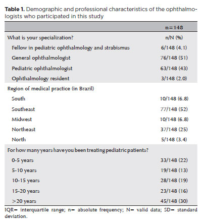
PURPOSE: This study aimed to identify the strategies adopted by Brazilian ophthalmologists to control myopia in clinical practice.
METHODS: This was a prospective cross-sectional study. Data were collected using an online questionnaire.
RESULTS: Responses from 148 participants were collected between March and May 2024. The majority of respondents were general ophthalmologists (51%) and pediatric ophthalmologists (43%). They came from all regions of Brazil, but more than half (52%) were from the Southeast region. Most participants (30%) had over 20 years of clinical practice experience. A significant proportion (89.2%) treated progressive myopia. The most requested complementary exams were optical biometry (83.78%) and corneal topography or tomography (69.59%). Behavioral measures were considered the most effective myopia treatment strategies by 41.2% of the respondents, followed by optical (33.8%) and pharmacological interventions (25%). Most recommended spending more time outdoors (94.59%) and reducing screen time (93.92%). Spectacle lenses for myopia (83.11%) and 0.025% atropine eye drops (54.73%) were the most prescribed treatments after the recommendation of environmental and behavioral changes.
CONCLUSION: This study presents a novel analysis of the clinical strategies for myopia control among Brazilian ophthalmologists. Understanding current clinical practices and identifying possible improvements are essential steps toward developing evidence-based guidelines and professional education aimed at improving patient care.
Keywords: Myopia/epidemiology; Refractive errors; Contact lenses; Myopia/drug therapy; Atropine/therapeutic use; Ophthalmologists; Practice patterns, physicians’; Surveys and questionnaires; Brazil/epidemiology
Arq. Bras. Oftalmol. 2022;85 (4 )
:359-363
| DOI: 10.5935/0004-2749.20220046
Abstract
Objetivo: Investigar os resultados pós-operatórios e avaliar os preditores de sucesso da facoemulsificação combinada à goniotomia com o Kahook Dual Blade para o tratamento da catarata e do glaucoma em olhos com glaucoma primário de ângulo aberto.
Métodos: Série de casos retrospectivos, não comparativos e intervencionistas, em que todos os pacientes com glaucoma primário de ângulo aberto submetidos ao procedimento de facoemulsificação combinada à goniotomia com o Kahook Dual Blade entre junho de 2018 e abril de 2019 foram inscritos. Todos os participantes tiveram um acompanhamento mínimo de 6 meses. Foram registrados os valores de pressão intraocular pré e pós-operatória (em 1, 3 e 6 meses), número de medicamentos antiglaucomatosos, melhor acuidade visual corrigida, complicações cirúrgicas e quaisquer eventos ou procedimentos subsequentes relacionados. A análise de regressão logística foi usada para investigar a associação entre diferentes variáveis e resultados cirúrgicos.
Resultados: Um total de 57 olhos de 47 pacientes foram incluídos (média de idade, 70,5 ± 7 anos). A pressão intraocular média reduziu de 15,5 ± 4,2 mmHg para 12,2 ± 2,4 mmHg na última visita de acompanhamento (p<0,001). O número médio de medicamentos antiglaucomatosos diminuiu significativamente de 1,9 ± 1,0 para 0,6 ± 1,0 durante o mesmo período (p<0,001). Com base no critério predefinido (redução da pressão intraocular ≥20% e/ou redução de ≥1 medicamento), a taxa de sucesso em 6 meses foi de 86%. Um valor de pressão intraocular pré-operatório mais alto (OR= 2,01; p=0,016) e maior porcentagem de redução da pressão intraocular inicial
(30 dias) (OR= 1,02; p=0,033) foram significativamente associados ao sucesso cirúrgico.
Conclusão: Nossos resultados sugerem que o procedimento de facoemulsificação combinada à goniotomia com o Kahook Dual Blade é uma alternativa eficaz e segura para o manejo da catarata em olhos com glaucoma primário de ângulo aberto, impactando positivamente no controle da pressão intraocular e no número de medicamentos. Olhos com pressão intraocular basal mais alta e resposta inicial mais pronunciada ao procedimento parecem apresentar melhores resultados em 6 meses. Mais estudos são necessários para avaliar a eficácia em longo prazo e o perfil de segurança.
Keywords: Glaucoma; Glaucoma de ângulo aberto; Catarata; Facoemulsificação; Pressão intraocular; Goniotomia
Arq. Bras. Oftalmol. 2023;86 (6 )
:1-6
| DOI: 10.5935/0004-2749.2021-0303
Abstract
Objetivos: Rever características epidemiológicas de crianças submetidas a cirurgia de catarata, em centro de referência no estado de São Paulo, Brasil, e fatos associados a atrasos no tratamento.
Métodos: Um total de 240 olhos submetidos a cirurgia de catarata, em 178 crianças, foram revisados neste estudo transversal observacional. Os seguintes aspectos foram analisados: características clínicas e epidemiológicas, sinais apontados pelos pais, teste do reflexo vermelho, olho operado e idade no diagnóstico e na cirurgia.
Resultados: A média de idades na primeira visita e cirurgia de catarata foi de 48.9 meses (DP=50,0 meses) e 64.5 meses (DP=55.4 meses), respectivamente. O sinal mais importante apontado pelos pais foi a leucocoria. O teste do reflexo vermelho foi realizado em dois terços das crianças com resultados anormais em 28%. Histórico familiar de catarata foi evidente em 30 (20,9%) crianças (n=144). Os achados mais prevalentes em termos de histórico de problemas oculares foram: cirurgias oculares prévias em 37 (16,6%) olhos (n= 223), alterações do segmento anterior em 20 (9,0%) olhos (n=221), estrabismo em 21 (9,5%) olhos (n=220) e nistagmo em 38 (24,4%) crianças (n=156).
Conclusões: Uma das causas para o atraso na admissão pode ter sido a falha em realizar o teste do reflexo vermelho, apesar de não ter sido possível verificar se todas as crianças foram submetidas ao exame. A hereditariedade foi o fator mais importante quanto à causa da catarata nessas crianças. A presença de estrabismo e nistagmo mais uma vez aponta para o diagnóstico tardio. Ausência de programas de referência e centros oftalmológicos especializados em crianças, além do número restrito de profissionais de apoio treinados na área e especialistas em oftalmologia pediátrica, foram as barreiras mais importantes para o tratamento adequado da catarata em crianças.
Keywords: Catarata/ congênito; Extração de catarata; Técnicas de diagnóstico oftalmológico ; Baixa visão; Atenção terciária à saúde; Criança
Arq. Bras. Oftalmol. 2026;89 (1 )
:1-5
| DOI: 10.5935/0004-2749.2025-0045
Abstract
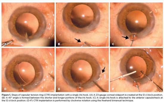
PURPOSE: To evaluate the effect of using a single iris retractor, affixed to the anterior capsulorhexis at the 12 o'clock position, on the ease of capsular tension ring implantation.
METHODS: This prospective comparative study comprised 37 patients with zonular weakness attributed to pseudoexfoliation syndrome who underwent capsular tension ring implantation during cataract surgery. In Group 1, a single iris retractor was inserted into the anterior capsulorhexis at the 12 o'clock position. Group 2 did not receive this intervention. Zonular weakness was graded on a scale of 1–5, and the subjective difficulty of capsular tension ring implantation was categorized as easy, medium, or difficult.
RESULTS: Group 1 and 2 comprised 20 and 17 patients, respectively. There were no significant differences between the groups in age, sex distribution, and presence of glaucoma (p=0.53, p=0.28, and p=1.00, respectively). The mean zonular weakness score was significantly higher in Group 1 (3.35 ± 0.45) than in Group 2 (2.71 ± 0.59; p=0.02). Capsular tension ring implantation was significantly easier in the iris retractor group (p<0.001).
CONCLUSIONS: Placement of a single iris retractor attached to the anterior capsulorhexis at the 12 o'clock position may facilitate easier capsular tension ring implantation, even in patients with greater zonular weakness. This technique could reduce the risk of capsular tension ring displacement into the iridocorneal angle or ciliary sulcus.
Keywords: Capsular tension ring; Cataract; Iris hook; Pseudoexfoliation syndrome; Zonular weakness; Cataract extraction; Phacoemulsification; Capsulorhexis.
Arq. Bras. Oftalmol. 2025;88 (6 )
:1-5
| DOI: 10.5935/0004-2749.2025-0085
Abstract
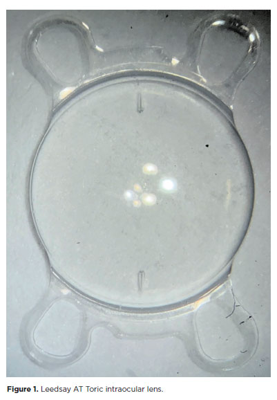
PURPOSE: The purpose of this study was to assess visual outcomes and patient satisfaction following cataract surgery involving the implantation of quad-loop intraocular lenses, including trifocal, bifocal, and toric variants.
METHODS: Information was obtained from both physical and electronic medical records of patients who underwent phacoemulsification cataract surgery with implantation of different intraocular lenses between January 1, 2022, and December 31, 2023. The study included individuals aged over 18 who received bilateral implantation of bifocal, trifocal, or monofocal toric intraocular lenses. Visual acuity was assessed at various postoperative time points using the logMAR scale. Quantitative variables were analyzed using mean and standard deviation.
RESULTS: A total of 92 eyes received premium intraocular lenses: 4 bifocal, 32 trifocal, 52 toric monofocal, and 4 trifocal toric lenses. The average preoperative corrected visual acuity was logMAR 0.478 ± 0.259. On the first postoperative day, the average uncorrected visual acuity was logMAR 0.301 ± 0.207. By day 30, 67.4% of eyes achieved uncorrected distance visual acuity of logMAR 0.2 or better. Patient satisfaction was high, with few reports of glare or halos.
CONCLUSION: Quad-loop intraocular lenses-including trifocal, bifocal, and toric models-demonstrated effective improvement in visual acuity and high levels of patient satisfaction. These lenses represent a suitable option for enhancing visual outcomes after cataract surgery. Additional studies with larger cohorts are recommended to confirm these results.
Keywords: Cataract extraction; Aberrometry/methods; Lenses, intraocular; Lens implantation, intraocular; Prosthesis design
Arq. Bras. Oftalmol. 2026;89 (2 )
:1-8
| DOI: 10.5935/0004-2749.2025-0175
Abstract
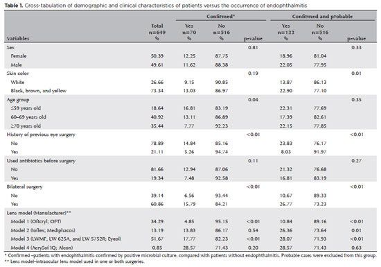
PURPOSE: Endophthalmitis is one of the most important adverse events after cataract surgery, as it can lead to total vision loss. This study aimed to describe the occurrence of endophthalmitis after phacoemulsification with intraocular lens implantation in patients treated in a community setting in Porto Velho, Rondônia, Brazil.
METHODS: This retrospective cohort study was conducted using a database of 649 medical records of patients who underwent surgery and were followed for three months. Poisson regression analysis was used to estimate relative risks and 95% confidence intervals (95% CIs).
RESULTS: The incidence of confirmed endophthalmitis was 11.94% (95% CI, 9.50-14.76), while the incidence of confirmed and probable cases was 20.50% (95% CI, 17.52-23.73). For confirmed cases, bilateral surgery and the use of lens model 3 were identified as risk factors for endophthalmitis, whereas age over 70 yr and preoperative antibiotic use were protective factors. For confirmed and probable cases, brown and yellow skin color, bilateral surgery, and the use of lens model 3 were also identified as risk factors. Gram-negative bacteria were the predominant etiological agents, and corneal edema was the main clinical manifestation. The mean duration of treatment was eight days, and 27.12% of patients used antibiotics.
CONCLUSION: The incidence observed was substantially higher than that reported in the literature, with a predominance of Gram-negative agents and an association with bilateral surgeries and the Eyeol intraocular lens model. These findings reinforce the need for continuous epidemiological surveillance and the implementation of specific biosafety and infection control protocols during cataract surgery campaigns.
Keywords: Endophthalmitis; Disease outbreaks; Phacoemulsification; Lens implantation, intraocular; Lenses, intraocular; Cataract; Risk factors; Anti-bacterial agents
Arq. Bras. Oftalmol. 2026;89 (1 )
:1-8
| DOI: 10.5935/0004-2749.2025-0109
Abstract
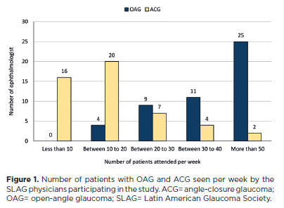
PURPOSE: To evaluate the preferred surgical practice patterns for glaucoma among members of the Latin American Glaucoma Society.
METHODS: A cross-sectional study was conducted using an electronic survey distributed in July 2023 via email to members of the Latin American Glaucoma Society. The questionnaire comprised four sections addressing the specialists' profiles, preferred surgical procedures for open-angle glaucoma, and choices in 10 different clinical scenarios, including congenital glaucoma.
RESULTS: Of the 63 members, 49 physicians (77.7%) responded – 13 women and 36 men – from nine Latin American countries. Thirty-one respondents (63.26%) had more than 20 yr of professional experience. For the surgical management of open-angle glaucoma, trabeculectomy was the most preferred procedure (48 physicians), followed closely by glaucoma drainage devices (47 physicians) and minimally invasive glaucoma surgery (29 physicians). Across the 10 clinical scenarios, glaucoma drainage devices were selected most frequently (203 preferences), followed by trabeculectomy (118), ciliary body laser procedures (107), and minimally invasive glaucoma surgery (40). However, minimally invasive glaucoma surgery was the preferred option for primary open-angle glaucoma with mild-to-moderate cataracts.
CONCLUSION: Among specialists of the Latin American Glaucoma Society, trabeculectomy and glaucoma drainage devices remain the most commonly performed surgical procedures. Minimally invasive glaucoma surgery is primarily used in combination with cataract surgery, while ciliary body laser procedures are generally reserved for cases of previous glaucoma drainage device failure or as an initial option for newly diagnosed glaucoma cases.
Keywords: Glaucoma; Ophthalmologic surgical procedures; Latin America; Practice patterns, physicians; Surveys and questionnaires
Arq. Bras. Oftalmol. 2025;88 (2 )
:1-8
| DOI: 10.5935/0004-2749.2023-0268
Abstract
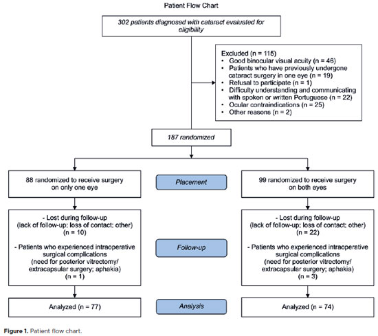
PURPOSE: This prospective, randomized, unmasked, clinical trial aimed to report the visual outcomes of cataract surgery on both eyes versus cataract surgery on one eye in Brazilian patients.
METHODS: This study included patients with bilateral cataracts and binocular visual acuity worse than or equal to 0.3 logarithm of the minimum angle of resolution. The patients were randomly assigned to undergo surgery on one (Control Group) or both eyes (one eye at a time; Intervention Group). Postoperatively, self-reported visual function using Catquest-9SF (primary outcome measure), binocular visual acuity, stereopsis, and ocular dominance (secondary outcome measures) were compared.
RESULTS: A total of 151 patients (77 and 148 eyes in the Control and Intervention Groups, respectively) completed the follow-up. Patients who underwent surgery on both eyes exhibited significantly better self-reported visual function (p=0.036) and stereopsis (p=0.026) than those who underwent surgery on one eye. Binocular visual acuity and ocular dominance did not affect the group comparisons.
CONCLUSIONS: Surgery on both eyes resulted in significantly better self-reported visual function and stereopsis than surgery on one eye.
Keywords: Cataract; Cataract extraction; Quality of life; Treatment outcome; Visual acuity; Binocular vision; Stereopsis
Arq. Bras. Oftalmol. 2026;89 (4 )
:1-5
| DOI: 10.5935/0004-2749.2026-0010
Abstract
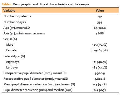
PURPOSE: To evaluate changes in scotopic pupil diameter before and after cataract surgery performed by phacoemulsification with intraocular lens implantation.
METHODS: This prospective longitudinal observational study included patients who underwent cataract surgery. Scotopic pupil diameter was measured preoperatively and 30-40 days postoperatively using an automated keratometer after a standardized dark-adaptation period under controlled ambient illumination. Each eye was considered an independent unit of observation. Because some participants contributed both eyes, intraindividual correlation was accounted for using a linear mixed-effects model with random patient intercepts. Time of assessment (preoperative versus postoperative), age, sex, and eye laterality were included as fixed effects.
RESULTS: A total of 354 eyes from 251 patients were analyzed. The mean patient age was 69.3±7.2 yr. Mean scotopic pupil diameter decreased from 5.3±0.9mm preoperatively to 4.8±0.8mm postoperatively, representing a mean reduction of 0.5mm (9.4%). In the linear mixed-effects model, cataract surgery was associated with a significant reduction in pupil diameter, with an adjusted mean difference of 0.45mm (95% confidence interval [95% CI], 0.39-0.51; p<0.001). Age (p=0.061), sex (p=0.920), and eye laterality (p=0.152) were not significantly associated with the magnitude of pupil diameter change.
CONCLUSION: Phacoemulsification with intraocular lens implantation was associated with a significant reduction in scotopic pupil diameter, independent of age, sex, and eye laterality. This finding should be considered during preoperative planning, particularly when selecting intraocular lenses whose optical performance depends on postoperative pupil size.
Keywords: Cataract; Pupil; Phacoemulsification; Lens implantation, intraocular; Lenses, intraocular; Pseudophakia
Arq. Bras. Oftalmol. 2025;88 (5 )
:1-7
| DOI: 10.5935/0004-2749.2024-0368
Abstract
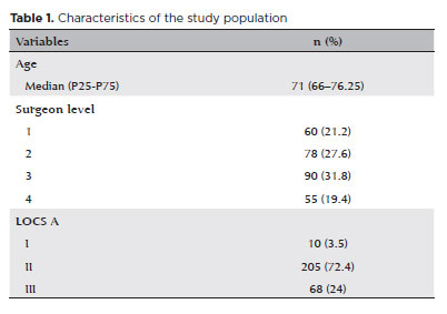
PURPOSE: To compare endothelial corneal cell changes following cataract surgery performed by phacoemulsification with intraocular lens implantation, conducted by surgeons with varying levels of experience.
METHODS: Two hundred and eighty-three eyes diagnosed with cataract were included. Lens opacity was classified into three categories (I, II, and III). Surgeons were categorized into four experience levels (1, 2, 3, and 4), based on years of practice and lifetime surgeries performed. Corneal endothelial characteristics were assessed using non-contact specular microscopy, with measurements taken before surgery and 30-60 days post-surgery.
RESULTS: Pre- and postoperative endothelial analysis showed no significant differences between surgeon levels regarding visual acuity achieved, corneal thickness, and endothelial hexagonality. However, the central endothelial cell density index showed a significantly greater reduction among level 1 surgeons (p=0.026). Grade II cataracts exhibited significant variations in the central endothelial cell density (p=0.011) and average cell size, with level 1 surgeons showing the largest increases (p=0.024).
CONCLUSIONS: The analysis revealed significant differences in visual acuity and endothelial indices between surgeon experience levels, with less experienced surgeons showing greater variations and poorer performance. Clinical protocols should consider these data to establish safer training protocols.
Keywords: Cataract extraction; Phacoemulsification; Endothelium; corneal; Lens implantation, intraocular; Visual acuity; Internship and residency; Surgeons
Arq. Bras. Oftalmol. 2024;87 (4 )
:1-6
| DOI: 10.5935/0004-2749.2023-0200
Abstract
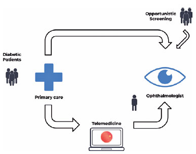
PURPOSE: Timely screening and treatment are essential for preventing diabetic retinopathy blindness. Improving screening workflows can reduce waiting times for specialist evaluation and thus enhance patient outcomes. This study assessed different screening approaches in a Brazilian public healthcare setting.
METHODS: This retrospective study evaluated a telemedicine-based diabetic retinopathy screening implemented during the COVID-19 pandemic and compared it with in-person strategies. The evaluation was conducted from the perspective of a specialized referral center in an urban area of Central-West Brazil. In the telemedicine approach, a trained technician would capture retinal images by using a handheld camera. These images were sent to specialists for remote evaluation. Patient variables, including age, gender, duration of diabetes diagnosis, diabetes treatment, comorbidities, and waiting time, were analyzed and compared.
RESULTS: In total, 437 patients with diabetes mellitus were included in the study (mean age: 62.5 ± 11.0 years, female: 61.7%, mean diabetes duration: 15.3 ± 9.7 years, insulin users: 67.8%). In the in-person assessment group, the average waiting time between primary care referral and specialist evaluation was 292.3 ± 213.9 days, and the referral rate was 73.29%. In the telemedicine group, the average waiting time was 158.8 ± 192.4 days, and the referral rate was 29.38%. The telemedicine approach significantly reduced the waiting time (p<0.001) and significantly lowered the referral rate (p<0.001).
CONCLUSION: The telemedicine approach significantly reduced the waiting time for specialist evaluation in a real-world setting. Employing portable retinal cameras may address the burden of diabetic retinopathy, especially in resource-limited settings.
Keywords: Telemedicine/methods; Diabetic retinopathy; Diagnostic screening programs; Vision screening; Practice patterns, physicians
 ABO is licensed under a Creative Commons Attribution-NonComercial 4.0 Internacional.
03-fig01.jpg)
08-tab01.jpg)
03-fig01.jpg)
16-tab01tb.jpg)
01-fig01.jpg)