Abstract
Objetivo: Examinar o efeito de infiltrados sub-epiteliais corneanos nas propriedades biomecânicas da córnea após ceratoconjuntivite epidêmica, em comparação com controles saudáveis.
Métodos: Este estudo transversal incluiu pacientes consecutivos com infiltrados sub-epiteliais corneanos bilaterais após ceratoconjuntivite epidêmica e controles saudáveis. Foram medidas a melhor acuidade visual corrigida, uma pontuação do infiltrado sub-epitelial da córnea, a escala de graduação de Fantes e a espessura central da córnea. A histerese da córnea, o fator de resistência da córnea, a pressão intraocular correlacionada à tonometria de Goldmann e a pressão intraocular compensada da córnea foram avaliados com o Ocular Response Analyzer.
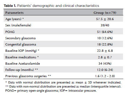
Resultados: Este estudo incluiu 66 olhos de 33 pacientes com infiltrados corneanos sub-epiteliais após ceratoconjuntivite epidêmica e selecionou aleatoriamente 37 olhos de 37 voluntários saudáveis. As pontuações médias da escala de Fantes e dos infiltrados sub-epiteliais corneanos nos primeiros olhos acometidos foram respectivamente de 1,8 ± 0,8 e 2,9 ± 1,3. Nos olhos contralaterais, foram respectivamente de 1,3 ± 1,1 e 1,9 ± 1,7 (p=0,009 e p=0,002, respectivamente). O primeiro e o segundo olhos envolvidos tinham córneas significativamente mais finas (respectivamente 526,1 ± 28,1 µm; p=0,005 e 523,4 ± 38,1 µm; p=0,044) em comparação com os controles saudáveis (557,0 ± 38,1 µm). Embora a acuidade visual melhor corrigida tenha mostrado uma correlação positiva com o fator de resistência da córnea (r=0,363, p=0,045) e com a histerese da córnea (r=0,414, p=0,021), a pontuação dos infiltrados sub-epiteliais corneanos mostrou uma correlação negativa com a pressão intraocular correlacionada à tonometria de Goldmann (r=-0,479, p=0,006) e com a pressão intraocular compensada da córnea (r=-0,413, p=0,021).
Conclusão: Os olhos com infiltrados corneanos sub-epiteliais tinham córneas significativamente mais finas em comparação com os controles saudáveis. Ao se medirem os valores de pressão intraocular em pacientes com infiltrados sub-epiteliais corneanos, deve-se levar em consideração tanto as correlações positivas do fator de resistência da córnea e da histerese da córnea com a melhor acuidade visual corrigida quanto as correlações negativas da pressão intraocular correlacionada à tonometria de Goldmann e da pressão intraocular compensada da córnea com a pontuação do infiltrado sub-epitelial da córnea.
Keywords: Ceratoconjuntivite; Pressão intraocular; Epitélio corneano; Corticosteroides; Ciclosporina;Tonometria ocular
 ABO is licensed under a Creative Commons Attribution-NonComercial 4.0 Internacional.
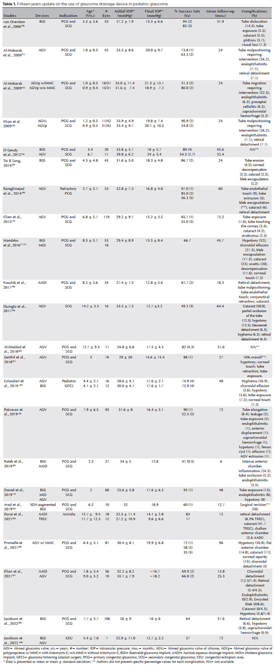
15-tab01.jpg)
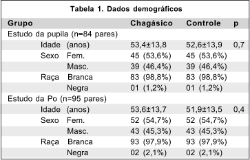
05-tab01tb.jpg)
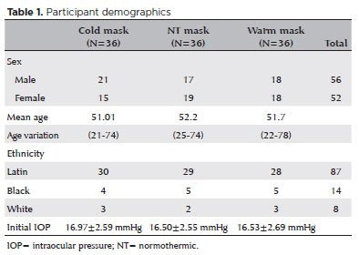
07-fig01.jpg)
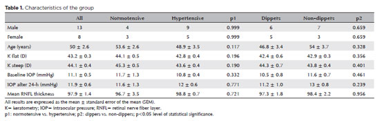
09-tab01tb.jpg)
11-tab01.jpg)