Arq. Bras. Oftalmol. 2024;87 (1 )
:1-7
| DOI: 10.5935/0004-2749.2021-0048
Abstract
Objetivo: Identificar parâmetros tomográficos de normalidade em córneas de crianças e adolescentes sem a presença de atopias sistêmicas e alergias oculares.
Métodos: Este estudo descritivo transversal avaliou pacientes com idade entre 8 e 16 anos que foram submetidos a exame biomicroscópico completo por lâmpada de fenda e avaliação tomográfica da córnea por tomógrafo dual Scheimpflug, excluindo pacientes com doença ocular (incluindo conjuntivite alérgica) ou prick test positivo para atopias sistêmicas.
Resultados: Cento e setenta pacientes foram avaliados e após cumpridos os critérios de exclusão, 34 (68 olhos) foram analisados. A média etária da amostra foi 10,76 ± 2,31 anos; 19 (55,9%) eram meninos e 15 (44,1%) meninas. A média da ceratometria em dioptrias (D) no meridiano mais plano (Kflat), mais curvo (Ksteep) e máxima (Kmax) foram 42,37 ± 1,63D, 43,53 ± 1,65D e 43,90 ± 1,73D, respectivamente. Os valores médios da asfericidade corneana (ε2) e do ponto mais fino da córnea foram 0,28 ± 0,11 e 550,20 ± 37,90 micras (μm). A assimetria corneana inferior-superior (I-S) e coma foi em média 0,74 ± 0,59D e 0,28 ± 0,12D, respectivamente.
Conclusão: O conhecimento dos valores médios e sua variação de parâmetros tomográficos da córnea em crianças e adolescentes sem atopias sistêmicas ou alergias oculares pode ser útil para o diagnóstico precoce do ceratocone e o seu tratamento em estágio inicial.
Keywords: Córnea; Topografia corneana; Astigmatismo; Conjuntivite alérgica; Tomografia; Ceratocone/diagnóstico; Humanos; Criança; Adolescente.
Arq. Bras. Oftalmol. 2026;89 (1 )
:1-6
| DOI: 10.5935/0004-2749.2025-0071
Abstract
PURPOSE: This study aimed to evaluate the outcomes of strabismus surgical correction in patients with Down syndrome.
METHODS: We conducted a retrospective chart review of patients with Down syndrome who underwent strabismus surgery between January 1997 and May 2024 at an Ophthalmology Outpatient Clinic in São Paulo, Brazil. The data collected included age, sex, medical and ocular history, surgical details, and follow-up outcomes. The patients were categorized by strabismus type into esotropia, fourth nerve palsy, and mixed groups. Surgical success was defined as final alignment within 10Δ of orthotropia and, where applicable, whether there was resolution of abnormal head posture of ocular origin. Patients with postoperative follow-up <6 months were excluded.
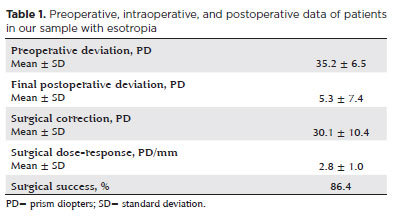
RESULTS: A total of 37 patients (21 females) were included. Of these, 22 (59.5%) were in the esotropia group, 10 (27.0%) in the fourth nerve palsy group, and 5 (13.5%) in the mixed group. The surgical success rate in the esotropia group was 86.4%, with a mean preoperative deviation of 35.2 (± 6.5)Δ, and mean surgical correction of 30.1 (± 10.4)Δ. The success rate in the fourth nerve palsy group was 40.0%, with a mean preoperative deviation of 10.4 (± 4.3)Δ. Overall, success was achieved with a single surgical procedure in 73.0% of the sample. No significant associations were found between surgical success and the clinical and demographic variables, including sex, age at surgery, oblique muscle overaction, pattern strabismus, visual acuity, amblyopia, preoperative deviation, or postoperative follow-up duration (p>0.05).
CONCLUSIONS: When standard surgical tables are applied, strabismus surgery in patients with Down syndrome appears to be safe and effective. We found high success rates, particularly among patients with esotropia. We observed no tendencies toward over- or under-correction. These findings support the use of conventional surgical protocols with this patient population.
Keywords: Down Syndrome/complications; Strabismus/surgery; Esotropia/surgery; Oculomotor nerve diseases/physiopathology; Vision disorders; Humans; Brazil.
Arq. Bras. Oftalmol. 2024;87 (4 )
:1-8
| DOI: 10.5935/0004-2749.2023-0054
Abstract
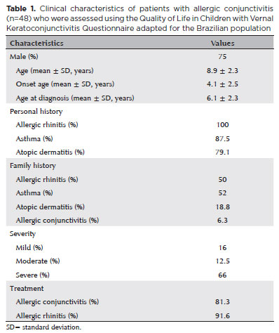
PURPOSE: The prevalence of ocular allergy varies according to the population and location of the study. Severe forms of ocular allergy are associated with compromised quality of life. In this study, we aimed to evaluate the application of the Brazilian-Portuguese version of the Quality of Life in Children with Keratoconjunctivitis questionnaire to children and adolescents with different subtypes of allergic conjunctivitis.
METHOD: A total of 48 patients (aged 5-12 years) with allergic conjunctivitis were included in this study. They were enrolled and monitored at a specialized center. After the clinical appointment, the children responded to the questionnaire on two occasions at an interval of 30 days. Individual scores (ranging from 0 to 3) of the 16 items were added.
RESULTS: The Brazilian-Portuguese version of the Quality of Life in Children with Keratoconjunctivitis questionnaire demonstrated good translation, adaptation, and intellectual properties, with substantial internal consistency (Cronbach's α coefficient = 0.702). There was no significant difference between the responses of the two interviews, revealing good reproducibility. The moderate/severe forms of allergic conjunctivitis had significantly higher quality of life scores (indicating a poorer quality of life) than the mild forms.
CONCLUSIONS: The Brazilian-Portuguese version of the Quality of Life in Children with Keratoconjunctivitis proved to be quick, reliable, and reproducible for assessing the quality of life in children with allergic conjunctivitis. However, its ability to detect changes resulting from symptom aggravation or treatment needs to be further evaluated.
Keywords: Conjunctivitis, allergic; Keratoconjunctivitis; Hypersensitivity; Quality of life; Child; Surveys and questionnaires
Arq. Bras. Oftalmol. 2024;87 (3 )
:1-6
| DOI: 10.5935/0004-2749.2022-0220
Abstract
PURPOSE: This study aimed to investigate the effect of upper eyelid blepharoplasty with the removal of the skin and a strip of orbicularis oculi muscle on the ocular surface, tear film, and dry eye-related symptoms.
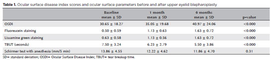
METHODS: Twenty-two eyes of 22 consecutive patients operated by a single surgeon (21 females; mean age, 61 years; age range, 41-75 years) were included. All subjects completed the Ocular Surface Disease Index questionnaire, underwent in vivo confocal microscopy, tear film breakup time measurements, the Schirmer test with anesthesia, and fluorescein and lissamine green staining measurements before, 1 month, and 6 months after upper blepharoplasty alone with preseptal orbicularis excision.
RESULTS: A significant increase in Ocular Surface Disease Index, and corneal fluorescein and lissamine green staining and a significant decrease in tear film breakup time were observed after 1 month (p=0.003, p=0.004, p=0.029, and p=0.024 respectively) and 6 months (p=0.001 for all findings). No significant difference in the Schirmer test score was observed during the follow-up. None of the in vivo confocal microscopy parameters showed significant changes during the study.
CONCLUSIONS: An increase in dry eye symptoms and a decrease in tear film stability along with ocular surface staining were observed in patients undergoing upper eyelid blepharoplasty.
Keywords: Blepharoplasty; Intravital microscopy; Lissamine green dyes; Fluorescein; Severity of illness index; Dry eye syndromes


 ABO is licensed under a Creative Commons Attribution-NonComercial 4.0 Internacional.


06-fig01.jpg)
09-fig01.jpg)
08-fig01.jpg)
13-fig01.jpg)
13-fig01.jpg)
14-fig01.jpg)
10-tab01tb.jpg)
02-fig01.jpg)