Arq. Bras. Oftalmol. 2023;86 (3 )
:1-4
| DOI: 10.5935/0004-2749.20230027
Abstract
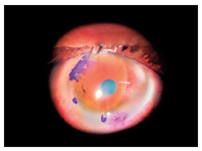
O objetivo é alertar a comunidade oftalmológica sobre uma manifestação atípica de neoplasia escamosa da superfície ocular (OSSN) que pode levar a um atraso no diagnóstico e tratamento, evoluindo com prognóstico reservado e significativas sequelas. Homem, imunocompetente, 61 anos com diagnóstico inicial de esclerite necrosante em olho direito há 3 meses, em tratamento com prednisona sistêmica porém com persistência da dor e baixa acuidade visual. Realizado biópsia conjuntival em região acometida e diagnosticado como neoplasia escamosa da superfície ocular invasiva. Evolui com invasão intraocular e orbital sendo submetido a exenteração. Assim sendo, deve-se suspeitar de síndrome mascarada frente a um paciente com lesões nódulo-ulcerativas da conjuntiva e esclera. Essa forma clínica pode ser mais agressiva, com maior chance de comprometimento intraocular e orbital. Quanto mais precoces o diagnóstico e o tratamento, melhor o prognóstico para o paciente.
Keywords: Neoplasias de células escamosas; Neoplasias da túnica conjuntiva; Doenças da córnea; Neoplasias oculares Esclerite; Interferon alfa-2
Arq. Bras. Oftalmol. 2023;86 (1 )
:79-82
| DOI: 10.5935/0004-2749.20220077
Abstract
Um homem de 53 anos, com história de 3 dias de edema periorbital e perda de visão no olho esquerdo, apresentou trombose séptica do seio cavernoso com envolvimento bilateral das veias orbitais, causando uma orbitopatia congestiva. O paciente foi tratado com uma cantotomia e cantólise de emergência, colírios para redução da pressão intraocular, antibióticos, anticoagulantes e exames seriados. A tomografia de coerência óptica finalmente demonstrou destruição isquêmica difusa de ambas as camadas da retina, sugerindo uma oclusão da artéria oftálmica ou das artérias ciliares posteriores curtas e da artéria retiniana central, com ausência de envolvimento do segmento intracavernoso da artéria carótida interna. O paciente permaneceu sem percepção luminosa no olho esquerdo.
Keywords: Trombose do corpo cavernoso; Doenças orbitárias; Tomografia de coerência óptica; Humanos; Relato de casos
Arq. Bras. Oftalmol. 2025;88 (5 )
:1-4
| DOI: 10.5935/0004-2749.2025-0020
Abstract
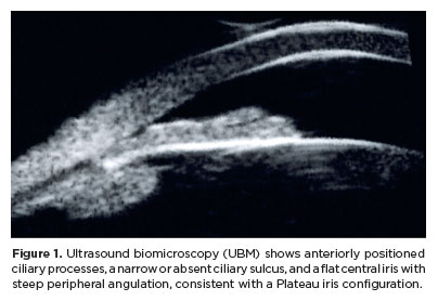
Angle-closure glaucoma is a major cause of visual impairment worldwide, with Plateau iris syndrome presenting management challenges. We present a case report of a 58-year-old woman with advanced, uncontrolled angle-closure glaucoma and Plateau iris. Her history included laser peripheral iridotomy and three glaucoma medications in both eyes. Different treatments were implemented. For the eye with lower intraocular pressure, fewer peripheral anterior synechiae, and milder disease: phacoemulsification with intraocular lens implantation. For the eye with more advanced disease, a two-step approach was used: slow-coagulation transscleral cyclophotocoagulation using the double-arc protocol, followed by phacoemulsification with intraocular lens implantation 2 months later. Both eyes achieved improved visual acuity and intraocular pressure control with fewer medications, without significant complications. This case highlights transscleral cyclophotocoagulation followed by phacoemulsification as an alternative to combined surgeries in uncontrolled angle-closure glaucoma with Plateau iris, offering a simpler technique, more predictable refractive and pressure-control outcomes, and more straightforward postoperative management.
Keywords: Glaucoma, angle-closure/surgery; Iris diseases/surgery; Laser coagulation/methods; Phacoemulsification; Lens implantation, intraocular; Case reports
Arq. Bras. Oftalmol. 2025;88 (2 )
:1-5
| DOI: 10.5935/0004-2749.2023-0078
Abstract
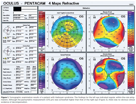
We present the case of a 37-year-old woman who underwent bilateral penetrating keratoplasty for congenital hereditary endothelial dystrophy at the age of 10 years. Over the subsequent 27 years, the patient's vision slowly deteriorated. Our examination revealed decompensation of the right corneal graft. We addressed this with regraft surgery. We then learned that the patient had been suffering from progressive hearing loss since adolescence. Tonal audiometry revealed hearing perceptive deafness of 25 dB, which was more prominent in the left ear. Because the patterns of progressive sensorineural hearing loss and congenital hereditary endothelial dystrophy have both been linked to the same gene, slc4a11, we tested our patient for mutations in this gene. The test was positive for a heterozygous slc4a11 gene fifth exon mutation on chromosome 20p13-p12, which causes a frameshift. A combined clinical and genetic evaluation confirmed a diagnosis of Harboyan syndrome. After the genetic diagnosis of the disease, she was evaluated for the need for a hearing aid due to her hearing loss. The patient was also informed about genetic counseling.
Keywords: Chromosomes; Corneal dystrophies; Exon; Genetic testing; Hearing loss; Hereditary/genetics; Mutation; Sensorineural; slc4a11 gene


 ABO is licensed under a Creative Commons Attribution-NonComercial 4.0 Internacional.


02-fig01.jpg)
02-fig01.jpg)
08-fig01.jpg)
12-fig01.jpg)
11-fig01.jpg)
08-fig01.jpg)
10-fig01.jpg)
02-fig01.jpg)
03-fig01.jpg)
01-fig01.jpg)
03-fig01.jpg)