
Ricardo Nunes Eliezer1; Niro Kasahara1; Cristiano Caixeta-Umbelino1; Renato Kingleufus Pinheiro1; Carmo Mandia Junior1; Roberto Freire Santiago Malta1
DOI: 10.1590/S0004-27492006000300005
ABSTRACT
PURPOSE: To compare the safety and efficacy of human preserved amniotic membrane (AM) in the trabeculectomy for treatment of primary open-angle glaucoma. METHODS: A prospective, randomized clinical trial compared primary trabeculectomy with amniotic membrane (study group) and without amniotic membrane (control group) in the treatment of the glaucoma. Intraocular pressure (IOP), number of glaucoma medications and appearance of the bleb were compared between the two groups. Thirty-two patients divided into two groups of 16 patients were followed for a period of 12 months. RESULTS: The difference of the mean postoperative intraocular pressure between groups was not statistically significant (15.19 ± 3.33 in the control group and 12.81 ± 2.48 in the study group p=0.297) at one year follow-up. Postoperative number of medications decreased in both groups (p<0.001and p=0.007, study group and control respectively). At the end of a 12-month follow-up period, nine eyes (56.25%) showed thin, avascular blebs in the study group as compared to only one eye (6.25%) in the control group. CONCLUSIONS: Trabeculectomy with amniotic membrane and standard trabeculectomy promote lower postoperative intraocular pressure although results showed no statistically significant difference between groups regarding postoperative intraocular pressure after one year follow-up.
Keywords: Glaucoma, open-angle; Trabeculectomy; Intraocular pressure; Biological dressings; Postoperative complications
RESUMO
OBJETIVO: Comparar a eficácia e a segurança do uso da membrana amniótica (MA) na trabeculectomia para o tratamento cirúrgico do glaucoma primário de ângulo aberto. MÉTODOS: Estudo prospectivo aberto, randomizado, grupos paralelos de tratamento. Trinta e dois pacientes com indicação de tratamento cirúrgico para glaucoma foram selecionados e aleatoriamente divididos em dois grupos. O primeiro grupo foi submetido a trabeculectomia com o uso intra-operatório da membrana amniótica (grupo estudo) e o segundo grupo foi submetido a trabeculectomia sem o uso da membrana amniótica (grupo controle) comparando o efeito redutor da pressão intra-ocular, número de medicações e aparência da bolha filtrante. Trinta e dois pacientes divididos em dois grupos de 16 pacientes foram acompanhados por 12 meses. RESULTADOS: A média das pressões pós-operatórias no grupo da membrana amniótica 12,81± 2,48 e no grupo controle 15,19±3,33 não apresentaram diferença estatisticamente significante no seguimento de um ano (p=0,297). O número de medicações pós-operatórias diminuiu nos dois grupos (p<0,001 e p=0,007 grupo estudo e grupo controle respectivamente). No final de 12 meses de pós-operatório nove olhos (56,25%) apresentaram bolhas finas e avasculares no grupo estudo comparando com apenas um olho (6,25%) do grupo controle. CONCLUSÃO: A trabeculectomia com membrana amniótica e a trabeculectomia simples mostraram redução da pressão intra-ocular no pós-operatório, embora a diferença entre elas não seja estatisticamente significante.
Descritores: Glaucoma de ângulo aberto; Trabeculectomia; Pressão intra-ocular; Curativos biológicos; Complicações pós-operatórias
ORIGINAL ARTICLE
Use of amniotic membrane in trabeculectomy for the treatment of glaucoma: a pilot study
Uso da membrana amniótica na trabeculectomia para o tratamento do glaucoma: estudo piloto
Ricardo Nunes Eliezer; Niro Kasahara; Cristiano Caixeta-Umbelino; Renato Kingleufus Pinheiro; Carmo Mandia Junior; Roberto Freire Santiago Malta
Assistente do serviço de glaucoma Department of Ophthalmology, Glaucoma Service, Faculdade de Ciências Médicas da Santa Casa de Misericórdia de São Paulo - FCMSCSP - São Paulo (SP) - Brazil
ABSTRACT
PURPOSE: To compare the safety and efficacy of human preserved amniotic membrane (AM) in the trabeculectomy for treatment of primary open-angle glaucoma.
METHODS: A prospective, randomized clinical trial compared primary trabeculectomy with amniotic membrane (study group) and without amniotic membrane (control group) in the treatment of the glaucoma. Intraocular pressure (IOP), number of glaucoma medications and appearance of the bleb were compared between the two groups. Thirty-two patients divided into two groups of 16 patients were followed for a period of 12 months.
RESULTS: The difference of the mean postoperative intraocular pressure between groups was not statistically significant (15.19 ± 3.33 in the control group and 12.81 ± 2.48 in the study group p=0.297) at one year follow-up. Postoperative number of medications decreased in both groups (p<0.001and p=0.007, study group and control respectively). At the end of a 12-month follow-up period, nine eyes (56.25%) showed thin, avascular blebs in the study group as compared to only one eye (6.25%) in the control group.
CONCLUSIONS: Trabeculectomy with amniotic membrane and standard trabeculectomy promote lower postoperative intraocular pressure although results showed no statistically significant difference between groups regarding postoperative intraocular pressure after one year follow-up.
Keywords: Glaucoma, open-angle/surgery; Trabeculectomy; Intraocular pressure; Biological dressings; Postoperative complications
RESUMO
OBJETIVO: Comparar a eficácia e a segurança do uso da membrana amniótica (MA) na trabeculectomia para o tratamento cirúrgico do glaucoma primário de ângulo aberto.
MÉTODOS: Estudo prospectivo aberto, randomizado, grupos paralelos de tratamento. Trinta e dois pacientes com indicação de tratamento cirúrgico para glaucoma foram selecionados e aleatoriamente divididos em dois grupos. O primeiro grupo foi submetido a trabeculectomia com o uso intra-operatório da membrana amniótica (grupo estudo) e o segundo grupo foi submetido a trabeculectomia sem o uso da membrana amniótica (grupo controle) comparando o efeito redutor da pressão intra-ocular, número de medicações e aparência da bolha filtrante. Trinta e dois pacientes divididos em dois grupos de 16 pacientes foram acompanhados por 12 meses.
RESULTADOS: A média das pressões pós-operatórias no grupo da membrana amniótica 12,81± 2,48 e no grupo controle 15,19±3,33 não apresentaram diferença estatisticamente significante no seguimento de um ano (p=0,297). O número de medicações pós-operatórias diminuiu nos dois grupos (p<0,001 e p=0,007 grupo estudo e grupo controle respectivamente). No final de 12 meses de pós-operatório nove olhos (56,25%) apresentaram bolhas finas e avasculares no grupo estudo comparando com apenas um olho (6,25%) do grupo controle.
CONCLUSÃO: A trabeculectomia com membrana amniótica e a trabeculectomia simples mostraram redução da pressão intra-ocular no pós-operatório, embora a diferença entre elas não seja estatisticamente significante.
Descritores: Glaucoma de ângulo aberto/cirurgia; Trabeculectomia; Pressão intra-ocular; Curativos biológicos; Complicações pós-operatórias
INTRODUCTION
Trabeculectomy is the procedure of choice for the surgical treatment of glaucoma until nowadays(1). However, recent studies have demonstrated a loss of efficacy and minor reduction of intraocular pressure in patients who underwent surgery over the years(1-3). This loss of efficacy of the trabeculectomy is related to the continuous process of healing and fibroblastic proliferation in the episcleral surface inside the filtering bleb. The use of 5-fluorouracil (5-FU) or mitomycin-C (MMC) can improve the results of trabeculectomy, but they have been associated with an increased incidence of postoperative complications(4).
The use of amniotic membrane in ophthalmology goes back to 1940(5), when some authors showed its beneficial effect in the treatment of ocular surface disorders(6-9).
Amniotic membrane can promote epitheliazation of ocular surface and act as an inhibitor of fibrosis(10-11).
Barton et al. used AM in twenty-four albino rabbits for glaucoma filtration surgery(12). Conjunctival biopsies were explanted for estimation of fibroblast outgrowth. In tissue culture, significantly less fibroblast outgrowth occurred from AM transplantation explants when compared with unoperated conjunctiva.
Other authors performed trabeculectomy in which MMC was applied to the first group, and AM were transplanted around the scleral flap in the second group; the third group was the control(13). Cell counts in the AM group were lower than those in the control group regarding fibroblasts and macrophages.
The purpose of this study was to evaluate the efficacy and safety of amniotic membrane in primary trabeculectomy.
METHODS
Thirty-two white patients with primary open-angle glaucoma (POAG) scheduled for glaucoma filtration surgery at the Glaucoma Service of the "Santa Casa de São Paulo" from August 2001 to August 2003 were randomly assigned to two groups. One group of trabeculectomy with intraoperative use of AM (study group) and the other group a trabeculectomy without AM (control group). We did not use mitomycin C in both groups. Surgery was indicated when progression had occurred despite maximal tolerated medical therapy, non-compliance or advanced glaucoma in which the target pressure was not achieved by medical and/or laser therapy.
The Ethics Committee approved the study and patients signed an informed consent.
Surgical principles:
(A) Preparation of amniotic membrane
Amniotic membrane is obtained under sterile conditions after elective cesarean delivery from a seronegative donor. Donors at risk of having human immunodeficiency virus (HIV), hepatitis B virus (HBV), hepatitis C virus (HCV) and Creutzfeldt-Jacob disease (CJD) must be excluded(10).
Under a lamellar flow hood, the placenta is first washed free of blood clots with balanced physiologic saline containing 50 µg/ml penicillin, 50 µg/ml streptomycin, 100 µg/ml neomycin, and 2.5 µg/ml amphotericin B. The inner amniotic membrane is separated from the rest of the chorion by blunt dissection through the potential spaces between these tissues. The membrane is then flattened onto a nitrocellulose paper, with the epithelium/basement membrane surface up. The membrane with the paper is cut into 4x4 cm pieces and placed in a sterile vial containing Dulbecco's modified Eagle's medium and glycerol at a ratio of 1:1. The vials are frozen at - 80°C. The membrane is defrosted immediately before use by warming the container to room temperature for 10 minutes(10).
(B) Surgical technique
Surgeries were performed by two of us (RNE, CMJ). After peribulbar anesthesia patients were prepared and draped. A limbus-based conjunctival flap was fashioned, followed by hemosthasis with bipolar wet field cautery and the rectangular scleral flap was dissected. A paracentesis was created with a 27-gauge needle, and scleral block was excised with Vanas scissor. Iridectomy was performed with Vanas scissor and the scleral flap was secured with at least two sutures. Then a 5x5 mm folded AM graft with the stroma in both sides was placed over the sclera and held in place with two 10-0 nylon sutures (Figure 1). Tenon's capsule and the conjuctiva were closed with 10-0 nylon running suture. Anterior chamber was filled with balanced saline solution and conjuctival suture checked for Seidel. At the end of the procedure, 1ml dexamethasone 1% and 1 ml gentamicin 80 mg were injected subconjunctivally in the inferior fornix.

Only one ophthalmologist examined all patients between 9:00 am and 12:00 am during one year follow-up. He used the same slit lamp and the same Goldmann tonometer. Postoperative management included application of topical dexamethasone 1% q4h, and ofloxacin 0.3 % bid, tapered over several weeks. Patients were evaluated 1 day, 1 week, 1 month, 2 months, 3 months, 4 months, 5 months, 6 months and 12 months after surgery.
The appearance of the bleb was classified at the last examination by the same ophthalmologist into one of the three categories: flat and vascularized, elevated but not avascular, or elevated thin and avascular.
Statistical analysis included paired Student t test to compare intraocular pressure (IOP) change. Non parametric Mann-Whitney test was used to compare visual acuity (VA), number of medication and IOP change from baseline between groups. Wilcoxon test was used to compare pre and postoperative VA and number of medication in each group. ANOVA was used to compare IOP in each visit. A p value of less than 0.05 was considered statistically significant.
RESULTS
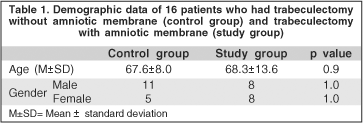
Patients demographics are presented in table 1.

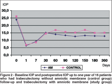
Figure 2 shows baseline IOP and postoperative mean IOP of thirty two patients who had trabeculectomy without amniotic membrane (control group) and thirty one patients who had trabeculectomy with amniotic membrane (study group) up to one year.

The mean postoperative IOP was 15.47±2.92 in the control group and 13.13±2.50 in the study group at one year follow up.
Table 2 shows mean preoperative visual acuity (VA) was 0.37±0.35 for study group and 1.31±1.38 for the control group (p=0.262).The mean postoperative VA was 0.60±0.83 for study group and 1.39±1.31 for the control group (p=0.134). The postoperative difference between groups was not significant. Table 3 shows the pre- and postoperative number of medications for the study group and control. Postoperative number of medications decreased in both groups (p<0.001 and p=0.007, study group and control respectively).The postoperative difference between groups was not significant (p=0.210).


At the end of a 12-month follow-up period, in the study group two of 31 eyes (6.25%) exhibited flat, vascularized bleb, 14 eyes (45.16%) had elevated but not avascular blebs and nine eyes (56.25%) showed thin, avascular blebs. In the control group 5 of 16 eyes (31.25%) exhibited flat, vascularized bleb, ten eyes (62.50%) had elevated but not avascular bleb and one eye (6.25%) showed a thin, avascular bleb. Complications were: one eye (6.25%) with encapsulated bleb in the study group, in the control group one eye (6.25%) presented with shallow anterior chamber after surgery, one eye (6.25%) had coroidal detachment and one eye (6.25%) developed encapsulated bleb.
DISCUSSION
In this study, we used AM in trabeculectomy to prevent healing in the subconjunctival space and promote long-living blebs. These effects are mediated via an influence on subconjunctival epithelial and subconjunctival fibroblast function, promoting epithelial maturation and down-regulating fibrogenic TGF-b signaling and myofibroblast differentiation(6-8).
Although the difference of mean postoperative IOP between groups was 15.19±3.33 in the control group and 12.81± 2.48 in the study group (p=0.297), our results show no statistical significant difference in postoperative IOP after one year follow-up.
The author performed trabeculectomy with MMC and amniotic membrane placed under the scleral flap. Among 14 eyes of 13 patients, IOP was controlled to less than 20 mmHg after surgery in 13 eyes(13). Unlike Fujishima et al.(13), MMC was not used together with AM. In our study, we aimed to evaluate only the effect of amniotic membrane in primary trabeculectomies without the additive effect of antimetabolites and we were the first to use the folded AM graft with the epithelium/basement membrane in both sides. The folded AM decreases the fibroses between the tissues and that is why we had more avascular blebs in the study group.
In the study group we found only one flat vascularized bleb as compared to 5 in the control group and 9 elevated avascular blebs as compared to one in the control group. These elevated avascular blebs resemble those of MMC trabeculectomy which are more long-lasting blebs.
Complications were present in only one eye (encapsulated bleb) in the study group and in 3 eyes (one shallow anterior chamber, one coroidal detachment and one encapsulated bleb) in the control group.
Other complications, such as infection and hypotonous maculophaty, were not seen in this study(14).
CONCLUSION
Despite the reduction in the IOP between the two groups failed to reach statistically significance, the avascular appearance of the blebs was shown. The conclusions were that trabeculectomy with amniotic membrane and standard trabeculectomy promote lower postoperative IOP although results showed no statistical significant difference between groups in postoperative IOP after one year follow-up.
This pilot study revealed promising results. A large randomized surgical clinical trial comparing AM trabeculectomies and standard trabeculectomies is in progress.
REFERENCES
1. Cairns JE. Trabeculectomy. Preliminary report of a new method. Am J Ophthalmol. 1968;66(4):673-9.
2. Molteno AC, Bosma NJ, Kittleson JM. Otago glaucoma surgery outcome study: long-term results of trabeculectomy - 1976 to 1995. Ophthalmology. 1999;106(9): 1742-50.
3. Three year follow-up the Fluorouracil Filtering Surgery Study. Am J Ophthalmol. 1993;115(1):82-92.
4. Chen CW, Huang HT, Bair JS, Lee CC. Trabeculectomy with simultaneous topical application of mitomycin-C in refractory glaucoma. J Ocul Pharmacol. 1990l;6(3):175-82.
5. Susanna R Jr, Costa VP, Malta RF, Barboza WL, Vasconcellos JP. Intraoperative mitomycin-C without conjunctival and Tenon's capsule touch in primary trabeculectomy. Ophthalmology. 2001;108(6):1039-42.
6. De Roth A. Plastic repair of conjunctival defects with fetal membrane. Arch Ophthalmol 1940;23:522-5.
7. Kim JC, Tseng SC. Transplantation of preserved human amniotic membrane for surface reconstruction in severely damaged rabbit corneas. Cornea. 1995; 14(5):473-84.
8. Dua HS, Gomes JA, King AJ, Maharajan VS. The amniotic membrane in ophthalmology. Surv Ophthalmol. 2004;49(1):51-77.
9. Shimazaki J, Kosaka K, Shimmura S, Tsubota K. Amniotic membrane transplantation with conjunctival autograft for recurrent pterygium. Ophthalmology. 2003;110(1):119-24.
10. Solomon A, Espana EM, Tseng SC. Amniotic membrane transplantation for reconstruction of the conjunctival fornices. Ophthalmology. 2003;110(1):93-100.
11. Tseng SC, Prabhasawat P, Lee SH. Amniotic membrane transplantation for conjunctival surface reconstruction. Am J Ophthalmol. 1997;124(6):765-74..
12. Barton K, Budenz DL, Khaw PT, Tseng SC. Glaucoma filtration surgery using amniotic membrane transplantation. Invest Ophthalmol Vis Sci. 2001;42(8): 1762-8.
13. Fujishima H, Shimazaki J, Shinozaki N, Tsubota K. Trabeculectomy with the use of amniotic membrane for uncontrollable glaucoma. Ophthalmic Surg Lasers. 1998;29(5):428-31.
14. Nuyts RM, Greve EL, Geijssen HC, Langerhorst CT. Treatment of hypotonous maculopathy after trabeculectomy with mitomycin C. Am J Ophthalmol. 1994;118(3):322-31.
 Correspondence to:
Ricardo Nunes Eliezer
Rua Baronesa de Itu nº 870 apto 72
São Paulo (SP)
Cep 01231-000
E-mail: [email protected]
Recebido para publicação em 27.04.2005
Versão revisada recebida em 10.02.2006
Aprovação em 12.02.2006
Nota Editorial: Depois de concluída a análise do artigo sob sigilo editorial e com a anuência dos Drs. Leopoldo Magacho e João Antonio Prata Jr. sobre a divulgação de seus nomes como revisores, agradecemos sua participação neste processo.