Arq. Bras. Oftalmol. 2025; 88 (4): 10.5935/0004-2749.2024-0330
Total: 1053
Sérgio Ferreira Alves Júnior1; Luiz Fernando Teixeira2; Paulo Gois Manso2; Diogo Goulart Corrêa3; Soraya Silveira Monteiro1; José Roberto Falco Fonseca1
DOI: 10.5935/0004-2749.2024-0330
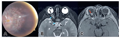
A 2-year-old patient had right ocular esotropia for 1.5 years. Right fundoscopyrevealed a large disc, central glial tuft (arrow), and a halo of pigmentary changes in the peripapillary area surrounding the nerve (arrowhead) consistent with methylglycinediacetic acid (MGDA) (Figure 1A). Funnel-shaped morphology of the optic disc (black arrow), adjacent retinal surface elevation and enhancement (red arrow), and irregular thickening of the nerve (blue arrowheads) were detected (Figure 1B, Figure 1C). Irregular thickness of optic nerve occurs in 89% MGDA, the nature being uncertain(1-3).
REFERENCES
1. Ellika S, Robson CD, Heidary G, Paldino MJ. Morning glory disc anomaly: characteristic MR imaging findings. AJNR Am J Neuroradiol. 2013;34(10):2010-4.
2. Nguyen DT, Boddaert N, Bremond-Gignac D, Robert MP. Optic nerve abnormalities in morning glory disc anomaly: an MRI study. J Neuroophthalmol. 2022;42(2):199-202.
3. Doneda C, Pinelli L, Scaramuzzi M, Galli J, Fazzi E, Parazzini C, et al. Morning glory disc anomaly associated with ipsilateral optic nerve and chiasm thickening: three cases and review of the literature. Neuropediatrics. 2017;48(6):463-6.
Submitted for publication:
December 3, 2024.
Accepted for publication:
January 16, 2025.
Funding: This study received no specific financial support.
Disclosure of potential conflicts of interest: The authors declare no potential conflicts of interest.

How to cite this article:
 ABO is licensed under a Creative Commons Attribution-NonComercial 4.0 Internacional.