Arq. Bras. Oftalmol. 2025; 88 (2): 10.5935/0004-2749.2024-0322
Total: 1319
Luiz Álvarez Cascos López; Blanca Benito Pascual; Laura Gil Amado; Nabil Dris Hassan; Santiago López García
DOI: 10.5935/0004-2749.2024-0322
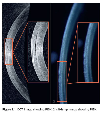
A 46-year-old woman who underwent myopic laser in situ keratomileusis (LASIK) and Ahmed valve surgery in the right eye was treated for an infiltrated ulcer in the same eye. After healing and achieving normal central intraocular pressure, she was prescribed topical corticosteroids for corneal haze reduction. Anterior segment optical coherence tomography was performed after 1 month, which showed fluid in the interface between the corneal flap and the stroma, leading to the diagnosis of pressure-induced stromal keratopathy (PISK)(1-3).
REFERENCES
Estopinal CB, Mian SI. LASIK flap: postoperative complications. Int Ophthalmol Clin. 2016;56(2):67-81.
Ravipati A, Pradeep T, Donaldson KE. Interface fluid syndrome after LASIK surgery: retrospective pooled analysis and systematic review. J Cataract Refract Surg. 2023;49(8):885-9.
Senthil S, Rathi V, Garudadri C. Misleading Goldmann applanation tonometry in a post-LASIK eye with interface fluid syndrome. Indian J Ophthalmol [Internet|. 2010[cited 2023 Nov 21]; 58(4):333-5. Available from: https://pmc.ncbi.nlm.nih.gov/articles/PMC2907040/
Submitted for publication:
October 25, 2024.
Accepted for publication:
November 19, 2024.
Informed consent was obtained from the patient included in this study.
Funding: This study received no specific financial support.
Disclosure of potential conflicts of interest: The authors declare no potential conflicts of interest.

How to cite this article:
 ABO is licensed under a Creative Commons Attribution-NonComercial 4.0 Internacional.