
Isabela Spinelli Mota1; Juliana Souza Gomes1; Fernanda Cunha1; Michelle Gantois1,2
DOI: 10.5935/0004-2749.2024-0148
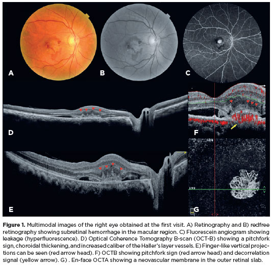
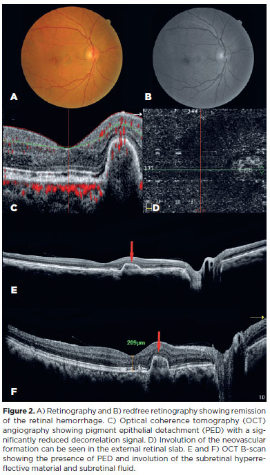
The pitchfork sign (PS) is a distinctive finding on optical coherence tomography (OCT) that is characteristic of type 2 macular neovascularization (MNV) secondary to punctate inner choroidopathy (PIC)(1-3). A 57-year-old male presented to us with complaints of blurring of vision in the right eye (OD) for one month. At the time of admission, the best-corrected visual acuity was 20/150. OCT and OCT angiography revealed a PS (Figure 1). Thus, the patient was diagnosed with type 2 MNV secondary to PIC. A loading dose of three aflibercept intravitreal injections (IVI) was administered. No other drugs were prescribed. A good response was observed in the treated eye. At the 1-year follow-up, no neovascularization reactivity was observed, and the visual acuity had improved to 20/20 (Figure 2). The first-choice treatment for MNV is anti-VEGF IVIs. Other reports have described good morphofunctional results in patients treated with bevacizumab or ranibizumab. However, they reported the reappearance of new lesions(2,4). In our patient, the visual acuity significantly improved and the retinal lesions regressed after the administration aflibercept IVIs, without the use of corticosteroids or immunosuppressants.


AUTHORS' CONTRIBUTIONS:
Significant contribution to conception and design - Isabela Spinelli Mota, Juliana Souza Gomes, Maria Fernanda Cunha, Michelle Gantois. Data acquisition: Isabela Spinelli Mota, Juliana Souza Gomes, Maria Fernanda Cunha, Michelle Gantois. Data analysis on interpretation: Isabela Spinelli Mota, Juliana Souza Gomes, Maria Fernanda Cunha, Michelle Gantois. Manuscript drafting: Isabela Spinelli Mota, Juliana Souza Gomes, Michelle Gantois. Significant intellectual content revision of the manuscript: Isabela Spinelli Mota, Juliana Souza Gomes, Maria Fernanda Cunha, Michelle Gantois. Final approve of the manuscript: Isabela Spinelli Mota, Juliana Souza Gomes, Maria Fernanda Cunha, Michelle Gantois. Statistical analysis: Not applicable. Obtaining funding: Not applicable. Supervision of administrative, technical, or material support: Michelle Gantois. Research group leader: Michelle Gantois.
REFERENCES
1. Watzke RC, Packer AJ, Folk JC, Benson WE, Burgess D, Ober RR. Punctate inner choroidopathy. Am J Ophthalmol.1984;98(5):572-84.
2. Ahnood D, Madhusudhan S, Tsaloumas MD, Waheed NK, Keane PA, Denniston AK. Punctate inner choroidopathy: A review. Surv Ophthalmol. 2017;62(2):113-26.
3. Hoang QV, Cunningham ET Jr, Sorenson JA, Freund KB. The “pitchfork sign” a distinctive optical coherence tomography finding in inflammatory choroidal neovascularization. Retina. 2013;33(5):1049-55.
4. Lupidi M, Muzi A, Castellucci G, Kalra G, Piccolino FC, Chhablani J, et al. The choroidal rupture: Current concepts and insights. Surv Ophthalmol. 2021;66(5):761-70.
Submitted for publication:
May 24, 2024.
Accepted for publication:
July 26, 2024.
Funding: This study received no specific financial support.
Disclosure of potential conflicts of interest: The authors declare no potential conflicts of interest.