INTRODUCTION
Benign familial fleck retina (BFFR) (OMIM 228980) is a congenital abnormality characterized by multifocal yellowish retinal infiltrates involving the post-equatorial retina(1,2). Aish and Dajani introduced this term in 1980 to distinguish the condition from similar disorders with distinctive clinical presentations, including fundus albipunctatus, fundus flavimaculatus, and others(2-5). Most of these are associated with an altered electroretinogram (ERG) and abnormal perimetry, with peripheral constriction to the central field(3). In contrast, the ERG is normal in BFFR, and the patient has no symptoms(2).
The flecks are located at the level of the retinal pigment epithelium (RPE), extending to the far periphery but sparing the macula(6,7). Here we report a young patient diagnosed with BFFR.
CASE REPORT
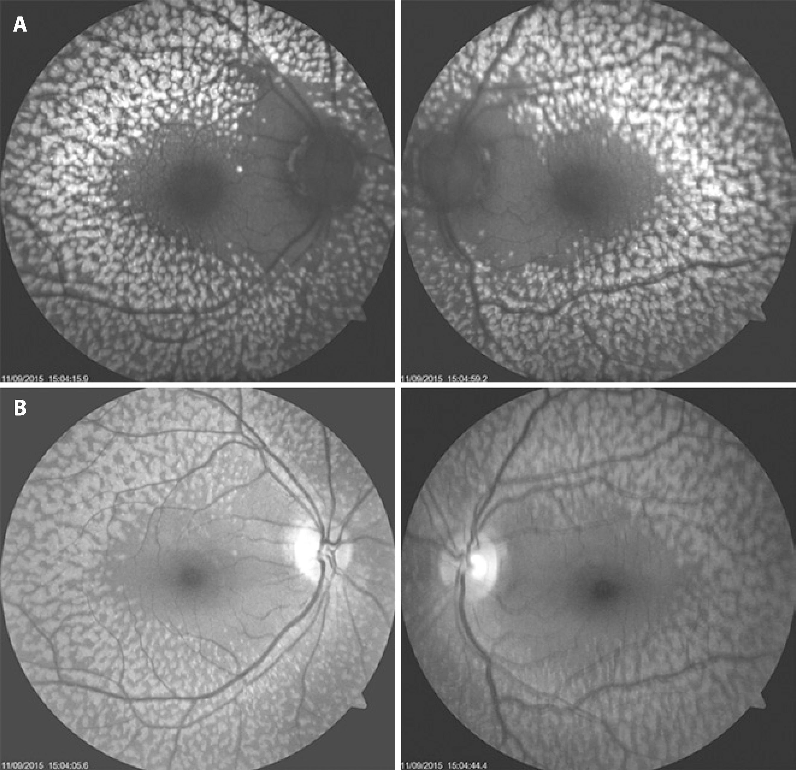
A 27-year-old woman was referred for a regular ophthalmic examination. Her visual acuity was 20/20 OU and there were no abnormalities in slit-lamp biomicroscopy. Fundus photographs in both eyes displayed multiple, small, bilateral, symmetrical retinal flecks that affected the post-equatorial retina but spared the macular region (Figure 1). No family members were affected, and there was no history of consanguinity between the parents.

Figure 1 A and B) Fundus photographs showing numerous yellowish flecks that affected the post-equatorial retina but spared the macula.
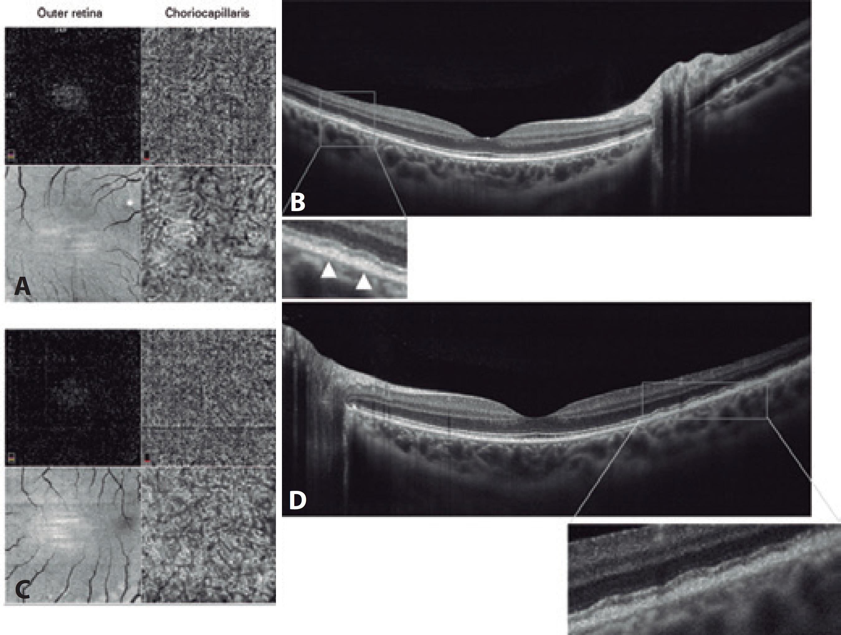
Increased autofluorescence corresponding to retinal flecks was observed in the infrared image (Figure 2). Fluorescein angiography showed a healthy macula with hypofluorescent spots related to pigment clumps; these extended to the far periphery but without macular injury. ERGs were recorded under scotopic and photopic conditions and were normal except for the selective reduced amplitude of oscillatory potentials (Figure 3). Electrooculograms revealed light peak/dark trough ratios (Arden ratios) of 1.9 in the right eye and 2.0 in the left eye (Figure 3). The patient’s macula was tested with a spectral domain (SD) optical coherence tomography (OCT) B-scan and SD-OCT angiography, using an Avanti RTVue XR with AngioVue® software (Optovue, Inc., Fremont, CA). The imaging data were obtained using split-spectrum amplitude-decorrelation angiography software. The testing demonstrated a normal foveal structure with increased thickness of the RPE in both eyes. The 3 × 3 mm SD-OCT angiography showed no macular injury from the outer retina to the choriocapillaris slab in both eyes. Projection artifacts were removed using a default proprietary algorithm (Figure 4). At a follow-up examination 18 months later, the patient continued to have no ocular symptoms.

Figure 2 A) Fundus autofluorescence demonstrating a symmetrical pattern of yellow-white fleck lesions that affected both fundi. B) Infrared imaging showing the same configuration.

Figure 3 A) Standard full-field electroretinogram elicited by various stimulus intensities in scotopic and photopic conditions with no abnormalities. B) Electrooculogram recordings. The light peak/dark trough ratio (Arden ratio) was 1.9 in the right eye and 2.0 in the left eye.

Figure 4 Optical coherence tomography (OCT) angiography with a split-spectrum amplitude decorrelation algorithm and structural en face OCT 3 × 3 mm (A and C), which showed no macular injury from the outer retina to the level of choroid capillary in both eyes. The OCT B-scan (B and D) demonstrated a slight increase in the thickness of the retinal pigment epithelium, leading to multiple small pigmented epithelium detachments. The enlarged images of the boxed regions show the outer retina and retinal pigment epithelium of both eyes in detail. The hyperreflective band corresponding to the photoreceptor inner/outer segment junction remained intact in both eyes.
DISCUSSION
The patients’ phenotype resembled that of BFFR described by Aish and Dajani in 1980(1), an extremely uncommon condition(2). Since the original description of a family with benign fleck retina, only three sporadic cases have been reported(2,7).
Some authors have postulates that flecks do not only represent a typical ophthalmic characteristic but may also correspond, in some patients, to retinal damage that contributes to loss of vision(4). It has been suggested that the flecks may be related to mutations in PLA2G5(8,9).
Flecked retina syndromes encompass a group of diseases that include Stargardt’s macular dystrophy, fundus albipunctatus, retinitis punctata albescens, Leber congenital amaurosis, pseudoxanthoma elasticum, Kjellin’s syndrome, Alport’s syndrome, Sjögren-Larsson syndrome, Bietti’s crystalline dystrophy, oxalosis, and cystinosis(8-9). It has been suggested that the flecks’ hyperautofluorescence in Stargadt’s macular dystrophy (OMIM 601691) may represent a precursor of photoreceptor death and RPE atrophy; this contradicts the definition of BFFR, but further studies are needed to confirm this in other diseases.
This report describes a sporadic case of BFFR and presents the standard full-field ERG, electrooculogram, fundus autofluorescence imaging, SD OCT B-scan, and SD OCT angiography findings for this condition.


 English PDF
 Print
Print
 Send this article by email
 How to cite this article
 Submit a comment
 Mendeley
 Scielo
 Pocket
 Share on Linkedin

