
Stella Maris Costa Castro1; Cintia Alves Salgado1; Fernando Portolani Andrade1; Sylvia Maria Ciasca2; Keila Miriam Monteiro Carvalho3
DOI: 10.1590/S0004-27492008000600014
ABSTRACT
PURPOSE: To assess binocular control in children with dyslexia. METHODS: Cross-sectional study with 26 children who were submitted to a set of ophthalmologic and visual tests. RESULTS: In the dyslexic children less eye movement control in voluntary convergence and unstable binocular fixation was observed. CONCLUSION: The results support the hypothesis that developmental dyslexia might present deficits which involve the magnocellular pathway and a part of the posterior cortical attentional network.
Keywords: Dyslexia; Attention; Vision, binocular; Visual perception; Ocular motility disorders; Learning disorders
RESUMO
OBJETIVO: Avaliar o controle binocular em crianças com dislexia. MÉTODOS: Estudo transversal do qual participaram 26 crianças, nas quais foram aplicadas uma série de exames oftalmológicos e visuais. RESULTADOS: Nas crianças com dislexia observou-se controle menor na convergência voluntária e na estabilidade da fixação binocular. CONCLUSÃO: Os resultados apóiam a hipótese de que na dislexia do desenvolvimento podem ocorrer déficits que envolvem a via visual magnocelular e uma parte da rede cortical posterior da atenção.
Descritores: Dislexia; Atenção; Visão binocular; Percepção visual; Transtornos da motilidade ocular; Transtornos de aprendizagem
ORIGINAL ARTICLE
Visual control in children with developmental dyslexia
Controle visual em crianças com dislexia do desenvolvimento
Stella Maris Costa CastroI; Cintia Alves SalgadoII; Fernando Portolani AndradeIII; Sylvia Maria CiascaIV; Keila Miriam Monteiro CarvalhoV
IOrthoptist, Post Graduation student in Medical Sciences, Universidade Estadual de Campinas - UNICAMP - Campinas (SP) - Brazil
IISpeech Therapist Master in Medical Sciences - UNICAMP - Campinas (SP) - Brazil
IIIResident student - UNICAMP - Campinas (SP) - Brazil
IVProfessor Participating in the Post-Graduation Program in Medical Sciences, Department of Neurology - UNICAMP - Campinas (SP) - Brazil
VMD. Professor Participating in the Post-Graduation Program in Medical Sciences, Department of Ophthalmology - UNICAMP - Campinas (SP) - Brazil
ABSTRACT
PURPOSE: To assess binocular control in children with dyslexia.
METHODS: Cross-sectional study with 26 children who were submitted to a set of ophthalmologic and visual tests.
RESULTS: In the dyslexic children less eye movement control in voluntary convergence and unstable binocular fixation was observed.
CONCLUSION: The results support the hypothesis that developmental dyslexia might present deficits which involve the magnocellular pathway and a part of the posterior cortical attentional network.
Keywords: Dyslexia/physiopathology; Attention; Vision, binocular/physiology; Visual perception; Ocular motility disorders; Learning disorders
RESUMO
OBJETIVO: Avaliar o controle binocular em crianças com dislexia.
MÉTODOS: Estudo transversal do qual participaram 26 crianças, nas quais foram aplicadas uma série de exames oftalmológicos e visuais.
RESULTADOS: Nas crianças com dislexia observou-se controle menor na convergência voluntária e na estabilidade da fixação binocular.
CONCLUSÃO: Os resultados apóiam a hipótese de que na dislexia do desenvolvimento podem ocorrer déficits que envolvem a via visual magnocelular e uma parte da rede cortical posterior da atenção.
Descritores: Dislexia/fisiopatologia; Atenção; Visão binocular/fisiologia; Percepção visual; Transtornos da motilidade ocular; Transtornos de aprendizagem
INTRODUCTION
Developmental dyslexia is a specific neurological condition affecting the reading learning process, with academic performance below expected levels in relation to chronological age, which is unexplainable by any kind of general intelligence deficit, lack of learning opportunities, general motivation or sensory dysfunction. Its origin is genetic, with anatomical studies demonstrating intrauterine neurological alterations(1). The most frequent signs include deficits in language acquisition, slow reading, difficulty in expressive language and in the ability to apprehend grapheme/phoneme correspondence, difficulty in understanding and memorizing reading content, inversions, omissions or substitution of letters and/or syllables in words while reading and writing(2-3).
The most accepted hypothesis to explain the problems derived from developmental dyslexia is the phonological deficit theory. It states that the core cognitive deficit lies in the ability to represent or recall speech sounds, but this theory does not explain visual, sensorial and motor coordination deficits that can occur in dyslexia(4). The visual theory does not exclude a phonological deficit, but it emphasizes a contribution of one of the visual pathways, namely the magnocellular(5-6). This theory contends that in people with dyslexia the magnocellular system is abnormal, causing difficulties in some aspects of visual perception and in binocular control, which may cause reading impairment.
The magnocellular pathway, also called dorsal or "where" pathway, directed toward parietal and frontal lobe, begins in the big ganglionary cells of the retina(7-9). Neurons in these pathways have distinct physiological properties from the small parvocellular cells in the ventral or "what" pathway. The neurons in the magnocellular way have larger receptive fields, respond in a transient and fast fashion, have broadband wavelength sensitivity, prefer low spatial frequencies and are sensitive to low-contrast stimuli(10). Because of its relationship with the posterior parietal cortex, the dorsal stream has been related to the process of space relations and visually guided movements, spatial attention and eye movement(11), binocular control(12), and visual-space attention(13-14). An important number of dyslexic individuals show anomalous standards of saccadic movements(15), instability of binocular fixation and reduced vergence(16-18).
The aim of this study was to evaluate visual processing in children with developmental dyslexia, using ophthalmologic tests, with emphasis on binocular control and, by doing so, observe any evidence of impaired magnocellular pathway performance.
METHODS
A transversal study was done involving 26 children, separated in to two different groups. Group 1: thirteen children with previous diagnosis of developmental dyslexia using the following criteria: intelligence coefficient compatible with normality, using Wechsler Intelligence Scale for Children (WISC), reading retardation of least 18 months, aged between 8 and 13 years, were recruited from the Laboratory of Learning Disabilities of College of Medical Sciences, of the State University of Campinas. Group 2: thirteen children age-matched, classified as normal readers with appropriate reading and academic level were invited from public schools in the area. Criteria of exclusion for both groups: auditory deficiency, significant neurological disease (epilepsy, head injury), ophthalmologic disorders such as strabismus or low vision and use of medication that interferes with the cognitive process. The research was approved by the Committee of Ethics on Research of the Faculty of Medical Sciences, State University of Campinas (UNICAMP) and all the responsible for the subjects included in the study read and signed an informed consent.
The dyslexic group (G1) consisted of 4 girls and 9 boys with an average age of 11 years, (mean age 11 years 2 months, SD 16 months, range: 8, 5-13, 2) and the control group (G2) was formed by 7 girls and 6 boys at an average age of 11 years (mean age 11 years 2 months, SD 19 months, range: 8, 1-13, 3) The procedures were carried out by the Laboratory of Learning Disabilities (DISAPRE) and the Department of Ophthalmology of UNICAMP.
Initially, both samples were submitted to ophthalmologic assessment: ocular refraction, biomicroscopy and fundoscopy. Refractive error measurement was carried out 40 minutes after cycloplegia with 1% cyclopentolate instilled two times (at 0 and 5 minutes) before refraction by retinoscopy: refractive error was considered significant when hypermetropy was above +2,00 SD, myopia above 0,75 SD, astigmatism above 1 CD between the prime meridians, and astigmatism in 0,75 interocular difference above 1 D. Vision assessment was performed on another day with refractive correction: uniocular visual acuity was tested for near and distant using the Lea Symbols tables for 40 cm and 3 meters (Good Lite 250800 and 257000®), contrast sensitivity test was performed using CSV - 1000 HGT® (Vector Vision, Dayton, OH, USA), colour vision with Ishihara® plates and stereoscopic acuity with Titmus® stereo test. Assessment of ocular dominance and handedness. Accommodation assessment: accommodative convergence/accommodation (AC/A) ratio was assessed using the gradient method and the near point of accommodation. Assessment of ocular alignment: prism and cover test performed at 4 meters and 33 centimeters. Assessment of eye movements: versions, near point of convergence, fusion range with isolated prisms and fusion range at synoptophore with large slides (7°) and small (2 1/2°) fusion targets and physiological diplopia.
Statistical data were verified by the SPSS program (Statistical Package for Social Sciences version 14), for two independent samples, Mann-Whitney U test, two tailed, corrected for ties. The level of significance required to support the hypothesis was established as P<0.05.
RESULTS
Ophthalmologic examination: Two participants were reported: a dyslexic participant with OD -0.50 CD 180º and OS +0.50 SD with -3.50 CD 180º, and one of the control group had hypermetropy +2.50 SD in one eye and 2.00 SD in the other eye. Biomicroscopy and fundoscopy were similar in both groups.
Visual assessment
Visual acuity (VA): Except for one dyslexic individual who presented anisometropia with VA distance OD 20/25, OS 20/25, near OD 20/25, OS 20/30, all others showed a monocular VA, distant or near, equal or better than 20/20.
Contrast sensitivity and color vision: the performance of both groups was similar, without alterations. Stereoscopic acuity: in both groups the results were normal (40 seconds of arc).
Accommodation assessment: The medians of the near point of accommodation were 9 centimeters in both groups (p=0,978 for the right eye and p=0,411 for the left eye). The medians of the AC/A ratio were 3D/SD for the dyslexic group, and 4D/SD for the control group.
Ocular and motor dominance
Ocular dominance and handedness: In both groups there was dominance of the right eye in 62% (8 participants in each group). Right-handedness occurred in 85% (11) of the dyslexic group and in 92% (12) of the control group. Crossed dominance (right eye/left hand or left eye/right hand) occurred in 40% (5) of the dyslexic group and in 31% (4) of the control group.
Assessment of ocular alignment
In the prism and cover test, only one participant of the dyslexic group presented exophoria for a distance of 4 meters (6D) the remaining ones did not show any deviation. For a distance of 33 centimeters, 3 participants in the dyslexic group presented exophoria to 6D and two exophorias from 10D to 12D. In the control sample, 4 children showed exophoria 2D and 1 with 8D and the remaining ones did not show any phoria.
Assessment of eye movements
Versions: only 1 participant, from the dyslexic group showed alteration: double hyperfunction of the inferior oblique muscle (+1).
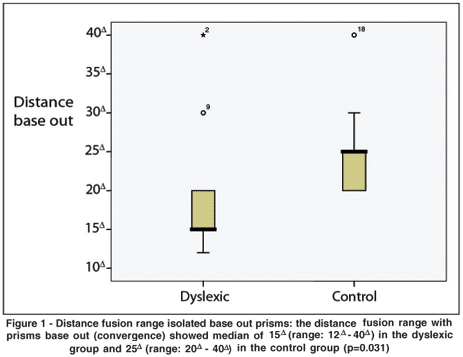
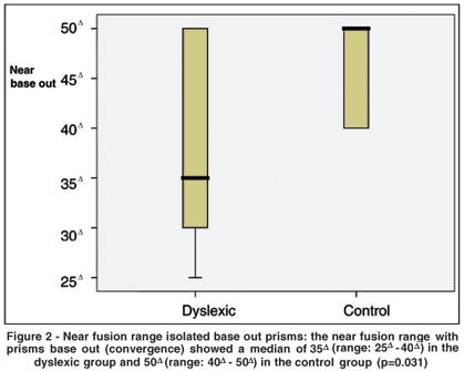
Fusion range: the evaluation was done with isolated prism using optotype acuity 20/40 as fixation target. In the fusion divergence, base in, there was no significant difference between the groups. Distance and near fusion convergence, base out (Figures 1 and 2), had different results.


Near point of convergence and fusion range at synotophore: were similar for both groups.
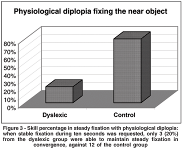
Physiological diplopia: all the participants were able to recognize the homonymous diplopia (fixing near target) and the crossed diplopia (fixing the distant target). Fixing the near target, dyslexic children were not able to maintain steady fixation (Figure 3).

DISCUSSION
The aim of this study was to analyze clinical visual assessment in children with dyslexia and those age-matched, with adequate reading skills. Almost all the tasks were not different for both groups, including local stereoscopic and color vision, which corroborates similar data(16,19). In this sample, predominance of left handedness was not found and crossed dominance was practically equal in both groups(20).
Nevertheless, two differences were found. In the assessment of convergence with isolated prisms the dyslexic group had a worse performance. The convergence serves not only to bring the eyes to adequate alignment, but also to keep such alignment(21) during which visual information is retrieved in order for the written text to be decoded. Low fusional amplitude has been found in dyslexic individuals(16-17,22). In one study(20), 12% of dyslexic individuals showed insufficient convergence against 2% in the control group. In another study(18), the group with dyslexia showed a poorer performance in fixation tests and vergencial control. Convergence fusion assessment with isolated prisms measures, at first, reflex vergence (when the bar of prisms was used), higher prismatic values demand superior voluntary effort(21). Probably the voluntary convergence is elicited by the frontal cortex in frontal eye field (junction of precentral and superior frontal sulcus)(23). This region receives visual inputs, produces movements of the eye and takes part in the dorsal stream for attention(24).
The physiological diplopia test evaluates visual system capacity to maintain ocular fixation and simultaneously pay attention to peripheral field stimulus. Attention is defined by the mental ability to select stimuli, responses or thoughts that are relevant to behavior from those that are not.
Psychological, functional, anatomical and neuronal analyses indicate that attentional processes are closely linked to oculomotor processes involving activation of common areas in the parietal, frontal and temporal lobes(11). Alterations in attention control in dyslexic children were demonstrated(25-26). Others authors(25) associate visual processing with visual attention. In visual attention tests worse performance was observed in dyslexic individuals than in controls. Learning to read involves training for rapid attentional shifts, associated with eye movements, along the sequential letters and words in a line(26). In this process the integrity of the parietal lobe seems essential.
These findings suggest that development dyslexia might involve impairments in a network of cortical areas, a weakness of the magnocellular pathway that provides input to the posterior cortical attentional network and, at the same time, are involved in eye movement control.
ACKNOWLEDGEMENTS
We are grateful to the dyslexic and control participants and their parents.
REFERENCES
1. Galaburda AM, Cestnick L. [Developmental dyslexia]. Rev Neurol. 2003; 36(Suppl 1):S3-9. Review. Spanish.
2. Shaywitz SE. Dyslexia. N Engl J Med. 1998;338(5):307-12. Review.
3. Habib M. The neurological basis of developmental dyslexia: an overview and working hypothesis. Brain. 2000;123 Pt 12:2373-99.
4. Ramus F, Rosen S, Dakin SC, Day BL, Castellote JM, White S, Frith U. Theories of developmental dyslexia: insights from a multiple case study of dyslexic adults. Brain. 2003;126(Pt 4):841-65.
5. Livingstone MS, Rosen GD, Drislane FW, Galaburda AM. Physiological and anatomical evidence for a magnocellular defect in developmental dyslexia. Proc Natl Acad Sci U S A. 1991;88(18):7943-7. Erratum in: Proc Natl Acad Sci U S A 1993;90(6):2556.
6. Stein J, Walsh V. To see but not to read; the magnocellular theory of dyslexia. Trends Neurosci. 1997;20(4):147-52.
7. Zeki SM. The functional organization of projections from striate to prestriate visual cortex in the rhesus monkey. Cold Spring Harb Symp Quant Biol. 1976;40:591-600.
8. Livingstone MS, Hubel DH. Psychophysical evidence for separate channels for the perception of form, color, movement, and depth. J Neurosci. 1987;7(11): 3416-68.
9. Mesulam MM, editor. Principles of behavioral and cognitive neurology. 2nd ed. Oxford; New York: Oxford University Press; c2000.
10. Boden C, Giaschi D. M-stream deficits and reading-related visual processes in developmental dyslexia. Psychol Bull. 2007;133(2):346-66.
11. Corbetta M. Frontoparietal cortical networks for directing attention and the eye to visual locations: identical, independent, or overlapping neural systems? Proc Natl Acad Sci U S A. 1998;95(3):831-8. Review.
12. Jaskowski P, Rusiak P. Posterior parietal cortex and developmental dyslexia. Acta Neurobiol Exp (Wars). 2005;65(1):79-94.
13. Felmingham KL, Jakobson LS. Visual and visuomotor performance in dyslexic children. Exp Brain Res. 1995;106(3):467-74.
14. Posner MI, Walker JA, Friedrich FJ, Rafal RD. Effects of parietal injury on covert orienting of attention. J Neurosci. 1984;4(7):1863-74.
15. Rayner K. Eye movements in reading and information processing: 20 years of research. Psychol Bull. 1998;124(3):372-422.
16. Buzzelli AR. Stereopsis, accommodative and vergence facility: do they relate to dyslexia? Optom Vis Sci. 1991;68(11):842-6.
17. Evans BJ, Drasdo N, Richards IL. Investigation of accommodative and binocular function in dyslexia. Ophthalmic Physiol Opt. 1994;14(1):5-19.
18. Eden GF, Stein JF, Wood MH, Wood FB. Verbal and visual problems in reading disability. J Learn Disabil. 1995;28(5):272-90. Comment in: J Learn Disabil. 1996;29(1):4-6.
19. Lennerstrand G, Ygge J, Jacobsson C. Control of binocular eye movements in normals and dyslexics. Ann N Y Acad Sci. 1993;682:231-9.
20. Latvala ML, Korhonen TT, Penttinen M, Laippala P. Ophthalmic findings in dyslexic schoolchildren. Br J Ophthalmol. 1994;78(5):339-43. Erratum in: Br J Ophthalmol 1994;78(8):662.
21. Burian HM, Von Noorden GK. Binocular vision and ocular motility; theory and management of strabismus. Saint Louis: Mosby; 1974.
22. Stein JF, Riddell PM, Fowler S. Disordered vergence control in dyslexic children. Br J Ophthalmol. 1988;72(3):162-6.
23. Fox MD, Corbetta M, Snyder AZ, Vincent JL, Raichle ME. Spontaneous neuronal activity distinguishes human dorsal and ventral attention systems. Proc Natl Acad Sci U S A. 2006;103(26):10046-51. Erratum in: Proc Natl Acad Sci U S A. 2006;103(36):13560.
24. Corbetta M, Shulman GL. Control of goal-directed and stimulus-driven attention in the brain. Nat Rev Neurosci. 2002;3(3):201-15.
25. Facoetti A, Paganoni P, Turatto M, Marzola V, Mascetti GG. Visual-spatial attention in developmental dyslexia. Cortex. 2000;36(1):109-23.
26. Vidyasagar TR. A neuronal model of attentional spotlight: parietal guiding the temporal. Brain Res Brain Res Rev. 1999;30(1):66-76.
 Address to correspondence:
Departamento de Oftalmologia
Rua Tessália Vieira de Camargo, 126 - Cidade Universitária "Zeferino Vaz"
Cx. Postal: 6111 - Campinas (SP) CEP 13083-887
E-mail: [email protected]
Recebido para publicação em 31.03.2008
Última versão recebida em 28.08.2008
Aprovação em 18.09.2008
Departments of Ophthalmology and Neurology, Faculty of Medical Sciences, Universidade Estadual de Campinas - UNICAMP - Campinas (SP) - Brazil.