Arq. Bras. Oftalmol. 2006; 69 (5): 10.1590/S0004-27492006000500020
Total: 1577
Leonardo Cunha Castro1; Carlos Eduardo Barbosa de Souza2; Eduardo Sone Soriano3; Luiz Alberto Soares Melo Jr.4; Augusto Paranhos Jr.5
DOI: 10.1590/S0004-27492006000500020
ABSTRACT
PURPOSE: To evaluate the influence of a blue light spectrum filter (BLSF), similar in light spectrum transmittance to the intraocular lens Acrysof NaturalTM, on standard automated perimetry (SAP) and short-wavelength automated perimetry (SWAP). METHODS: Twenty young individuals (<30 y.o.), without any systemic or ocular alterations (twenty eyes) underwent a random sequence of four Humphrey visual field tests: standard automated perimetry (SAP) and short-wavelength automated perimetry (SWAP) with and without a blue light spectrum filter. All patients had intraocular pressure lower than 21 mmHg, normal fundus biomicroscopy, and no crystalline lens opacity. Foveal threshold (FT), mean deviation (MD), and pattern standard deviation (PSD) indexes obtained from the visual field tests and the difference caused by eccentricity in short-wavelength automated perimetry examinations were analyzed using paired t test. Interindividual variability (standard deviation) was calculated using Pitman's test for correlated samples. RESULTS: Statistically significant reductions in the mean deviation (p<0.001) and in the foveal threshold (p<0.001) measured by short-wavelength automated perimetry with the use of the blue light spectrum filter in comparison to short-wavelength automated perimetry without the use of the blue light spectrum filter were observed, but not in standard automated perimetry exams. No other parameters showed statistically significant differences in the short-wavelength automated perimetry and standard automated perimetry tests. Interindividual standard deviation of the test points in the short-wavelength automated perimetry exams increased with eccentricity both with and without the use of the blue light spectrum filter, as sensitivity for inferior and superior hemifields (inferior hemifield minus superior hemifield), but no statistically significant difference in the variability when comparing the use or not of the blue light spectrum filter was noted. When comparing only the four most inferior points and the four most superior points, the inferior-superior difference increases in both situations - without and with the use of the blue light spectrum filter. The difference between without and with the use of the blue light spectrum filter was not statistically significant. CONCLUSION: Statistically significant reductions in mean deviation and foveal threshold in the short-wavelength automated perimetry with the use of the blue light spectrum filter were observed, but not in standard automated perimetry examinations. Additional studies are necessary to determine the influence of intraocular lenses with short-wavelength light filter after cataract extraction on short-wavelength automated perimetry.
Keywords: Cataract extraction; Lens implant; intraocular; Visual field; Perimetry; Lens; crystaline; Sensitivity and Specificity; Macular degeneration; Visual perception; Macula lutea
RESUMO
OBJETIVO: Avaliar a influência de um filtro para o espectro azul da luz, semelhante à lente intra-ocular Acrysof Natural®, nos exames de perimetria automatizada padrão (branco-no-branco) e de comprimento de onda curto (azul-no-amarelo). MÉTODOS: Vinte pacientes jovens sem alterações oculares (20 olhos) realizaram seqüência de 4 exames de campo visual: perimetria automatizada padrão e azul-no-amarelo com e sem o filtro para o espectro azul da luz. Os índices de limiar foveal (FT), desvio médio (MD) e desvio-padrão (PSD) obtidos em todos os exames e a diferença causada pela excentricidade nos exames de perimetria automatizada azul-no-amarelo foram analisados. Variabilidade interindivíduos (desvio-padrão dos pontos testados) foi calculada. RESULTADOS: Observou-se redução estatisticamente significante no desvio médio (p<0.001) e no limiar foveal (p<0.001) medidos pela perimetria automatizada azul-no-amarelo com o uso do filtro para o espectro azul da luz comparado quando realizado sem o filtro. Nenhum outro índice avaliado apresentou diferença estatisticamente significante nos exames de perimetria automatizada padrão ou azul-no-amarelo. Foi notado aumento da variabilidade interindivíduos com a excentricidade nos exames de perimetria automatizada azul-no-amarelo com e sem o uso do filtro para o espectro azul da luz, assim como a diferença de sensibilidade entre os hemisférios inferior e superior (hemisfério inferior menos superior), mas não houve diferença estatisticamente significante quando comparados os exames com e sem o uso do filtro. Quando foram comparados os 4 pontos mais inferiores e os 4 pontos mais superiores, a diferença inferior-superior aumentou com e sem o uso do filtro para o espectro azul da luz. CONCLUSÃO: Observou-se redução estatisticamente significante no desvio médio e limiar foveal nos exames de perimetria automatizada azul-no-amarelo com o uso do filtro para o espectro azul da luz, mas não nos exames de perimetria automatizada padrão.
Descritores: Extração de catarata; Implante de lente intra-ocular; Campo visual; Perimetria; Cristalino; Sensibilidade e especificidade; Degeneração macular; Percepção visual; Macula lutea
ORIGINAL ARTICLE
Influence of blue light spectrum filter on short-wavelength and standard automated perimetries
Influência de filtro para o espectro azul da luz na perimetria computadorizada branco-branco e azul-amarelo
Leonardo Cunha CastroI; Carlos Eduardo Barbosa de SouzaII; Eduardo Sone SorianoIII; Luiz Alberto Soares Melo Jr.IV; Augusto Paranhos Jr.V
IMédico Estagiário do Setor de Retina e Vítreo da Universidade Federal de São Paulo - UNIFESP - São Paulo (SP) - Brasil
IIMédico Pós-graduando do Instituto da Catarata da UNIFESP - São Paulo (SP) - Brasil
IIIChefe do Instituto da Catarata da UNIFESP - São Paulo (SP) - Brasil
IVMédico Pós-graduando do Setor de Glaucoma da UNIFESP - São Paulo (SP) - Brasil
VChefe do Setor de Glaucoma da UNIFESP - São Paulo (SP) - Brasil
ABSTRACT
PURPOSE: To evaluate the influence of a blue light spectrum filter (BLSF), similar in light spectrum transmittance to the intraocular lens Acrysof NaturalTM, on standard automated perimetry (SAP) and short-wavelength automated perimetry (SWAP).
METHODS: Twenty young individuals (<30 y.o.), without any systemic or ocular alterations (twenty eyes) underwent a random sequence of four Humphrey visual field tests: standard automated perimetry (SAP) and short-wavelength automated perimetry (SWAP) with and without a blue light spectrum filter. All patients had intraocular pressure lower than 21 mmHg, normal fundus biomicroscopy, and no crystalline lens opacity. Foveal threshold (FT), mean deviation (MD), and pattern standard deviation (PSD) indexes obtained from the visual field tests and the difference caused by eccentricity in short-wavelength automated perimetry examinations were analyzed using paired t test. Interindividual variability (standard deviation) was calculated using Pitman's test for correlated samples.
RESULTS: Statistically significant reductions in the mean deviation (p<0.001) and in the foveal threshold (p<0.001) measured by short-wavelength automated perimetry with the use of the blue light spectrum filter in comparison to short-wavelength automated perimetry without the use of the blue light spectrum filter were observed, but not in standard automated perimetry exams. No other parameters showed statistically significant differences in the short-wavelength automated perimetry and standard automated perimetry tests. Interindividual standard deviation of the test points in the short-wavelength automated perimetry exams increased with eccentricity both with and without the use of the blue light spectrum filter, as sensitivity for inferior and superior hemifields (inferior hemifield minus superior hemifield), but no statistically significant difference in the variability when comparing the use or not of the blue light spectrum filter was noted. When comparing only the four most inferior points and the four most superior points, the inferior-superior difference increases in both situations - without and with the use of the blue light spectrum filter. The difference between without and with the use of the blue light spectrum filter was not statistically significant.
CONCLUSION: Statistically significant reductions in mean deviation and foveal threshold in the short-wavelength automated perimetry with the use of the blue light spectrum filter were observed, but not in standard automated perimetry examinations. Additional studies are necessary to determine the influence of intraocular lenses with short-wavelength light filter after cataract extraction on short-wavelength automated perimetry.
Keywords: Cataract extraction; Lens implant, intraocular; Visual field; Perimetry/instrumentation; Lens, crystaline/surgery; Sensitivity and Specificity; Macular degeneration; Visual perception; Macula lutea
RESUMO
OBJETIVO: Avaliar a influência de um filtro para o espectro azul da luz, semelhante à lente intra-ocular Acrysof Natural®, nos exames de perimetria automatizada padrão (branco-no-branco) e de comprimento de onda curto (azul-no-amarelo).
MÉTODOS: Vinte pacientes jovens sem alterações oculares (20 olhos) realizaram seqüência de 4 exames de campo visual: perimetria automatizada padrão e azul-no-amarelo com e sem o filtro para o espectro azul da luz. Os índices de limiar foveal (FT), desvio médio (MD) e desvio-padrão (PSD) obtidos em todos os exames e a diferença causada pela excentricidade nos exames de perimetria automatizada azul-no-amarelo foram analisados. Variabilidade interindivíduos (desvio-padrão dos pontos testados) foi calculada.
RESULTADOS: Observou-se redução estatisticamente significante no desvio médio (p<0.001) e no limiar foveal (p<0.001) medidos pela perimetria automatizada azul-no-amarelo com o uso do filtro para o espectro azul da luz comparado quando realizado sem o filtro. Nenhum outro índice avaliado apresentou diferença estatisticamente significante nos exames de perimetria automatizada padrão ou azul-no-amarelo. Foi notado aumento da variabilidade interindivíduos com a excentricidade nos exames de perimetria automatizada azul-no-amarelo com e sem o uso do filtro para o espectro azul da luz, assim como a diferença de sensibilidade entre os hemisférios inferior e superior (hemisfério inferior menos superior), mas não houve diferença estatisticamente significante quando comparados os exames com e sem o uso do filtro. Quando foram comparados os 4 pontos mais inferiores e os 4 pontos mais superiores, a diferença inferior-superior aumentou com e sem o uso do filtro para o espectro azul da luz.
CONCLUSÃO: Observou-se redução estatisticamente significante no desvio médio e limiar foveal nos exames de perimetria automatizada azul-no-amarelo com o uso do filtro para o espectro azul da luz, mas não nos exames de perimetria automatizada padrão.
Descritores: Extração de catarata; Implante de lente intra-ocular; Campo visual; Perimetria/instrumentação; Cristalino/cirurgia; Sensibilidade e especificidade: Degeneração macular; Percepção visual; Macula lutea
INTRODUCTION
Cataract extraction may be associated with an increased risk for developing age-related macular degeneration(1-4). A possible mechanism for this association is the increase in the susceptibility of the eye to phototoxic (light) damage after crystalline lens extraction(4). Absorption of the blue light spectrum by endogenous and exogenous photosensitizers generates electronically excited reactive species in the macula lutea(5). These reactive species cause oxidative stress on the retina, which could lead to the development of age-related macular degeneration(5).
A new intraocular lens (IOL) with a short-wavelength light filter up to the blue spectrum (Acrysof NaturalTM, Alcon Laboratories Inc., Fort Worth, Texas, USA) was recently approved by the US Food and Drug Administration (FDA) for routine use in cataract extraction surgery. This IOL may, theoretically, reduce the incidence of age-related macular degeneration. However, a short-wavelength light filter up to the blue spectrum also could interfere with visual field tests, leading to misinterpretation of results. Therefore, we conducted this study to evaluate the effect of a blue light spectrum filter (BLSF), with the same light spectral transmittance of the Acrysof Natural®, on the standard automated perimetry (SAP) and short-wavelength automated perimetry (SWAP) tests in healthy individuals.
METHODS
Twenty volunteers recruited from the staff of the Department of Ophthalmology of the Federal University of São Paulo, not more than 30 years old, were included in the study. Exclusion criteria included visual acuity of 20/30 or worse (Snellen chart), crystalline lens or other ocular opacity, high intraocular pressure (>21 mmHg), optic nerve abnormality, history of diabetes mellitus or neurological disease, retinal disease and color vision anomaly. Color vision testing was performed using Ishihara color plates. The study protocol was in compliance with the Declaration of Helsinki and an informed consent was obtained from each participant.
Visual field examination were performed using the Humphrey Field Analyzer model 750 (Carl Zeiss Meditec, Dublin, CA, USA). As the participants did not have previous experience with any visual field test, they underwent an initial 24-2 SITA Fast test to learn how the examination was performed. The results of this test were not considered in the analysis. After this initial visual field test, each participant was submitted to a sequence of four tests: SAP and SWAP, with and without the BLSF. The sequence of the examination was randomized using a computer-generated randomization table. Patients underwent no more than two visual field tests per day, always in the afternoon and supervised by one of the authors (LCC or CEBS).
All visual field tests were performed with the right eyes. An interval of at least 15 minutes was allowed between the examination. Visual field tests were performed using the program 30-2 (Sita-Standard and Full-Threshold strategies on SAP and SWAP, respectively). A minimum of four minutes of light adaptation was allowed for SWAP. A yellow background luminance of 100 cd/m2 and a narrow band 440-nm blue stimulus and Goldmann size V target with duration of 200 ms were employed for this test. Goldmann size III target was used for SAP.
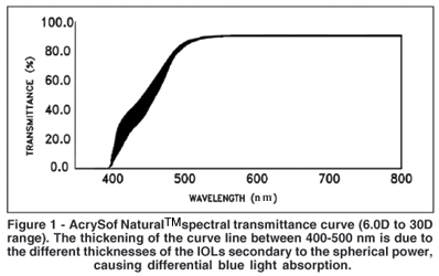
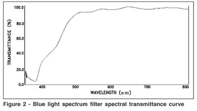
The BLSF was manufactured using an organic resin lens (CR 39, Essilor do Brasil, Manaus, AM, Brazil) colored with a yellow coloration lens tint (BPI Filter Vision 450- U.V. blocker - code BPI#37870, Brain Power Inc., Miami, FL, USA). The spectral transmittance of the BLSF was measured using a spectrometer (S2000, Ocean Optics Inc., Dunedin, FL, USA), and the light transmittance was in compliance with the spectral transmittance curve of the Acrysof NaturalTM IOL (Figures 1 and 2).
Visual field tests were completed within a two-month period. Mean deviation (MD), pattern standard deviation (PSD), and foveal threshold (FT) indexes were analyzed. These analyses were performed using paired t test. In addition, the variance of each point of the SWAP test was analyzed. The two test points located vertically in the blind spot area were excluded from all calculations. Linear regression analysis was performed to estimate age slopes of sensitivity for each test point location. The slopes were then used to calculate age-corrected thresholds. Interindividual variability (standard deviation) then was calculated for each test point location in the SWAP examination. Comparisons between the standard deviations of the test points were made using Pitman's test for correlated samples(6). The difference caused by eccentricity in SWAP (inferior hemifield mean threshold minus superior hemifield mean threshold, and the four most inferior points minus the four most superior points) was also evaluated using the paired t test. In light of the multiplicity of tests performed, we adopted a value of p<0.01 rather than p<0.05 as the criterion of statistical significance.
RESULTS
Five men and fifteen women were included in this study. The mean age of the participants was 24.2 years (SD, 3.8 years; range, 16 to 30 years old).
When comparing SWAP with the BLSF to SWAP without the use of the BLSF, the MD was reduced in all patients (mean, 3.31 dB; 99% confidence interval (CI), 2.46 dB to 4.17 dB; p<0.001), and the FT was reduced in 16 (80%) patients (mean, 2.85 dB; 99% CI, 1.19 dB to 4.51 dB; p<0.001). The PSD difference between these two examinations was not statistically significant (mean, 0.02 dB, 99% CI, -0.26 dB to 0.22 dB; p=0.79; Table 1).
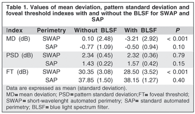
When comparing SAP with and without the BLSF, there were no statistically significant differences regarding MD, FT and PSD indexes (Table 1).
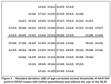
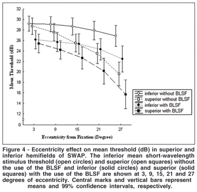
Interindividual standard deviation of the test points in the SWAP examination increased with eccentricity both with and without the use of the BLSF (p<0.01), but no statistically significant difference in the variability between the two examination was noted (p>0.01) (Figure 3).
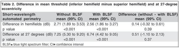
Results of SWAP sensitivity for inferior and superior hemifields showed that the difference in mean threshold (inferior hemifield minus superior hemifield) was 2.71 dB (99% CI, 1.89 dB to 3.53 dB, p<0.001) without using the BLSF and 2.56 dB (99% CI, 1.86 dB to 3.27 dB, p<0.001) with the use of the BLSF. The mean difference of inferior and superior hemifields without and with the use of the BLSF (mean, 0.14 dB; 99% CI, -0.32 to 0.61 dB, p=0.39) was not statistically significant.
When comparing only the four most inferior points and the four most superior points, the inferior-superior difference increases in both situations - without and with the use of the BLSF. The difference between without and with the use of the BLSF did not show statistically significant difference. These results are summarized in table 2.
DISCUSSION
We observed a reduction in FT and MD indexes when comparing SWAP examination with and without the use of the BLSF. These reductions may be explained by the fact that the BLSF reduces the transmission of the blue light stimulus used by SWAP. Nevertheless, the BLSF did not reduce the transmission of the light stimulus emitted by the SAP to the level that could alter the MD and FT indexes in this test.
Although the FT index presented lower sensitivity reduction than the MD index (2.85 dB and 3.31 dB, respectively) in the SWAP tests (with and without using the BLSF), this difference was not statistically significant. The lower sensitivity reduction in FT might be partially explained by the fact the foveal sensitivity is tested first when the patients have a greater attention span and are not visually fatigued(7-8). Wild and Hudson demonstrated that macular pigments are associated with a decrease in the foveal sensitivity that declines to zero at 5.5° of eccentricity(9). Therefore, it is unlikely that macular pigments would have a meaningful influence on the overall results in the SWAP.
The PSD index did not show any difference in either the SWAP or SAP tests. This finding reflects the fact that the reduction of light transmission by the BLSF was homogeneous and generalized. To minimize the influence of a learning-effect bias on the visual field data, we randomized the sequence of the SAP and SWAP with and without using the BLSF.
In SWAP examination, interindividual variability increased with eccentricity, which is in agreement with previous studies(10-11). In addition, the variation was similar with and without the use of the BLSF.
In SWAP examination, an inferior-superior hemifield asymmetry that increased with the eccentricity, both with and without the use of the BLSF was observed (Figure 4). These results are in agreement with a previous study(12). The use of the BLSF did not increase this asymmetry (Table 2).
The participants included in this study were younger than patients that have age-related cataracts. Carotenoid derivatives (luthein and zeaxanthin) are present in the macular region and act to reduce the transmission of blue light to the outer segment of the retina(13). As carotenoid derivatives in the macula are reduced with aging, different results in visual field indexes could be obtained in older patients.
The crystalline lens filters the light that enters the eye(4). In our study, we added another filter (BLSF) that further reduces light transmission. The extent to which the presence of these two light filters (crystalline lens and BLSF) differs from having only BLSF might not be determined by the study design employed here. Thus, the results from this study cannot be generalized to patients that previously underwent cataract extraction with intraocular lens implantation.
CONCLUSION
In summary, the alterations in the SWAP results due to the use of the BLSF, in which the diffuse component is mainly reduced, could lead to a misinterpretation of the results. The SWAP test has been considered an important tool for early identification of glaucomatous damage and has shown good correlation with structural abnormalities of the optic nerve head. Alterations in the SWAP test may reduce its sensitivity for the early diagnosis of glaucoma, since glaucomatous lesions may exhibit diffuse and localized components. Thus, studies involving patients having undergone cataract surgery and Acrysof NaturalTM implantation are necessary to verify the possible changes induced by this IOL in SWAP and SAP test results.
REFERENCES
1. Pollack A, Marcovich A, Bukelman A, Oliver M. Age-related macular degeneration after extracapsular cataract extraction with intraocular lens implantation. Ophthalmology. 1996;103(10):1546-54. Comment in: Ophthalmology. 1997; 104(6):900.
2. Klein R, Klein BE, Wong TY, Tomany SC, Cruickshanks KJ. The association of cataract and cataract surgery with the long-term incidence of age-related maculopathy: the Beaver Dam eye study. Arch Ophthalmol. 2002;120(11):1551-8.
3. Freeman EE, Munoz B, West SK, Tielsch JM, Schein OD. Is there an association between cataract surgery and age-related macular degeneration? Data from three population-based studies. Am J Ophthalmol. 2003;135(6):849-56. Comment in: Am J Ophthalmol. 2003;136(5):961.
4. Wang JJ, Klein R, Smith W, Klein BE, Tomany S, Mitchell P. Cataract surgery and the 5-year incidence of late-stage age-related maculopathy: pooled findings from the Beaver Dam and Blue Mountains eye studies. Ophthalmology. 2003t;110(10):1960-7.
5. Landrum JT, Bone RA, Kilburn MD. The macular pigment: a possible role in protection from age-related macular degeneration. Adv Pharmacol. 1997;38: 537-56.
6. Pitman EG. A note on normal correlation. Biometrika. 1939;31:9-12.
7. Heijl A. Time changes of contrast thresholds during automatic perimetry. Acta Ophthalmol (Copenh). 1977;55(4):696-708.
8. Hudson C, Wild JM, O'Neill EC. Fatigue effects during a single session of automated static threshold perimetry. Invest Ophthalmol Vis Sci. 1994;35(1): 268-80.
9. Wild JM, Hudson C. The attenuation of blue-on-yellow perimetry by the macular pigment. Ophthalmology. 1995;102(6):911-7.
10. Bengtsson B, Heijl A. Normal intersubject threshold variability and normal limits of the SITA SWAP and full threshold SWAP perimetric programs. Invest Ophthalmol Vis Sci. 2003;44(11):5029-34.
11. Wild JM, Cubbidge RP, Pacey IE, Robinson R. Statistical aspects of the normal visual field in short-wavelength automated perimetry. Invest Ophthalmol Vis Sci. 1998;39(1):54-63.
12. Sample PA, Irak I, Martinez GA, Yamagishi N. Asymmetries in the normal short-wavelength visual field: implications for short-wavelength automated perimetry. Am J Ophthalmol. 1997;124(1):46-52.
13. Krinsky NI, Landrum JT, Bone RA. Biologic mechanisms of the protective role of lutein and zeaxanthin in the eye. Annu Rev Nutr. 2003;23:171-201.
14. Teesalu P, Airaksinen PJ, Tuulonen A. Blue-on-yellow visual field and retinal nerve fiber layer in ocular hypertension and glaucoma. Ophthalmology. 1998; 105(11):2077-81.
15. Sample PA, Weinreb RN. Color perimetry for assessment of primary open-angle glaucoma. Invest Ophthalmol Vis Sci. 1990;31(9):1869-75.
16. Johnson CA, Adams AJ, Casson EJ, Brandt JD. Blue-on-yellow perimetry can predict the development of glaucomatous visual field loss. Arch Ophthalmol. 1993;111(5):645-50.
17. Johnson CA, Adams AJ, Casson EJ, Brandt JD. Progression of early glaucomatous visual field loss as detected by blue-on-yellow and standard white-on-white automated perimetry. Arch Ophthalmol. 1993;111(5):651-6.
 Address to correspondence:
Leonardo Cunha Castro
Rua Borges Lagoa, 1209/2201
São Paulo (SP)
Cep 04038-033
E-mail: [email protected]
Recebido para publicação em 25.10.2005
Última versão recebida em 03.02.2006
Aprovação em 27.02.2006
Department of Ophthalmology, Vision Institute, Universidade Federal de São Paulo - São Paulo, Brazil.

How to cite this article:
 ABO is licensed under a Creative Commons Attribution-NonComercial 4.0 Internacional.