Arq. Bras. Oftalmol. 2021;84 (5 )
:436-441
| DOI: 10.5935/0004-2749.20210068
Abstract
Objetivo: A Escala Bayley de Desenvolvimento Infantil (Bayley-III) é uma ferramenta que avalia o desenvolvimento de crianças nos 3 primeiros anos de vida, incluindo os domínios cognitivo e motor. Este estudo tem como objetivo correlacionar a acuidade visual de grades e a funcionalidade visual em crianças saudáveis usando a Bayley-III.
Métodos: A acuidade visual binocular de grades foi medida usando o teste dos Cartões de Acuidade de Teller seguido pela Bayley-III em crianças saudáveis com idade entre 1-42 meses. Os escores da acuidade visual (logMAR) e da Bayley-III para habilidades cognitivas e motoras (grossa e fina) foram comparados.
Resultados: Um grupo de 40 crianças (20 meninos) com idades entre 1,2-42,1 meses foi testado e a média da acuidade visual foi de 0,39 ± 0,27 logMAR, sendo que todas estavam dentro dos limites normais para a idade. Houve uma forte correlação negativa e significante entre acuidade visual e idade (r=-0,83; p<0,001). A média do escore cognitivo foi de 49,92 ± 18,93 pontos, com forte correlação positiva e significante entre o escore cognitivo e a idade (r=0,81; p<0,001). A média do escore motor grosso foi de 41,72 ± 16,23 pontos, com forte correlação positiva e significante entre o escore motor grosso e a idade (r=0,75; p<0,001). A média do escore motor fino foi de 39,75 ± 14,63 pontos, com uma forte correlação positiva e significante entre o escore motor fino e a idade (r=0,77; p<0,001). A regressão linear múltipla mostrou que maior idade e melhor acuidade visual foram significantemente associadas à escores cognitivo e motor mais altos.
Conclusões: Neste estudo foi encontrada alta correlação entre a acuidade visual de grades medida pelos cartões de acuidade de Teller e os escores cogninitivo e motor medidos pela Bayley-III em crianças saudáveis. A Bayley-III pode ser uma ferramenta útil para avaliar a repercussão da deficiência visual no desenvolvimento cognitivo e motor de crianças.
Keywords: Desenvolvimento infantil; Acuidade visual; Cognição; Destreza motora; Transtornos da visão; Testes neuropsicológicos; Criança
Arq. Bras. Oftalmol. 2025;88 (6 )
:1-9
| DOI: 10.5935/0004-2749.2024-0411
Abstract
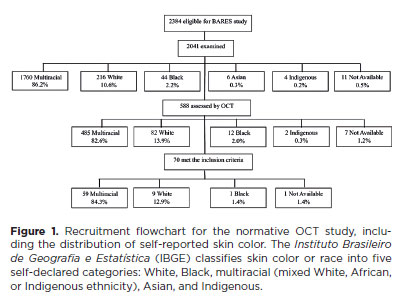
PURPOSE: This study evaluated macular thickness using spectral-domain optical coherence tomography in healthy participants from a population-based eye survey.
METHODS: The Brazilian Amazon Region Eye Survey was a population-based study assessing the prevalence and causes of visual impairment, blindness, and ocular diseases in adults aged ≥45 years from urban and rural areas of Parintins. A subgroup was selected based on inclusion criteria for both eyes: best-corrected visual acuity ≥20/32, normal eye examination results, and no prior ocular surgery. Scans were performed using the iVue optical coherence tomography device. Measurements were taken from the nine subfields defined by the Early Treatment Diabetic Retinopathy Study, examining the full retina as well as the inner and outer retinal layers. Associations of retinal thickness with age and sex were also analyzed. Statistical significance was set at p≤0.05.
RESULTS: In total, 70 healthy participants (25 males), aged 45–65 years (mean=52 ± 5), were included. Mean central foveal thickness was 248.71 ± 18.73 μm. A significant age-related reduction in macular thickness was observed, particularly in the inner superior parafovea (p=0.036), nasal perifovea (p=0.001), superior perifovea (p=0.028), outer layer of inferior parafovea (p=0.049), and the inferior perifovea of the full retina (p=0.029). Males showed significantly greater thickness in the outer layer, especially in the outer parafovea (p=0.004) and perifovea (p<0.0001).
CONCLUSIONS: This study established normative macular thickness values for healthy older adults in the Brazilian Amazon region using spectral-domain optical coherence tomography. Age and sex were found to significantly influence macular thickness and should be considered when interpreting measurements. These data will support future studies of retinal diseases in this population.
Keywords: Retinal diseases/diagnosis; Macula lutea/pathology; Macular degeneration/diagnosis; Diabetic retinopathy/diagnosis; Vision, low; Vision tests; Tomography, optical coherence/methods; Young adult; Cross-sectional studies; Brazil/epidemiology
Arq. Bras. Oftalmol. 2025;88 (6 )
:1-8
| DOI: 10.5935/0004-2749.2025-0053
Abstract
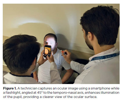
PURPOSE: This pilot study evaluated the diagnostic accuracy of a deep learning model for detecting pterygium in anterior segment photographs taken using smartphones in the Brazilian Amazon. The model’s performance was benchmarked against assessments made by experienced ophthalmologists, considered the clinical gold standard.
METHODS: In this cross-sectional study, 38 participants (76 eyes) from Barcelos, Brazil, were enrolled. Trained nonmedical health workers captured high-resolution anterior segment images using smartphones. These images were analyzed using a deep learning model based on the MobileNet-V2 convolutional neural network. Diagnostic metrics–including sensitivity, specificity, accuracy, positive predictive value, negative predictive value, and area under the receiver operating characteristic curve–were calculated and compared with the ophthalmologists’ evaluations.
RESULTS: The deep learning model achieved a sensitivity of 91.43%, specificity of 90.24%, positive predictive value of 88.46%, negative predictive value of 92.79%, and an area under the curve of 0.91. Logistic regression revealed no statistically significant association between pterygium and demographic variables such as age or gender.
CONCLUSIONS: The deep learning model demonstrated high diagnostic performance in identifying pterygium in a remote Amazonian population. These preliminary findings support the potential use of artificial intelligence–based tools to facilitate early detection and screening in underserved regions, thereby enhancing access to ophthalmic care.
Keywords: Pterygium/diagnostic imaging; Smartphone; Diagnostic techniques, ophthalmological; Deep learning; Telemedicine; Artificial intelligence; Cross-sectional studies; Brazil/epidemiology
 ABO is licensed under a Creative Commons Attribution-NonComercial 4.0 Internacional.
03-tab01.jpg)